

Toward in vivo two-photon analysis of mouse aqueous outflow structure and function
- PMID: 27179411
- PMCID: PMC5403624
- DOI: 10.1016/j.exer.2016.05.009
Toward in vivo two-photon analysis of mouse aqueous outflow structure and function
Abstract
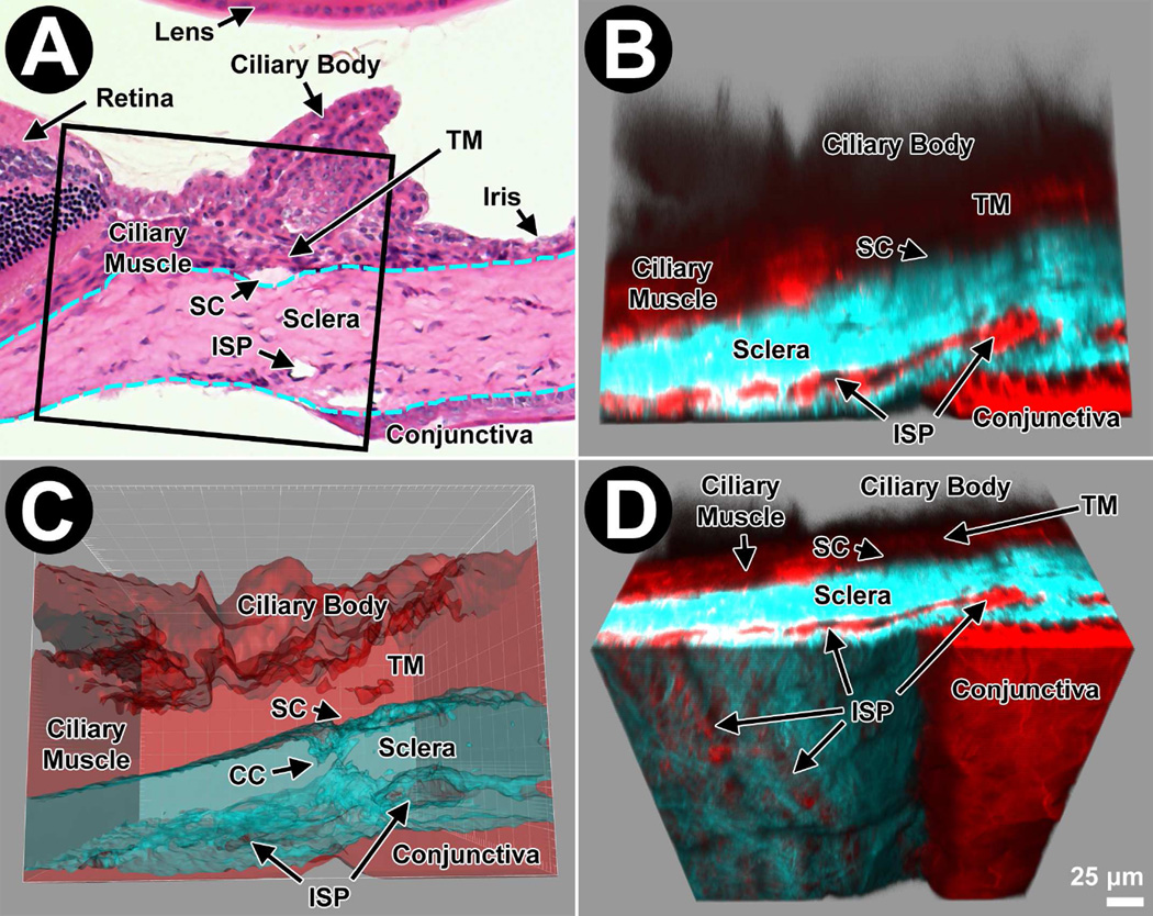
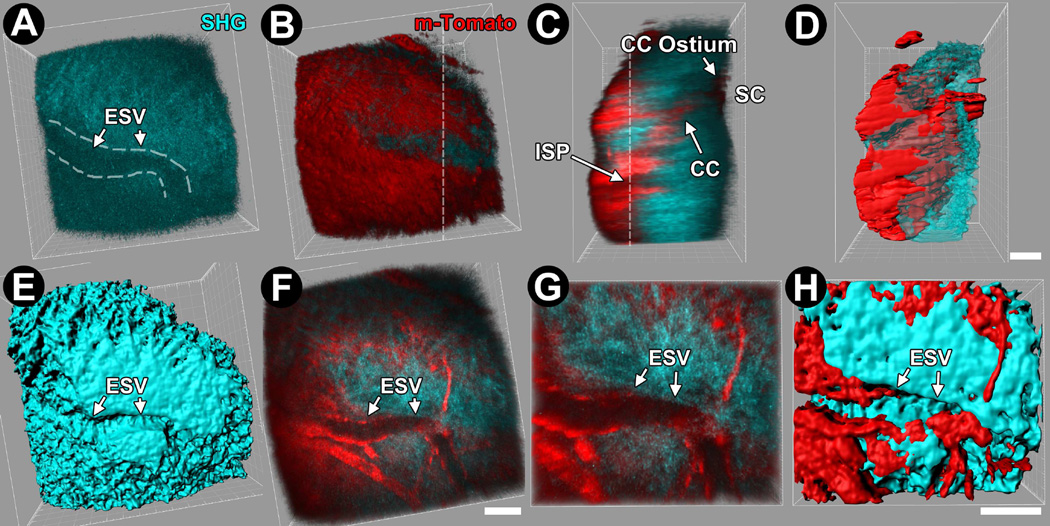
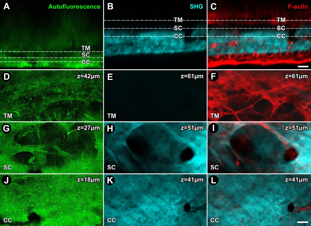
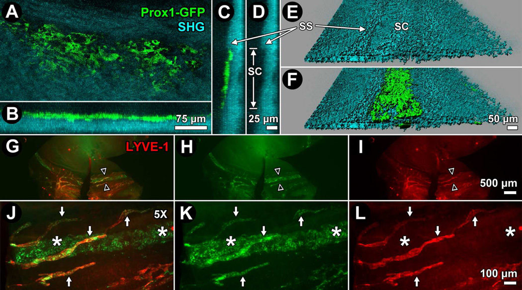
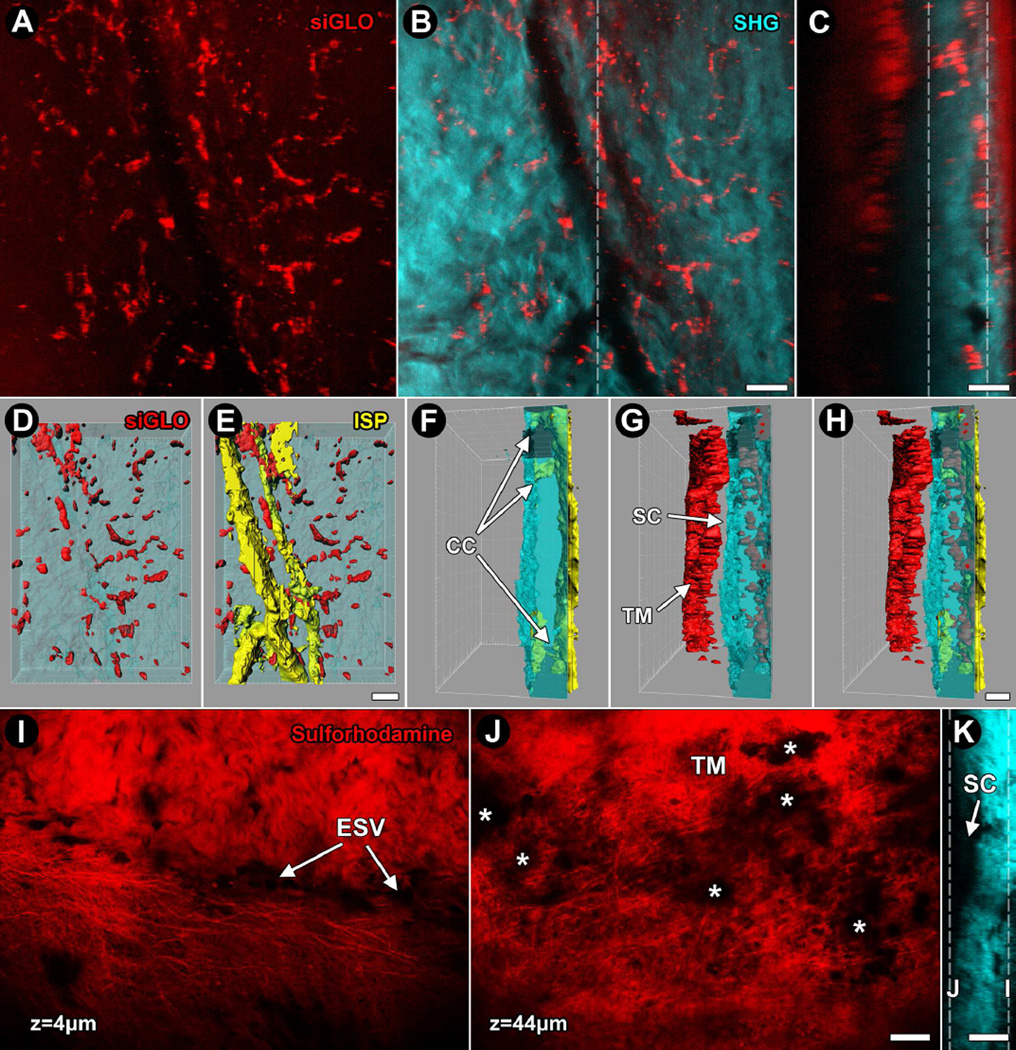
The promise of revolutionary insights into intraocular pressure (IOP) and aqueous humor outflow homeostasis, IOP pathogenesis, and novel therapy offered by engineered mouse models has been hindered by a lack of appropriate tools for studying the aqueous drainage tissues in their original 3-dimensional (3D) environment. Advances in 2-photon excitation fluorescence imaging (TPEF) combined with availability of modalities such as transgenic reporter mice and intravital dyes have placed us on the cusp of unlocking the potential of the mouse model for unearthing insights into aqueous drainage structure and function. Multimodality 2-photon imaging permits high-resolution visualization not only of tissue structural organization but also cells and cellular function. It is possible to dig deeper into understanding the cellular basis of aqueous outflow regulation as the technique integrates analysis of tissue structure, cell biology and physiology in a way that could also lead to fresh insights into human glaucoma. We outline recent novel applications of two-photon imaging to analyze the mouse conventional drainage system in vivo or in whole tissues: (1) collagen second harmonic generation (SHG) identifies the locations of episcleral vessels, intrascleral plexuses, collector channels, and Schlemm's canal in the distal aqueous drainage tract; (2) the prospero homeobox protein 1-green fluorescent protein (GFP) reporter helps locate the inner wall of Schlemm's canal; (3) Calcein AM, siGLO™, the fluorescent reporters m-Tomato and GFP, and coherent anti-Stokes scattering (CARS), are adjuncts to TPEF to identify live cells by their membrane or cytosolic locations; (4) autofluorescence and sulforhodamine-B to identify elastic fibers in the living eye. These tools greatly expand our options for analyzing physiological and pathological processes in the aqueous drainage tissues of live mice as a model of the analogous human system.
Keywords: Aqueous humor outflow; Collector channel; Conventional outflow; Glaucoma; Multiphoton microscopy; Schlemm’s canal; Second harmonic generation.
Copyright © 2016 Elsevier Ltd. All rights reserved.
Conflict of interest statement
Conflict of interest
The authors do not have conflicting relationships to report.
Figures
Similar articles
-
Collector Channels: Role and Evaluation in Schlemm's Canal Surgery.Curr Eye Res. 2020 Oct;45(10):1181-1187. doi: 10.1080/02713683.2020.1773866. Epub 2020 Jun 1. Curr Eye Res. 2020. PMID: 32449380 Review.
-
[Imaging of the Intrascleral Aqueous Drainage System - New Insights for Glaucoma Surgery Targeting the Trabecular Meshwork].Klin Monbl Augenheilkd. 2018 Mar;235(3):309-314. doi: 10.1055/s-0042-123193. Epub 2017 Feb 10. Klin Monbl Augenheilkd. 2018. PMID: 28187473 Review. German.
-
In-vivo imaging of the conventional aqueous outflow system.Curr Opin Ophthalmol. 2021 May 1;32(3):275-279. doi: 10.1097/ICU.0000000000000751. Curr Opin Ophthalmol. 2021. PMID: 33653980 Review.
-
Aqueous outflow regulation: Optical coherence tomography implicates pressure-dependent tissue motion.Exp Eye Res. 2017 May;158:171-186. doi: 10.1016/j.exer.2016.06.007. Epub 2016 Jun 11. Exp Eye Res. 2017. PMID: 27302601 Free PMC article. Review.
-
Detailed 3D micro-modeling of rat aqueous drainage channels based on two-photon imaging: simulating aqueous humor through trabecular meshwork and Schlemm's canal by two-way fluid structure interaction approach.Med Biol Eng Comput. 2022 Jul;60(7):1915-1927. doi: 10.1007/s11517-022-02580-6. Epub 2022 May 6. Med Biol Eng Comput. 2022. PMID: 35524088
Cited by
-
Fibrillin-1 mutant mouse captures defining features of human primary open glaucoma including anomalous aqueous humor TGF beta-2.Sci Rep. 2022 Jun 23;12(1):10623. doi: 10.1038/s41598-022-14062-8. Sci Rep. 2022. PMID: 35739142 Free PMC article.
-
Deep tissue analysis of distal aqueous drainage structures and contractile features.Sci Rep. 2017 Dec 6;7(1):17071. doi: 10.1038/s41598-017-16897-y. Sci Rep. 2017. PMID: 29213129 Free PMC article.
References
Publication types
MeSH terms
Substances
Grants and funding
LinkOut - more resources
Full Text Sources
Other Literature Sources
Medical
Research Materials
