. 2016 May 14:35:80.
doi: 10.1186/s13046-016-0360-9.

Transfer of malignant trait to BRCA1 deficient human fibroblasts following exposure to serum of cancer patients

Affiliations

Transfer of malignant trait to BRCA1 deficient human fibroblasts following exposure to serum of cancer patients

Dana Hamam et al. J Exp Clin Cancer Res. .

Abstract

Background: It was reported that metastases might occur via transfer of biologically active blood circulating molecules from the primary tumor to distant organs rather than only migration of cancer cells. We showed in an earlier study that exposure of immortalized human embryonic kidney cells (HEK 293) to cancer patient sera, induce their transformation into undifferentiated cancers due to a horizontal transfer of malignant traits. In the present work, we tested the hypothesis that even other human cells as long as they are deficient for a single oncosuppressor gene might undergo malignant transformation when exposed to human cancer serum.

Methods: We used the CRISPR/Cas9 system to establish a stable BRCA1 knockout (KO) in human fibroblasts. The BRCA1-KO fibroblasts were exposed to cancer patients' sera or healthy patients' sera for 2 weeks. Treated cells were analyzed for cell proliferation and transformation to study their susceptibility to the oncogenic potential of cancer patients' sera and to determine the possible mechanisms underlying their hypothesized transformation.

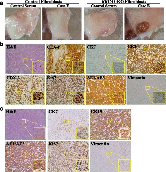
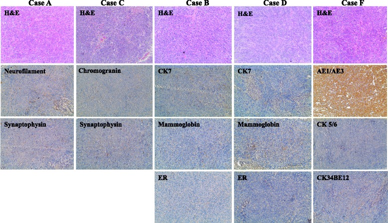
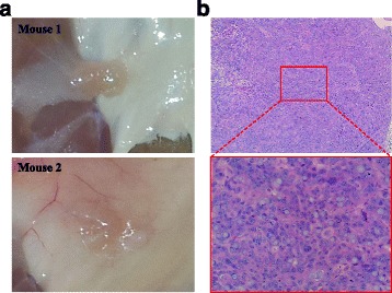
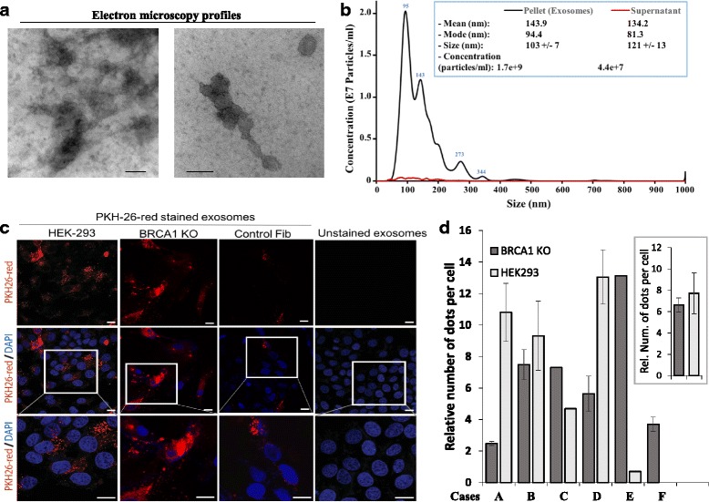
Results: BRCA1-KO fibroblasts treated with cancer patients' sera displayed higher proliferation and underwent malignant transformation as opposed to wild type control fibroblasts, which were not affected by exposure to cancer patients' sera. The malignant transformation was not seen when BRCA1-KO fibroblasts were treated with healthy human sera. Histological analysis of tumors generated by BRCA1-KO fibroblasts showed that they were carcinomas with phenotypical characteristics related to the cancers of the blood donor patients. Interestingly, BRCA1-KO fibroblasts were significantly more prone to internalize serum-derived exosomes, when compared to wild type fibroblasts. This suggests that oncosuppressor genes might protect the integrity of the cell genome also by blocking integration of cancer-derived exosomes.

Conclusion: These data support the hypothesis that any human cells carrying a single oncosuppressor mutation is capable of integrating cancer factors carried in the blood and undergo complete malignant transformation. Oncosuppressor genes might protect the cell genome by impeding the integration inside the cells of these mutating factors.

Keywords: BRCA1; Exosomes; Fibroblasts; Genometastasis; Metastasis; Transformation; Tumor suppressor genes.


Figures

Fig. 1
Fig. 1
Cancer patient sera increased BRCA1-KO fibroblasts growth. Cells were cultured for 2 weeks in control human serum, or cancer patient sera. Cell growth was then analyzed by counting the number of viable cells at every passage (5 days duration for every passage). a Line graph shows the population doublings capability and (b) column graph represents cumulative population doublings at the end of the treatment periods. Data are mean ± SD of 2 control sera vs. 6 cancer patient sera. (P = 0.027)
Fig. 2
Fig. 2
Cancer patient sera induced the transformation of BRCA1-KO fibroblasts. a NOD/SCID mice were injected with either control or BRCA1-KO fibroblasts, that were treated for 2 weeks with healthy individual (Control) or cancer patient sera. 4 weeks after cells transplantation, mice were euthanized and the tumors photographed. Representative pictures are shown. Tumor masses were only observed when mice were injected with BRCA1-KO fibroblasts treated with cancer patient sera. (n = 3 mice per group). (b and c) Formalin-fixed paraffin-embedded tumors generated following injection of BRCA1-KO fibroblasts treated with CRC-LM patient serum (b: Case E) or pancreatic cancer patient serum (c: Case J) were processed for H&E staining, or immunolabeled with antibodies against tumor specific markers
Fig. 3
Fig. 3
Tumors generated with cancer patient sera-treated BRCA1-KO did not all display differentiation characteristics. BRCA1-KO fibroblasts were treated with cancer patient sera for 2 weeks (Cases A, B, C, D, and F). Treated cells were injected into NOD/SCID mice that were followed for 4 weeks for tumors growth. Developing tumors were excised and photographed (See Additional file 4: Figure S3). Formalin-fixed paraffin-embedded xenotransplant samples were processed for H&E staining, or immunolabeled with antibodies against tumor specific markers
Fig. 4
Fig. 4
Cancer patient sera permanently transformed BRCA1-KO fibroblasts. a BRCA1-KO fibroblasts were treated with cancer patient serum for 2 weeks. Cells were then transferred to regular culture medium (10 % FBS-supplemented DMEM-F12 medium with 1 % antibiotics) for 2 weeks. Cells were injected subcutaneously into NOD/SCID mice. 4 weeks after cells transplantation, mice were euthanized and the tumors photographed. Representative pictures are shown. b Formalin-fixed paraffin-embedded xenotransplants samples were processed for H&E staining
Fig. 5
Fig. 5
BRCA1-KO fibroblasts internalized exosomes more efficiently than control cells. a Exosomes were isolated as described under Methods. Representative micrographs of transmission electron microscopy on cancer patient sera exosome preparations. The image showed small vesicles of approximately 50–120 nm in diameter. Scale bars 100 nm. b NanoSight analysis of samples prepared as in (a). The size was centered around 94 nm in diameter. c Confocal microscopy monitoring of PKH-26-labeled exosome uptake in vitro into BRCA1-KO fibroblasts, control fibroblasts, and HEK293 cells. Note that exosomes internalized more in BRCA1-KO cells. They were dispersed in the cytoplasm and tended to form aggregates in the perinuclear regions. d The number of PKH-26-labeled exosomes (dots) was counted. Data are expressed as relative number (Rel. Num.) of exosomes per cell compared to that in control fibroblasts. In the insert, data are expressed as mean +/− SD (n = 6 exosome preparations; P = 0.032 for HEK293 cells compared to control fibroblasts, P = 0.028 for BRCA1-KO fibroblasts compared to control fibroblasts, and P = 0.35 for HEK293 cells compared to BRCA1-KO fibroblasts)

References

    1. Eccles SA, Welch DR. Metastasis: recent discoveries and novel treatment strategies. Lancet. 2007;369(9574):1742–57. doi: 10.1016/S0140-6736(07)60781-8. - DOI - PMC - PubMed
    1. Jemal A, Bray F, Center MM, Ferlay J, Ward E, Forman D. Global cancer statistics. CA Cancer J Clin. 2011;61:69–90. doi: 10.3322/caac.20107. - DOI - PubMed
    1. Nguyen DX, Bos PD, Massagué J. Metastasis: from dissemination to organ specific colonization. Nat Rev Cancer. 2009;9:274–84. doi: 10.1038/nrc2622. - DOI - PubMed
    1. Skog J, Wurdinger T, van Rijn S. Glioblastoma microvesicles transport RNA and proteins that promote tumour growth and provide diagnostic biomarkers. Nat Cell Biol. 2008;10:1470–81. doi: 10.1038/ncb1800. - DOI - PMC - PubMed
    1. Hood JL, San RS, Wickline SA. Exosomes released by melanoma cells prepare sentinel lymph nodes for tumor metastasis. Cancer Res. 2011;71:3792–801. doi: 10.1158/0008-5472.CAN-10-4455. - DOI - PubMed

Publication types