. 2016 May 14;14(1):37.
doi: 10.1186/s12951-016-0190-0.

Iron oxide nanoparticles for neuronal cell applications: uptake study and magnetic manipulations

Affiliations

Iron oxide nanoparticles for neuronal cell applications: uptake study and magnetic manipulations

Michal Marcus et al. J Nanobiotechnology. .

Abstract

Background: The ability to direct and manipulate neuronal cells has important potential in therapeutics and neural network studies. An emerging approach for remotely guiding cells is by incorporating magnetic nanoparticles (MNPs) into cells and transferring the cells into magnetic sensitive units. Recent developments offer exciting possibilities of magnetic manipulations of MNPs-loaded cells by external magnetic fields. In the present study, we evaluated and characterized uptake properties for optimal loading of cells by MNPs. We examined the interactions between MNPs of different cores and coatings, with primary neurons and neuron-like cells.

Results: We found that uncoated-maghemite iron oxide nanoparticles maximally interact and penetrate into cells with no cytotoxic effect. We observed that the cellular uptake of the MNPs depends on the time of incubation and the concentration of nanoparticles in the medium. The morphology patterns of the neuronal cells were not affected by MNPs uptake and neurons remained electrically active. We theoretically modeled magnetic fluxes and demonstrated experimentally the response of MNP-loaded cells to the magnetic fields affecting cell motility. Furthermore, we successfully directed neurite growth orientation along regeneration.

Conclusions: Applying mechanical forces via magnetic mediators is a useful approach for biomedical applications. We have examined several types of MNPs and studied the uptake behavior optimized for magnetic neuronal manipulations.

Keywords: Cell positioning; Guidance; Magnetic field; Magnetic nanoparticles; Neuronal cells; Neuronal regeneration; Uptake.


Figures
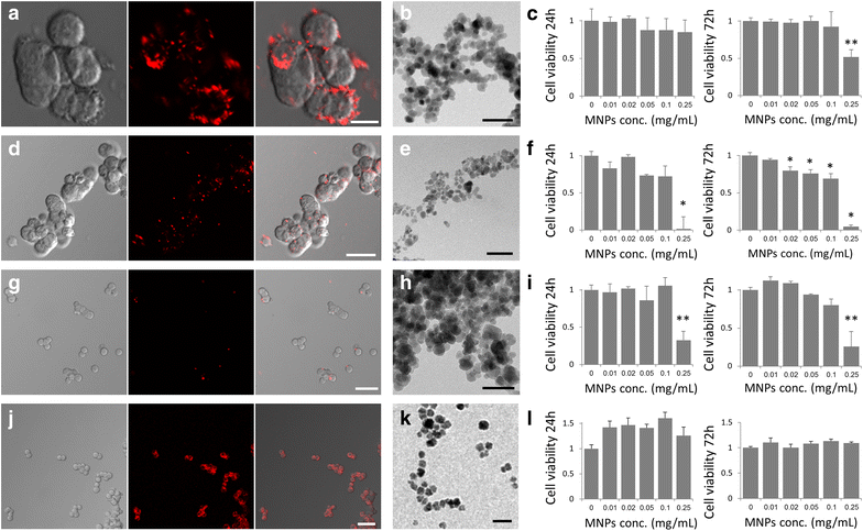
Fig. 1
Fig. 1
Characterization of the interactions of magnetic nanoparticles with PC12 cells: ac uncoated-magnetite MNPs, d–f starch-magnetite MNPs, gi dextran-magnetite MNPs, jl uncoated-maghemite MNPs. Left panel: Confocal images of PC12 cells incubated with MNPs. Scale bar = 10, 25, 50 and 50 µm, respectively. Middle panel: TEM images of particles. Scale bar = 50 nm. Right panel: Cytotoxicity assay of cells incubated with increasing concentrations of MNPs after 24 and 72 h of incubation (n = 3). T test, *p < 0.05 and **p < 0.01
Fig. 2
Fig. 2
Confocal microscopy images of PC12 cells at a single focal plane after 24 h of incubation with the fluorescent uncoated-maghemite MNPs. MNPs labeled with rhodamine (red) enter the cells. a Phase contrast image. b Fluorescent image. c Merged image. Scale bar = 10 µm
Fig. 3
Fig. 3
Cellular uptake of MNPs by PC12 cells; a Fluorescence intensity measurements from FACS of PC12 cells incubated with MNPs for 1, 2, 3 and 24 h. c Fluorescence intensity measurements from FACS of PC12 cells incubated with MNPs, ranging from 0.01 mg/ml to 0.5 mg/ml, for 24 h. b, d Average of fluorescence intensity normalized to control upon incubation of cells with MNPs. Reported values are an average of measurements (n = 3) of approximately 10,000 cells in each triplicate tested sample
Fig. 4
Fig. 4
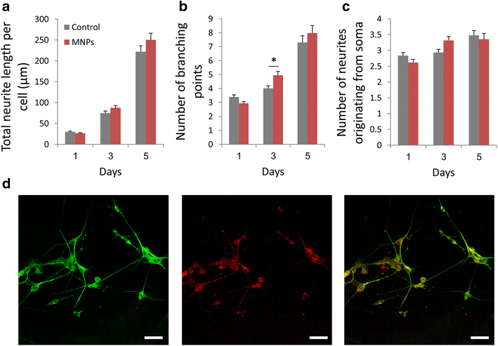
Morphological analysis of PC12 cells incubated with uncoated-maghemite MNPs (0.25 mg/ml) 1, 3 and 5 days after NGF treatment; a Total neurite length per cell; b Number of branching points; c Number of neurites originating from soma. T test, *p < 0.05. d Fluorescent confocal images of MNP-loaded cells after 5 days of NGF treatment; Left image: α-tubulin immunostaining of cells. Middle image: fluorescent MNPs uptaken by the cells. Right image: merged image. Scale bar = 50 µm
Fig. 5
Fig. 5
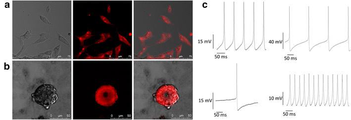
a Confocal microscopy images of SH-SY5Y cells at a single focal plane after 24 h of incubation with uncoated-maghemite MNPs. MNPs labeled with rhodamine (red) enter the cells; b Confocal microscopy images of primary leech neuron at a single focal plane after 24 h of incubation with the fluorescent MNPs. c Electrophysiological measurements of primary leech neurons in culture
Fig. 6
Fig. 6
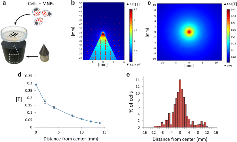
Magnetic positioning of PC12 cells. a Schematic illustration of the magnetic manipulation. Cells were incubated with MNPs and seeded on a plate placed above a Hiperco 50A magnetic tip. b Simulation of magnetic field lines and intensity in Comsol software. The image presents a side view of magnetic flux density of tip. Red arrows represent field direction, intensity is color coded (low intensity in dark blue, high intensity in red). c Top view simulation of magnetic flux density 0.9 mm above magnetic tip (thickness of plastic plate culture). d Actual measurements of magnetic field produced by single tip. Error bars represent standard deviation (n = 3). e Graph representing the cell distribution throughout the plate
Fig. 7
Fig. 7
Positioning of PC12 cells using two magnetic tips. a Simulation of magnetic field intensity in Comsol software. b Actual measurements of magnetic field produced by two tips. Error bars represent standard deviation (n = 3). c Distribution of the cells throughout the plate culture
Fig. 8
Fig. 8
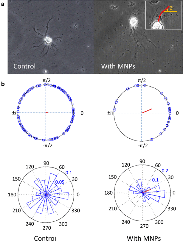
a Phase contrast images of leech neurons in culture in the presence of a magnetic field, either with MNPs or without (control). The neurites were traced and the direction of their tips was measured by calculating their angle relative to the direction of the magnetic field (inset). b Analysis of the direction of neurites outgrowth in the absence (control) or in the presence of MNPs, under an external magnetic field. The resultant vector length (marked in red) in the control group is 0.074 and the resultant vector length in the experiment group is 0.386 (p = 0.116, n = 312 and p = 0.009, n = 29, respectively, according to Rayleigh test for uniformity)

References

    1. Nayak MS, Kim Y-S, Goldman M, Keirstead HS, Kerr DA. Cellular therapies in motor neuron diseases. Biochim Biophys Acta. 2006;1762:1128–1138. doi: 10.1016/j.bbadis.2006.06.004. - DOI - PubMed
    1. Cores J, Caranasos TG, Cheng K. Magnetically targeted stem cell delivery for regenerative medicine. J Funct Biomater. 2015;6:526–546. doi: 10.3390/jfb6030526. - DOI - PMC - PubMed
    1. Kilgus C, Heidsieck A, Ottersbach A, Roell W, Trueck C, Fleischmann BK, Gleich B, Sasse P. Local gene targeting and cell positioning using magnetic nanoparticles and magnetic tips: comparison of mathematical simulations with experiments. Pharm Res. 2012;29:1380–1391. doi: 10.1007/s11095-011-0647-7. - DOI - PubMed
    1. Slaughter GE, Bieberich E, Wnek GE, Wynne KJ, Guiseppi-Elie A. Improving neuron-to-electrode surface attachment via alkanethiol self-assembly: an alternating current impedance study. Langmuir. 2004;20:7189–7200. doi: 10.1021/la049192s. - DOI - PubMed
    1. Jungblut M, Knoll W, Thielemann C, Pottek M. Triangular neuronal networks on microelectrode arrays: an approach to improve the properties of low-density networks for extracellular recording. Biomed Microdevices. 2009;11:1269–1278. doi: 10.1007/s10544-009-9346-0. - DOI - PMC - PubMed

MeSH terms