Review
. 2016;33(3):272-321.
doi: 10.14573/altex.1603161. Epub 2016 May 15.

Biology-inspired microphysiological system approaches to solve the prediction dilemma of substance testing

Affiliations
Review

Biology-inspired microphysiological system approaches to solve the prediction dilemma of substance testing

Uwe Marx et al. ALTEX. 2016.

Abstract

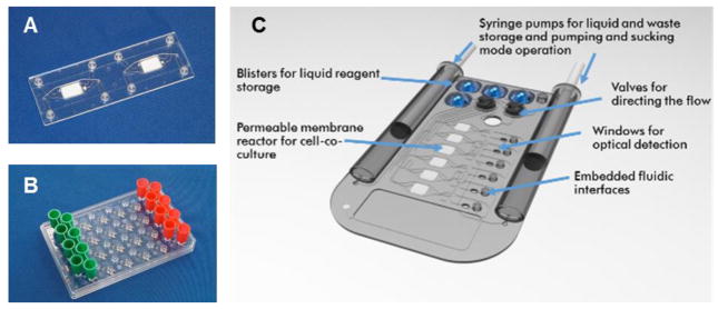
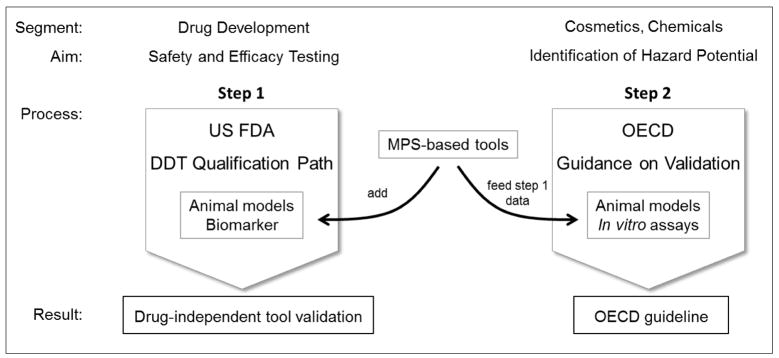
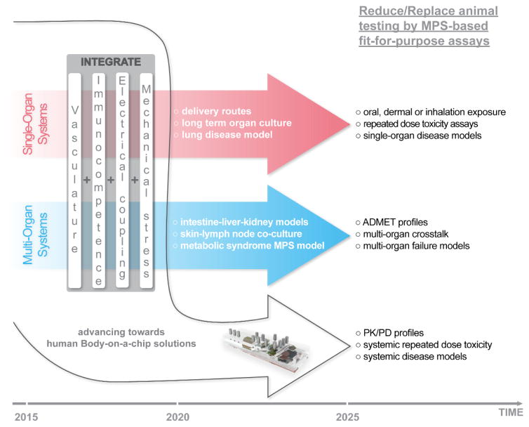
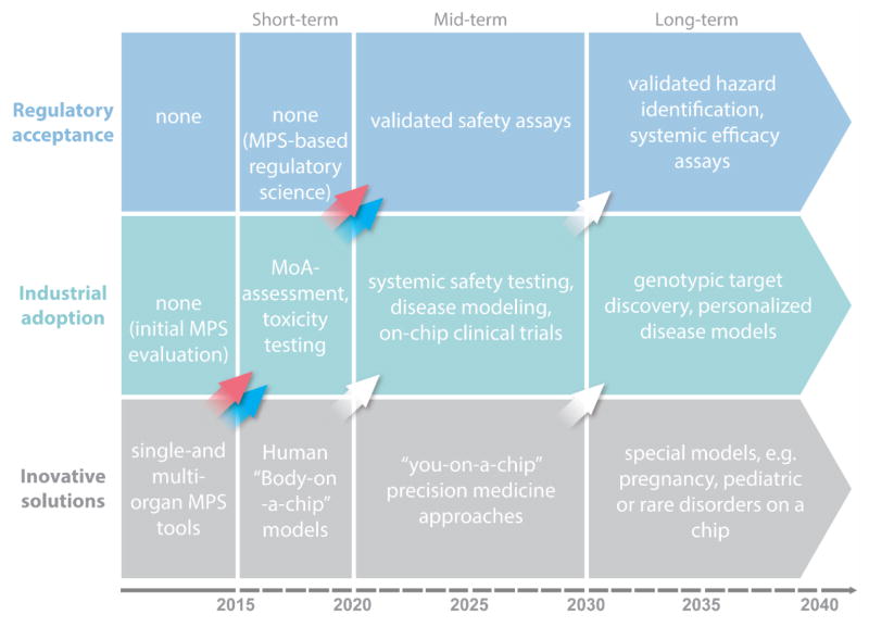
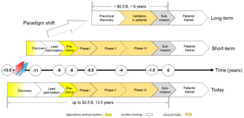
The recent advent of microphysiological systems - microfluidic biomimetic devices that aspire to emulate the biology of human tissues, organs and circulation in vitro - is envisaged to enable a global paradigm shift in drug development. An extraordinary US governmental initiative and various dedicated research programs in Europe and Asia have led recently to the first cutting-edge achievements of human single-organ and multi-organ engineering based on microphysiological systems. The expectation is that test systems established on this basis would model various disease stages, and predict toxicity, immunogenicity, ADME profiles and treatment efficacy prior to clinical testing. Consequently, this technology could significantly affect the way drug substances are developed in the future. Furthermore, microphysiological system-based assays may revolutionize our current global programs of prioritization of hazard characterization for any new substances to be used, for example, in agriculture, food, ecosystems or cosmetics, thus, replacing laboratory animal models used currently. Thirty-six experts from academia, industry and regulatory bodies present here the results of an intensive workshop (held in June 2015, Berlin, Germany). They review the status quo of microphysiological systems available today against industry needs, and assess the broad variety of approaches with fit-for-purpose potential in the drug development cycle. Feasible technical solutions to reach the next levels of human biology in vitro are proposed. Furthermore, key organ-on-a-chip case studies, as well as various national and international programs are highlighted. Finally, a roadmap into the future is outlined, to allow for more predictive and regulatory-accepted substance testing on a global scale.

Keywords: drug testing; in vitro models; microphysiological systems; organ-on-a-chip; predictive toxicology.


Conflict of interest statement

Conflict of Interest Statement

Uwe Marx is founder of TissUse GmbH which commercializes MPS platforms.

Thomas Hartung is cofounder of Organome LLC which aims to make mini-brains and other organotypic organ models commercially available.

Figures

Fig. 1
Fig. 1. Changes in drug development over the last seventy years
Number of drugs approved by the U.S. FDA (FDA, 2013; FDA, 2014a) are plotted against the pharmaceutical research and development spending of the members of the Pharmaceutical Research and Manufacturers of America (PhARMA, 2015). (lightening) – drug or substance failures with detrimental outcome for humans (see Table 1) (brackets) – average costs to develop one new drug including costs of failures within the corresponding decade
Fig. 2
Fig. 2. Drug development cycle: test throughput and cost profile
The vertical axis illustrates approximate numbers of tests performed (grey) and related spending (blue). The horizontal axis illustrates the development time in years (Paul et al., 2010). Animal tests are used in early discovery for mechanistic mode of action research and for toxicity and ADME profiling in the preclinical phase, while conventional in vitro assays are largely used in discovery for target validation, target-to-lead translation and lead optimization steps.
Fig. 3
Fig. 3. Types of MPS used for emulation of human biology in vitro
The top left figure shows the MIMETAS OrganoPlate for 3D perfused cell culture in microtiter format, the middle left shows the Lung-on-a-Chip that was developed by the Wyss institute, top centre depicts hanging drop microtiter plate by ETH Basel with microfluidic channel connecting multiple spheroids, Centre bottom shows an artist’s impression of the four organ system developed by TissUse, Right shows artist’s impressions of a human Body-on-a-chip platform. (Courtesy of MIMETAS, The Netherlands; Wyss Institute, USA; ETH, Switzerland; and TissUse GmbH, Germany, respectively.)
Fig. 4
Fig. 4. Human cell sources for in vitro formation of organoids at a glance
The expansion potential of different human cell and tissue sources used in MPS are plotted against their appearance in the human life-span (Grey arrows) – indicate differentiation potential of the respective stem cell pool (Red arrow) – illustrates induction of pluripotency using primary cells (Blue arrow) – highlights unlimited expansion potential of immortalized cell lines
Fig. 5
Fig. 5. Single-organ microcassette bioreactor device with hollow-fiber-based perfusion
Integration of electromechanically controlled peristaltic pumps (A). Controller for ten microcassettes (B). Micro-bioreactor in operation in an incubator for temperature, humidity and CO2 control (C). (Courtesy of ProBioGen AG, Germany.)
Fig. 6
Fig. 6. Biology-inspired increasing complexity of MPS-based liver models
Inspired by the lobulus architecture of human liver lobules, MPS have evolved from bile canaliculi forming cord-like liver cultures (A) through sinusoid-like arrangements supporting functional space Disse structures (B) towards tissue slice cultures maintaining functional organoid liver structures (C).
Fig. 7
Fig. 7. Biology-inspired Microfluidic alveolar models applying mechanical stretch
Design of a lung-on-a-chip system. (A) Cross-sectional view of the device in the native and stretched state. Human alveolar epithelial cells are cultivated on the top of the membrane and human pulmonary microvascular endothelial cells are cultivated on the bottom of the membrane. (B) View of the lung showing the stretch and resulting distribution of air during inhalation. Applying a cyclic vacuum through the side chambers causes the cell layer to stretch, mimicking the natural stretching during inhalation. Reprinted from Huh et al. (2010).
Fig. 8
Fig. 8. A prime example of a plate-based multi-organ system
(A) A 96-well format multi-tissue interaction testing chip (close-up shows spheroid compartment with loading port). Ten parallel microfluidic channels interconnect six culturing compartments, in which spheroids of different types can be loaded. (B) Platform operated in a standard incubator tilting the chips back and forth producing a gravity-induced flow between the different culturing compartments. (Courtesy of ETH, Switzerland.)
Fig. 9
Fig. 9. Scheme of the flow diagram of a μCCA
The chip is 25 by 25 mm and flow channels are 20 – 100 μm wide. Flow is laminar and typically more than 10,000 cells populate each tissue culture compartment. The design is based on the Hagen Poiseulle Law which allows matching human-like fluid velocity in each channel and liquid residence time in each compartment with the respective PKPD model in silico. (Modified from Marx et al. 2012.)
Fig. 10
Fig. 10. Multi-organ chip platform
(A) A PDMS chip (yellow), 3 mm high, bonded onto a microscopic slide hosts two independent microcircuits with a circulation channel of 100 × 500 μm. Each channel connects two tissue culture compartments, supporting the integration of 3D tissues, such as cell spheroids, and standard 96-well inserts for reconstructed barrier organ models. A peristaltic on-chip micropump (black) enables pulsatile unidirectional fluid flow at physiological frequencies. (B) Represents a worm’s-eye view of two blood-perfused circuits. (Reprinted from Marx et al. 2012.)
Fig. 11
Fig. 11. A prime example of MPS instrumentation
(A) BioChip-D (24 × 24 mm2) with microsensors for pH, dissolved oxygen, impedance and temperature. (B) The sensors are manufactured on a transparent substrate allowing additional optoanalytical methods. Computer-controlled 6xIMOLA-IVD arrangement with six measurement units and an automated fluidic system pre-mounted in an incubator (B). (Courtesy of cellasys GmbH, Germany.)
Fig. 12
Fig. 12
Technology chain for scalable industrial manufacturing of polymer-based MPS
Fig. 13
Fig. 13. Hard micro-machining of microfluidic plates and chips
MPS examples of a chip and a plate manufactured using thermoplastic polymers. Microscopy slide format chip with integrated fluidic interfaces (A) with cross-flow operating membranes for on-chip co-culture of cells. A microtiter plate-sized platform with on-plate reservoirs for passive hydrodynamic driven fluid flow (design HepaChip project, FKZ 031A121D) (B). Animation of an integrated MPS with parallel on-chip co-culture of different organ equivalents and on-chip liquid supply, including windows optical imaging of metabolites (C). (Courtesy of Microfluidic ChipShop, Germany.)
Fig. 14
Fig. 14. Impact of MPS-based models on drug discovery screening strategies
A model of how to transform drug discovery screening strategies by including disease mimicking multi-organ system approaches and tools is proposed. The model can be used to analyze and unravel disease mechanisms and define treatment interventions.
Fig. 15
Fig. 15. Target segments of various categories of MPS along the drug development cycle
Single-organ systems are envisioned to add significant value from target selection toward lead optimization (red box) with plate-based systems serving primarily the high throughput demand of earlier discovery and chip-based systems serving primarily the demand of lead identification and early lead optimization. Multi-organ systems are supposed to enable predictive measurements in the late lead optimization phase and early preclinical evaluation (blue box). All categories of MPS might add relevant data to the post-approval extension or restriction of indication to specific genotypic subpopulations (red-blue box). The vertical axis illustrates approximate numbers of tests performed (grey) and related spending (blue). The horizontal axis illustrates the development time in years.
Fig. 16
Fig. 16. First prototypes towards a human body-on-a-chip
Images illustrate four MPS prototypes in experimental evaluation aiming to advance multi-organ systems into miniature organisms on a chip. (A) The German “GO-Bio-MOC” program-derived 10-organ prototype of TissUse GmbH, Germany (Courtesy of TissUse GmbH, Germany); (B) The Russian “Homunculus” program-derived 6-organ prototype design of Bioclinicum, Russia (Courtesy of Bioclinicum, Russia); (C) The US 10-organ prototype of Cornell University, USA (Courtesy of Cornell University, USA); and (D) a US 4-way PhysioMimetics prototype of the Human Physiome on a Chip Program, MIT, USA (Courtesy of MIT, USA), developed within the framework of the US DARPA/NIH/FDA MPS initiative.
Fig. 17
Fig. 17
A possible stepwise validation roadmap for upcoming MPS-based tools
Fig. 18
Fig. 18. Next MPS developments meeting industrial needs
The next hurdles to overcome in order to achieve significant reduction and replacement of animal testing are further integration of vasculature, immunocompetence, electrical signaling and mechanical forces into the respective organ models. These developments are expected to lead to a further reduction of industrial substance testing in animals and will strongly support the development of human “Body-on-a-chip” solutions aiming to replace systemic testing and disease modeling in laboratory animals.
Fig. 19
Fig. 19. A roadmap toward the reduction and replacement of animals and patients
Red blue and white arrows – translational impact of single-organ, multi-organ and human Body-on-a-chip MPS-based approaches, respectively.
Fig. 20
Fig. 20. MPS-based approaches and tools can shift the current drug testing paradigm
The substance testing paradigm shift envisioned (top) compares the drug development phases and related costs with the current situation (below)

References

    1. Adler S, Basketter D, Creton S, et al. Alternative (non-animal) methods for cosmetics testing: current status and future prospects – 2010. Arch Toxicol. 2011;85:367–485. - PubMed
    1. Agarwal A, Goss JA, Cho A, et al. Microfluidic heart on a chip for higher throughput pharmacological studies. Lab Chip. 2013;13:3599–3608. - PMC - PubMed
    1. Ahadian S, Ramón-Azcón J, Ostrovidov S, et al. Interdigitated array of Pt. electrodes for electrical stimulation and engineering of aligned muscle tissue. Lab Chip. 2012;12:3491–3503. - PubMed
    1. Alépée N, Bahinski A, Daneshian M, et al. State-of-the-art of 3D cultures (organs-on-a-chip) in safety testing and pathophysiology. ALTEX. 2014;31:441–477. - PMC - PubMed
    1. Alrifaiy A, Lindahl O, Ramser K. Polymer-based microfluidic devices for pharmacy, biology and tissue engineering. Polymers. 2012;4:1349–1398.

Substances