Congenital Glioblastoma multiforme and eruptive disseminated Spitz nevi
- PMID: 27180150
- PMCID: PMC4868014
- DOI: 10.1186/s13052-016-0260-9
Congenital Glioblastoma multiforme and eruptive disseminated Spitz nevi
Abstract
Background: Glioblastoma multiforme (GBM) is the deadliest malignant primary brain tumor in adults. GBM develops primarily in the cerebral hemispheres but can develop in other parts of the central nervous system. Its congenital variant is a very rare disease with few cases described in literature.
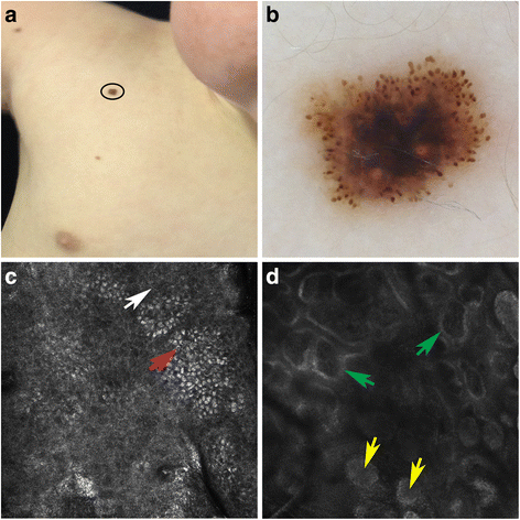
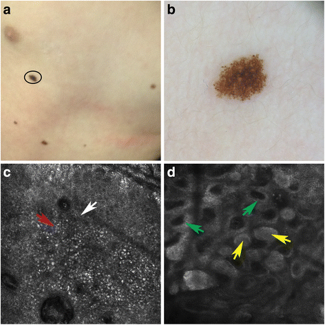
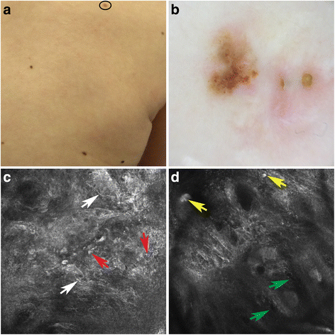
Case presentation: We describe the case of a patient with congenital GBM who developed eruptive disseminated Spitz nevi (EDSN) after chemotherapy. Few cases of EDSN have been described in literature and this rare clinical variant, which occurs predominantly in adults, is characterized by multiple Spitz nevi in the trunk, buttocks, elbows and knees. There is no satisfactory treatment for EDSN and the best therapeutic choice is considered the clinical observation of melanocytic lesions.
Conclusion: We recommend a close follow-up of these patients with clinical observation, dermoscopy and reflectance confocal microscopy (RCM). However, we suggest a surgical excision of the lesions suspected of being malignant.
Keywords: Chemotherapy; Congenital glioblastoma multiforme; Eruptive disseminated Spitz nevi; Reflectance confocal microscopy; Spitz nevi.
Figures


References
-
- Moscarella E, Lallas A, Kyrgidis A, Ferrara G, Longo C, Scalvenzi M, Staibano S, Carrera C, Díaz MA, Broganelli P, Tomasini C, Cavicchini S, Gianotti R, Puig S, Malvehy J, Zaballos P, Pellacani G, Argenziano G. Clinical and dermoscopic features of atypical Spitz tumors: A multicenter, retrospective, case–control study. J Am Acad Dermatol. 2015;73(5):777–874. doi: 10.1016/j.jaad.2015.08.018. - DOI - PMC - PubMed
-
- Pellacani G, Longo C, Ferrara G, Cesinaro AM, Bassoli S, Guitera P, Menzies SW, Seidenari S. Spitz nevi: In vivo confocal microscopic features, dermatoscopic aspects, histopathologic correlates, and diagnostic significance. J Am Acad Dermatol. 2009;60(2):236–247. doi: 10.1016/j.jaad.2008.07.061. - DOI - PubMed
Publication types
MeSH terms
LinkOut - more resources
Full Text Sources
Other Literature Sources
Medical

