. 2016 Dec;2(1):43.
doi: 10.1186/s40792-016-0171-4. Epub 2016 May 15.

Infective endocarditis of an aorto-right atrial fistula caused by asymptomatic rupture of a sinus of Valsalva aneurysm: a case report

Affiliations

Infective endocarditis of an aorto-right atrial fistula caused by asymptomatic rupture of a sinus of Valsalva aneurysm: a case report

Akihiko Ikeda et al. Surg Case Rep. 2016 Dec.

Abstract

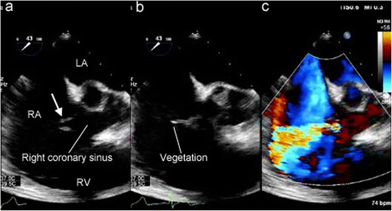
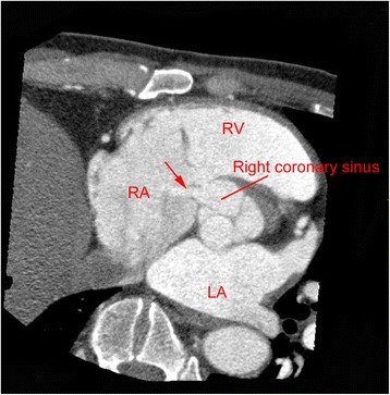
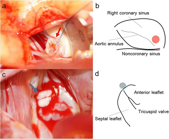
Asymptomatic rupture of a sinus of Valsalva aneurysm is rare. A fistula following rupture of a sinus of Valsalva aneurysm may cause infective endocarditis. Here, we report a case of infective endocarditis of an aorto-right atrial fistula caused by asymptomatic rupture of a sinus of Valsalva aneurysm. A 45-year-old male, who was first diagnosed with a heart murmur at the age of 37 years, presented with fever. Blood culture was positive for Streptococcus gordonii. Ultrasound echocardiography revealed an aorto-right atrial fistula caused by rupture of a sinus of Valsalva aneurysm. After the infective endocarditis was healed by antibiotics, we successfully performed surgical repair of the aorto-right atrial fistula. Although asymptomatic rupture of a sinus of Valsalva aneurysm is uncommon, it should be recognized as a possible cause of infective endocarditis.

Keywords: Aorto-right atrial fistula; Infective endocarditis; Sinus of Valsalva aneurysm.


Figures

Fig. 1
Fig. 1
a Preoperative transesophageal echocardiography showing a windsock-like fistula tract originating in the right coronary sinus protruding into the right atrium (arrow). b String-shaped vegetation was attached to the tip of the windsock-like fistula. c An L-R shunt via the fistula tract was seen. LA left atrium, RA right atrium, RV right ventricle
Fig. 2
Fig. 2
Preoperative computed tomography showing a defect between the right coronary sinus and the right atrium (arrow). LA left atrium, RA right atrium, RV right ventricle
Fig. 3
Fig. 3
a An intraoperative photograph showing the windsock-like aneurysm being pulled into the sinus of Valsalva (red arrow). b A schema showing the position of the aneurysm, which is located just above the aortic annulus of the right coronary cusp (red circle). c An intraoperative photograph showing the exit into the right atrium, which was surrounded by thickened excessive tissue (blue arrow). d A schema showing the position of the exit into the right atrium, which is located near the anteroseptal commissure of the tricuspid valve, which corresponds with the membranous septum (blue circle)

References

    1. Dong C, Wu QY, Tang Y. Ruptured sinus of Valsalva aneurysm: a Beijing experience. Ann Thorac Surg. 2002;74:1621–4. doi: 10.1016/S0003-4975(02)03987-5. - DOI - PubMed
    1. Lee SH, Kim JB, Park NH, Kim HS, Keum DY. Asymptomatic ruptured sinus of Valsalva aneurysm combined with perimembranous ventricular septal defect, and bicuspid aortic valve in adult patient. Thorac Cardiovasc Surg. 2013;61:327–9. - PubMed
    1. Moustafa S, Mookadam F, Connelly MS. Reticent uneventful rupture of right coronary sinus of Valsalva aneurysm into right ventricle. Heart Lung Circ. 2013;22:390–1. doi: 10.1016/j.hlc.2012.09.008. - DOI - PubMed
    1. Medina HM, Vazquez J, Pritchett A, Lakkis N, Dokainish H. Comprehensive imaging including three-dimensional echocardiography of an infected, ruptured sinus of valsalva aneurysm. Echocardiography. 2007;24:1096–8. doi: 10.1111/j.1540-8175.2007.00545.x. - DOI - PubMed
    1. Hirapur I, Veeranna RM, Agrawal N. Classical windsock deformity of ruptured sinus of Valsalva: an unusual appearance on transthoracic echocardiography. BMJ Case Rep. 2014; doi:10.1136/bcr-2014-204493. - PMC - PubMed

LinkOut - more resources