

Sterculic Acid and Its Analogues Are Potent Inhibitors of Toxoplasma gondii
- PMID: 27180571
- PMCID: PMC4870972
- DOI: 10.3347/kjp.2016.54.2.139
Sterculic Acid and Its Analogues Are Potent Inhibitors of Toxoplasma gondii
Abstract
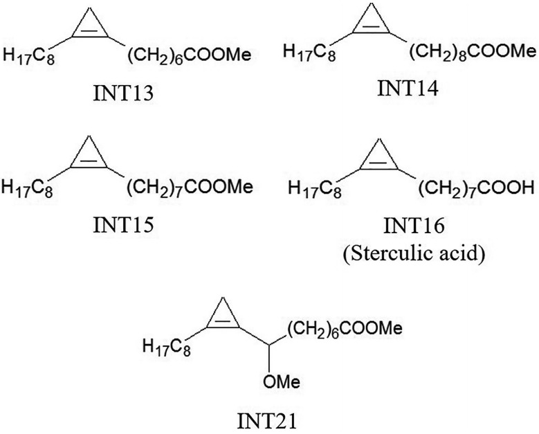
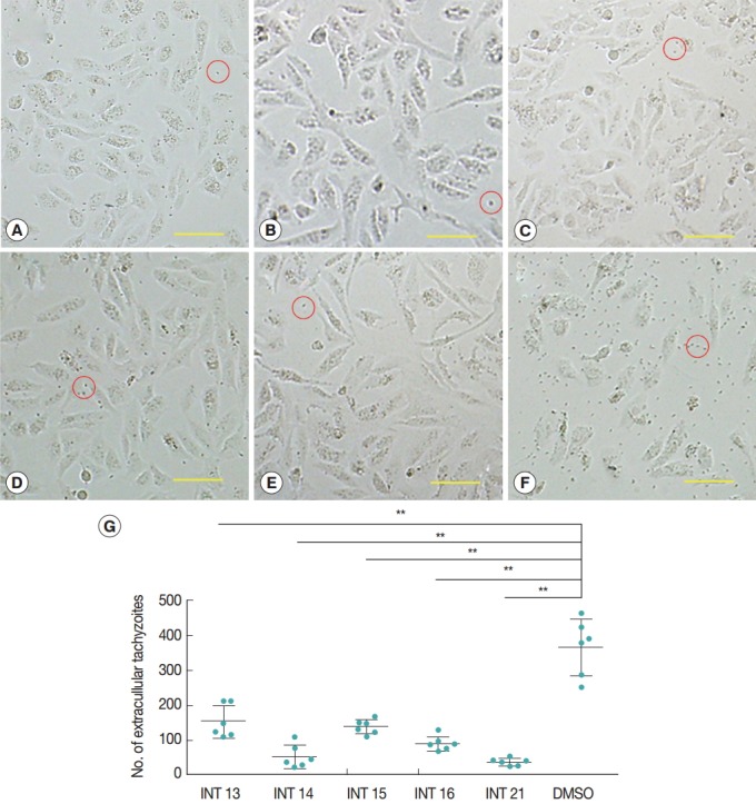
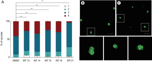
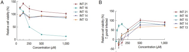
Toxoplasmosis is a serious disease caused by Toxoplasma gondii, one of the most widespread parasites in the world. Lipid metabolism is important in the intracellular stage of T. gondii. Stearoyl-CoA desaturase (SCD), a key enzyme for the synthesis of unsaturated fatty acid is predicted to exist in T. gondii. Sterculic acid has been shown to specifically inhibit SCD activity. Here, we examined whether sterculic acid and its methyl ester analogues exhibit anti-T. gondii effects in vitro. T. gondii-infected Vero cells were disintegrated at 36 hr because of the propagation and egress of intracellular tachyzoites. All test compounds inhibited tachyzoite propagation and egress, reducing the number of ruptured Vero cells by the parasites. Sterculic acid and the methyl esters also inhibited replication of intracellular tachyzoites in HFF cells. Among the test compounds, sterculic acid showed the most potent activity against T. gondii, with an EC50 value of 36.2 μM, compared with EC50 values of 248-428 μM for the methyl esters. Our study demonstrated that sterculic acid and its analogues are effective in inhibition of T. gondii growth in vitro, suggesting that these compounds or analogues targeting SCD could be effective agents for the treatment of toxoplasmosis.
Keywords: Toxoplasma gondii; anti-Toxoplasma gondii effect; stearoyl-CoA desaturase; sterculic acid.
Conflict of interest statement
We have no conflict of interest related to this work.
Figures
Similar articles
-
Sterculic Acid: The Mechanisms of Action beyond Stearoyl-CoA Desaturase Inhibition and Therapeutic Opportunities in Human Diseases.Cells. 2020 Jan 7;9(1):140. doi: 10.3390/cells9010140. Cells. 2020. PMID: 31936134 Free PMC article. Review.
-
Identification and characterization of stearoyl-CoA desaturase in Toxoplasma gondii.Acta Biochim Biophys Sin (Shanghai). 2019 Jun 20;51(6):615-626. doi: 10.1093/abbs/gmz040. Acta Biochim Biophys Sin (Shanghai). 2019. PMID: 31139819 Free PMC article.
-
A protein extract and a cysteine protease inhibitor enriched fraction from Jatropha curcas seed cake have in vitro anti-Toxoplasma gondii activity.Exp Parasitol. 2015 Jun;153:111-7. doi: 10.1016/j.exppara.2015.03.011. Epub 2015 Mar 25. Exp Parasitol. 2015. PMID: 25816973
-
Effects of sterculic acid on stearoyl-CoA desaturase in differentiating 3T3-L1 adipocytes.Biochem Biophys Res Commun. 2003 Jan 10;300(2):316-26. doi: 10.1016/s0006-291x(02)02842-5. Biochem Biophys Res Commun. 2003. PMID: 12504086
-
Adenosine metabolism in Toxoplasma gondii: potential targets for chemotherapy.Curr Pharm Des. 2007;13(6):581-97. doi: 10.2174/138161207780162836. Curr Pharm Des. 2007. PMID: 17346176 Review.
Cited by
-
Sterculic Acid: The Mechanisms of Action beyond Stearoyl-CoA Desaturase Inhibition and Therapeutic Opportunities in Human Diseases.Cells. 2020 Jan 7;9(1):140. doi: 10.3390/cells9010140. Cells. 2020. PMID: 31936134 Free PMC article. Review.
-
The Mechanism of Action of Ursolic Acid as a Potential Anti-Toxoplasmosis Agent, and Its Immunomodulatory Effects.Pathogens. 2019 May 9;8(2):61. doi: 10.3390/pathogens8020061. Pathogens. 2019. PMID: 31075881 Free PMC article.
-
3-O-Methyl-Alkylgallates Inhibit Fatty Acid Desaturation in Mycobacterium tuberculosis.Antimicrob Agents Chemother. 2019 Aug 23;63(9):e00136-19. doi: 10.1128/AAC.00136-19. Print 2019 Sep. Antimicrob Agents Chemother. 2019. PMID: 31209015 Free PMC article.
-
Genome-Wide Transcriptomic Analysis Identifies Pathways Regulated by Sterculic Acid in Retinal Pigmented Epithelium Cells.Cells. 2020 May 11;9(5):1187. doi: 10.3390/cells9051187. Cells. 2020. PMID: 32403229 Free PMC article.
-
Identification and characterization of stearoyl-CoA desaturase in Toxoplasma gondii.Acta Biochim Biophys Sin (Shanghai). 2019 Jun 20;51(6):615-626. doi: 10.1093/abbs/gmz040. Acta Biochim Biophys Sin (Shanghai). 2019. PMID: 31139819 Free PMC article.
References
-
- Lüder CG, Bohne W, Soldati D. Toxoplasmosis: a persisting challenge. Trends Parasitol. 2001;17:460–463. - PubMed
-
- Martins-Duarte ES, Urbina JA, de Souza W, Vommaro RC. Antiproliferative activities of two novel quinuclidine inhibitors against Toxoplasma gondii tachyzoites in vitro. J Antimicrob Chemother. 2006;58:59–65. - PubMed
-
- Ferra B, Holec-Gasior L, Kur J. Serodiagnosis of Toxoplasma gondii infection in farm animals (horses, swine, and sheep) by enzyme-linked immunosorbent assay using chimeric antigens. Parasitol Int. 2015;64:288–294. - PubMed
-
- Jeffcoat R, Pollard MR. Studies on the inhibition of the desaturases by cyclopropenoid fatty acids. Lipids. 1977;12:480–485. - PubMed
MeSH terms
Substances
LinkOut - more resources
Full Text Sources
Other Literature Sources
Medical
