. 2016 May 16:6:25960.
doi: 10.1038/srep25960.

Differential neuronal vulnerability identifies IGF-2 as a protective factor in ALS

Affiliations

Differential neuronal vulnerability identifies IGF-2 as a protective factor in ALS

Ilary Allodi et al. Sci Rep. .

Abstract

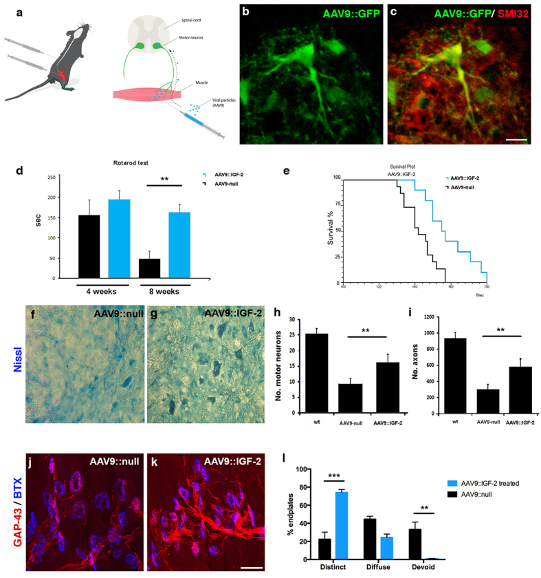
The fatal disease amyotrophic lateral sclerosis (ALS) is characterized by the loss of somatic motor neurons leading to muscle wasting and paralysis. However, motor neurons in the oculomotor nucleus, controlling eye movement, are for unknown reasons spared. We found that insulin-like growth factor 2 (IGF-2) was maintained in oculomotor neurons in ALS and thus could play a role in oculomotor resistance in this disease. We also showed that IGF-1 receptor (IGF-1R), which mediates survival pathways upon IGF binding, was highly expressed in oculomotor neurons and on extraocular muscle endplate. The addition of IGF-2 induced Akt phosphorylation, glycogen synthase kinase-3β phosphorylation and β-catenin levels while protecting ALS patient motor neurons. IGF-2 also rescued motor neurons derived from spinal muscular atrophy (SMA) patients from degeneration. Finally, AAV9::IGF-2 delivery to muscles of SOD1(G93A) ALS mice extended life-span by 10%, while preserving motor neurons and inducing motor axon regeneration. Thus, our studies demonstrate that oculomotor-specific expression can be utilized to identify candidates that protect vulnerable motor neurons from degeneration.


Figures

Figure 1
Figure 1. IGF-2 was persistently expressed in oculomotor neurons in health and ALS, in mouse and man.
(a) Schematic of the central nervous system of the mouse and connected muscles, highlighting the location of the oculomotor neurons (CNIII, in blue) in the midbrain and their targets the extraocular muscles, which are relatively resistant to degeneration in ALS. Also depicted are the vulnerable hypoglossal motor neurons (CNXII, in yellow) and the tongue muscles they innervate and vulnerable spinal motor neurons in the ventral horn (in green), which innervate limb muscles (by Mattias Karlen). Neurofilament and SV2a stainings were used to visualize the presynaptic motor nerve and α-bungarotoxin (BTX) to label acetylcholine receptors (AChRs) on the muscle, showing that extraocular muscles (b) were fully innervated in symptomatic P126 SOD1G93A mice, while tongue (c) and lumbrical muscles (d) showed partial (arrow head) or complete (*) denervation. (e) Weight curve of the fALS SOD1G93A mouse model, showing the decrease in weight compared to wild-type littermates, characteristic for disease (P < 0.01 to P < 0.001, t(17) = 3.88–5.19), n = 8 control mice, n = 10 SOD1G93A mice multiple t test). In P126 control mice, insulin-like growth factor-2 (IGF-2) protein was preferential to oculomotor neurons (f,i), with 2.7-fold higher levels than in hypoglossal (g,i) and 20-fold higher than spinal (h,i) motor neurons (F(2, 185) = 61.69, P < 0.0001, n = 4, ANOVA). IGF-2 levels were 7.6-fold higher in hypoglossal than in spinal motor neurons (P < 0.05, ANOVA). Analysis of P126 symptomatic SOD1G93A mice showed that IGF-2 levels remained preferential to oculomotor neurons (j,m) with levels 4.1-fold higher than in hypoglossal (k,m) and 3.3-fold higher than in spinal motor neurons (l,m) (F(2, 241) = 36.05, P < 0.0001, n = 3, ANOVA). Analysis of non-demented control patient tissues showed that IGF-2 protein levels were higher in oculomotor motor neurons (n,q) compared to hypoglossal motor neurons ((o,q) F(2, 346) = 22.67, P < 0.05, n = 5, ANOVA; P < 0.0001, Kruskal-Wallis) and spinal motor neurons ((p,q) P < 0.0001, ANOVA; P < 0.0001, Kruskal-Wallis). IGF-2 remained preferential to oculomotor motor neurons in end-stage ALS patient tissue ((ru) F(2, 500) = 98.78, P < 0.0001, n = 5, ANOVA; P < 0.0001, Kruskal-Wallis). Values in the graphs represent means ± SEM. Scale bar in (d) 30 μm (applicable to (b,c)), (l) 50 μm (applicable to (fh,j,k), (t) 30 μm (applicable to (ns)).
Figure 2
Figure 2. IGF-1R and IGF-2R expression was predominant in resistant oculomotor neurons and extraocular muscles.
Phosphorylated IGF-1R (pIGF-1R) protein was present in oculomotor motor neurons in wild-type and SOD1G93A mice (a,c,e), with signficantly lower levels in motor neurons in spinal cord in control (b) arrow heads, (e) t(113) = 8.69, n = 4 mice, P < 0.0001, Student’s t test, values represent means ± SEM) and SOD1G93A mice ((d) arrow heads, (e) t(141) = 4.30, P < 0.0001, n = 5 mice, Student’s t test, values represent means ± SEM). IGF-1R protein was strongly expressed and co-localized with endplates in extraocular muscles in wild-type and SOD1G93A mice (f,h), while it was barely detectable in lumbrical muscles (g,i). Western blot analysis confirmed that the pIGF-1R protein level was 4-fold higher in extraocular muscles than in lumbrical muscles ((j) t(8) = 8.20, n = 5 mice, P < 0.0001, Student’s t test, values represent means ± SEM). Phosphorylated IGF-2R (pIGF-2R) protein was present at comparable levels in oculomotor (k,m) and spinal (l,n) motor neurons in both wild-type ((o) t(92) = 0.6829, P = 0.4964, n = 3 mice, Student’s t test) and SOD1G93A mice ((o) t(102) = 1.880, P = 0.0630, n = 3 mice, Student’s t test). Peripherally, IGF-2R protein was barely detectable in extraocular muscles (p,r) and undetectable in lumbrical muscles (q,s) of wild-type and SOD1G93A mice using immunofluorescence. Western blot analysis showed that IGF-2R was indeed present in extraocular muscles at 3.8-fold higher levels than in lumbrical muscles ((t) t(8) = 2.86, P = 0.021, n = 5 mice/group, Student’s t test, values represent means ± SEM). Scale bars: (p) 50 μm (applicable to (ad) and (mo)), (q) 40 μm (applicable to (eh) and (nq)).
Figure 3
Figure 3. IGF-2 protected human spinal motor neurons from ALS-like toxicity in vitro.
Human spinal motor neurons generated from iPSCs expressed Islet-1/2 (a) and ChAT (b) (photos of iPSC clone AM/ALS1.1 shown, see supplementary Table 1). The number of motor neurons was significantly decreased after co-culture with SOD1G93A astrocytes ((ce), F(8, 126) = 231.02, P < 0.0001, ANOVA). Addition of IGF-2 (50 ng/ml) 24–48 after initiation of toxicity could rescue motor neurons ((d,e), F(8, 126) = 231.02, P < 0.0001, ANOVA). Exposure of cultures to glutamate also induced motor neuron degeneration ((f,h), F(8, 126) = 125.87, P < 0.001, ANOVA), which could be rescued by adding IGF-2 (treatment initiated 24–48h after addition of glutamate) ((g,h), F(8, 126) = 125.87, P < 0.0001, ANOVA). Values represent means ± SD from 5 independent experiments performed in triplicate. Addition of IGF-2 induced phosphorylation of GSK-3 on the S9 residue p-GSK-(S9), thus inhibiting enzyme activity in a dose-dependent way (i). Representative images showing p-GSK-(S9) levels in the absence (j,k) or presence (l,m) of IGF-2. Representative images depicting AKT activation by phosphorylation on residue S473 (p-AKT-(S473)), in the absence (n,o) or presence (p,q) of IGF-2. IGF-2 induced p-AKT-(S473) levels in iPSC motor neurons ((r), t(8) = 5.99, P < 0.001, n = 5, Student’s t test, values represent means ± SD). Immunofluorescent analysis of β-catenin levels in control (s,t) and IGF-2 treated (u,v) motor neurons. IGF-2 induced β-catenin levels in iPSC motor neurons ((z), t(8) = 6.36, P < 0.001, n = 5, Student’s t test, values represent means ± SD). Scale bars: (a) 60 μm, (b) 50 μm, (g) 75 μm (applicable to (c,d,f)), (k) 75 μm (applicable to (j,l,m)), (o) 50 μM (applicable to (n,p,q), (t) 50 μM (applicable to (s,u,v)).
Figure 4
Figure 4. IGF-2 protected SMA patient spinal motor neurons in culture.
SMA patient motor neurons in long-term culture without (a) or with (b) IGF-2 (photos of iPSC clone SMA 1.1 shown, see supplementary Table 2). (c) The number of SMA motor neurons in culture was significantly decreased compared to wild-type cells (F(5, 84) = 135,65, P < 0.0001, ANOVA). Treatment of the cultures with IGF-2 (50–100 ng/ml, added at 4 weeks) was protective to motor neurons (8 weeks, grey and black bars, F(5, 84) = 135.65, P < 0.0001, ANOVA). Values represent means ± SD from 5 independent experiments performed in triplicate. (d) At 8 weeks, untreated SMA iPSC motor neurons (shown in yellow) showed shorter axon lengths than wild-type cells (shown in black). SMA motor neurons treated with IGF-2 (shown in teal and red) had longer axons than untreated SMA motor neurons (P < 0.001, Kolmogorov-Smirnov test, 5 independent experiments performed in triplicate).
Figure 5
Figure 5. IGF-2 prolonged the survival of SOD1G93A fALS mice by preserving motor neurons and inducing nerve regeneration.
(a) Schematic drawing of the IGF-2 in vivo delivery (by Mattias Karlen) where SOD1G93A mice were injected into the hindlimb quadriceps and thoracic muscles at 80 days of age with AAV9::GFP, AAV9::IGF-2 or AAV9::null at a total dose of 11 × 1011 vg. (b) Injection of AAV9::GFP resulted in GFP expression within the spinal cord 2 weeks post-injection. (c) Colocalization of GFP (green) with SMI32 (red) demonstrated that motor neurons were efficiently transduced (n = 5 mice). (d) Rotarod performance of AAV9::IGF-2 treated mice (n = 10) was significantly improved compared to AAV9::null mice (n = 10) in particular at 8 weeks after treatment (F(1, 36) = 18.66, P < 0.0001, two-way ANOVA; at 8 weeks: T(16) = 4.778, P < 0.001, Student’s t test). Error bars indicate mean ± SEM). (e) Kaplan-Meier survival curves demonstrated a significantly extended survival by 14 days in AAV9::IGF-2 mice (n = 10) compared to AAV9::null animals (n = 15) (χ2  =  5.3, P = 0.02). Representative motor neuron pools in the lumbar segment of the spinal cords of AAV9::null (f) and AAV9::IGF-2 (g) treated mice at P140. Quantification of motor neurons (h) and axons (i) in the lumbar spinal cords of AAV9::IGF-2 and AAV9-null treated mice (mean ± SD) at P140. Motor neuron and axon counts significantly increased in the AAV9::IGF-2 treatment group compared to the AAV9::null group (motor neurons: F(2, 87) = 397.75, P < 0.0001; axons: F(2, 33) = 230.28, P < 0.0001, two-way ANOVA, n = 3/group). (j–l) Analysis of GAP-43 in NMJs of lumbrical muscles showed that AAV9::IGF-2 treatment significantly increased the proportion of endplate with distinct GAP-43 expression in SOD1G93A mice compared to AAV9::null treatment F(2, 25) = 38.07, P < 0.0001, n = 5 muscles (3 mice) AAV9::null, n = 6 muscles (3 mice) AAV9::IGF-2, ANOVA, values shown as mean ± SEM). (jl) AAV::IGF-2 treated mice also had significantly fewer endplates which were devoid of GAP-43 staining (P < 0.001, ANOVA, values shown as mean ± SEM). Scale bar: (c) 50 μm (applicable to (b)), (k) 40 μm (applicable to (j)).

References

    1. Al-Chalabi A. et al.. The genetics and neuropathology of amyotrophic lateral sclerosis. Acta Neuropathol 124, 339–352 (2012). - PubMed
    1. Renton A. E., Chio A. & Traynor B. J. State of play in amyotrophic lateral sclerosis genetics. Nat Neurosci 17, 17–23 (2014). - PMC - PubMed
    1. Boillee S. et al.. Onset and progression in inherited ALS determined by motor neurons and microglia. Science 312, 1389–1392 (2006). - PubMed
    1. Jaarsma D., Teuling E., Haasdijk E. D., De Zeeuw C. I. & Hoogenraad C. C. Neuron-specific expression of mutant superoxide dismutase is sufficient to induce amyotrophic lateral sclerosis in transgenic mice. J Neurosci 28, 2075–2088 (2008). - PMC - PubMed
    1. Corti S. et al.. Genetic correction of human induced pluripotent stem cells from patients with spinal muscular atrophy. Sci Transl Med 4, 165ra162 (2012). - PMC - PubMed

Publication types

MeSH terms