

Cystic Lung Diseases: Algorithmic Approach
- PMID: 27180915
- PMCID: PMC7534033
- DOI: 10.1016/j.chest.2016.04.026
Cystic Lung Diseases: Algorithmic Approach
Abstract
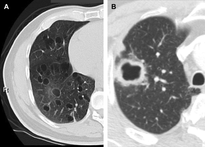
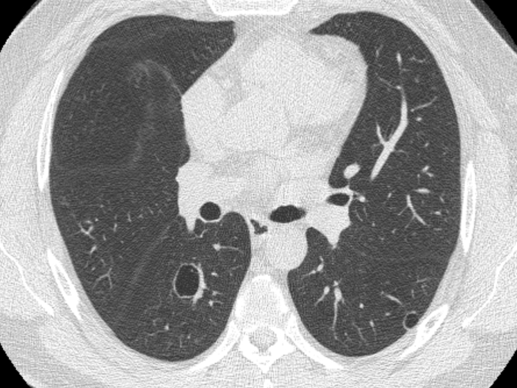
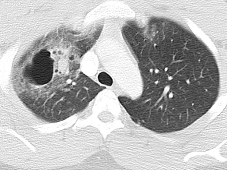
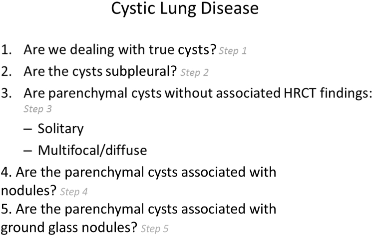
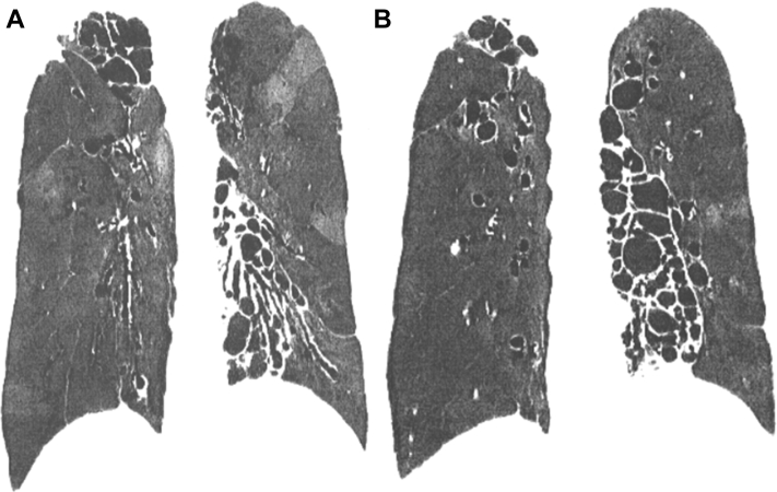
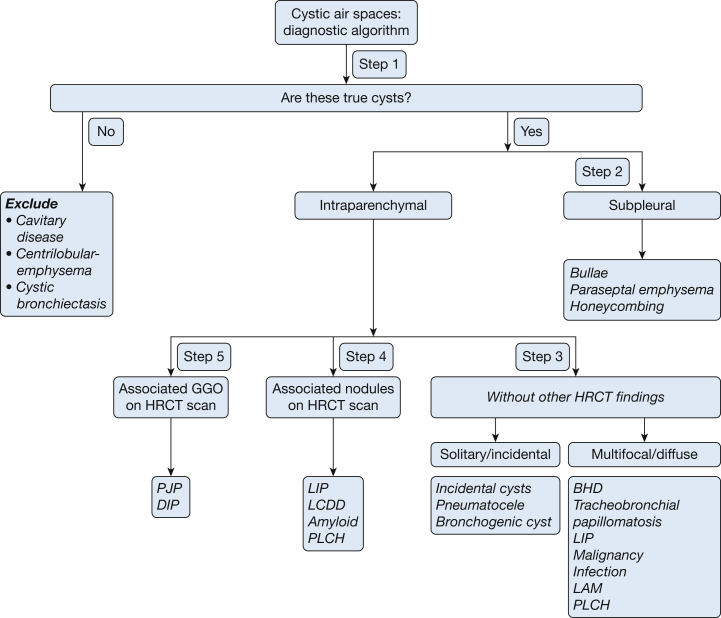
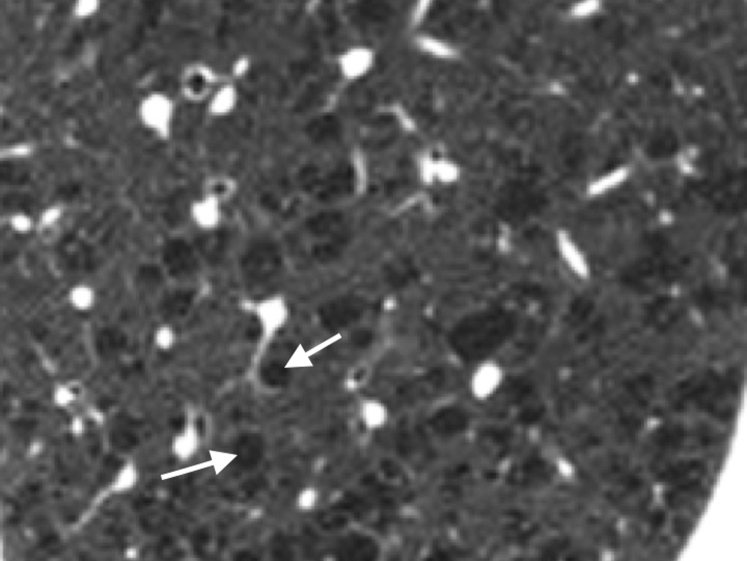
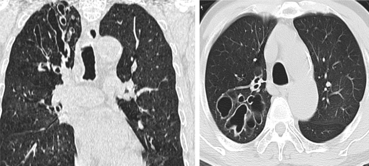
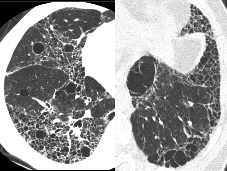
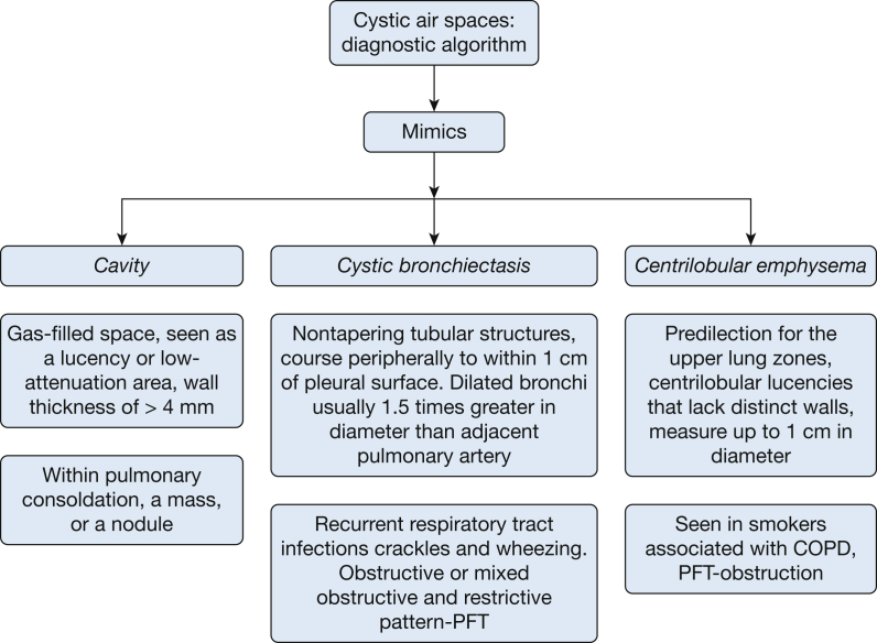
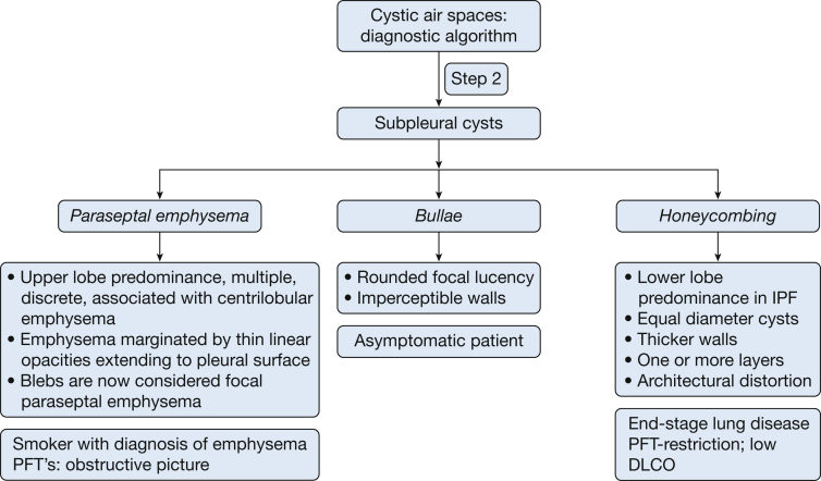
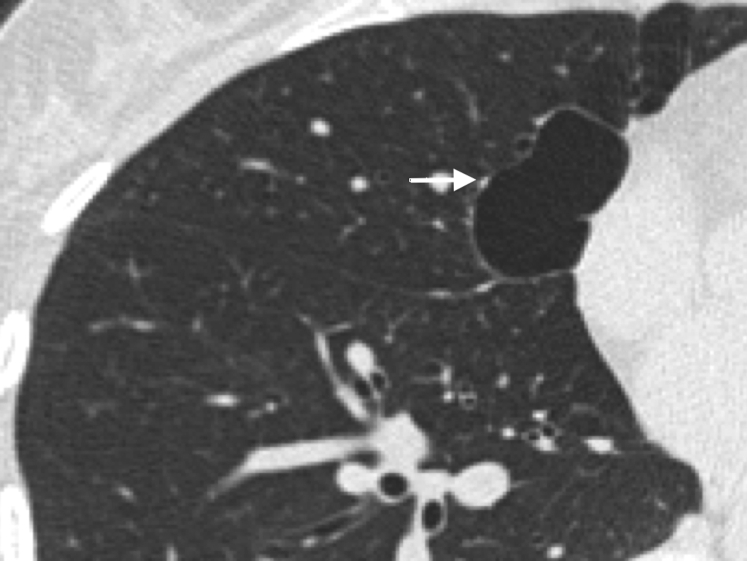
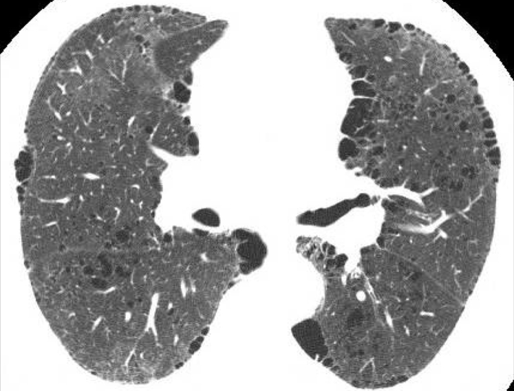
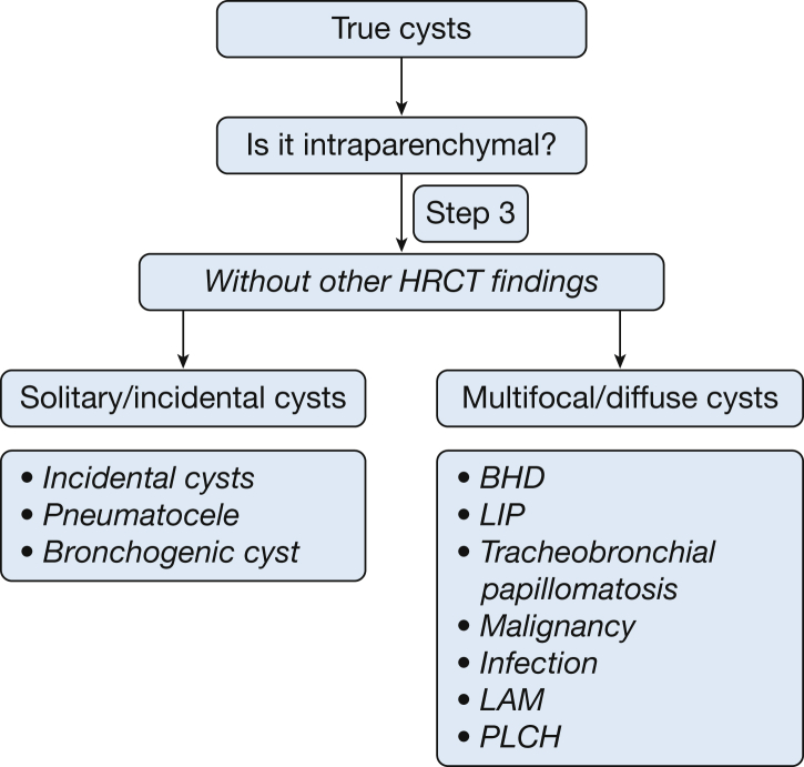
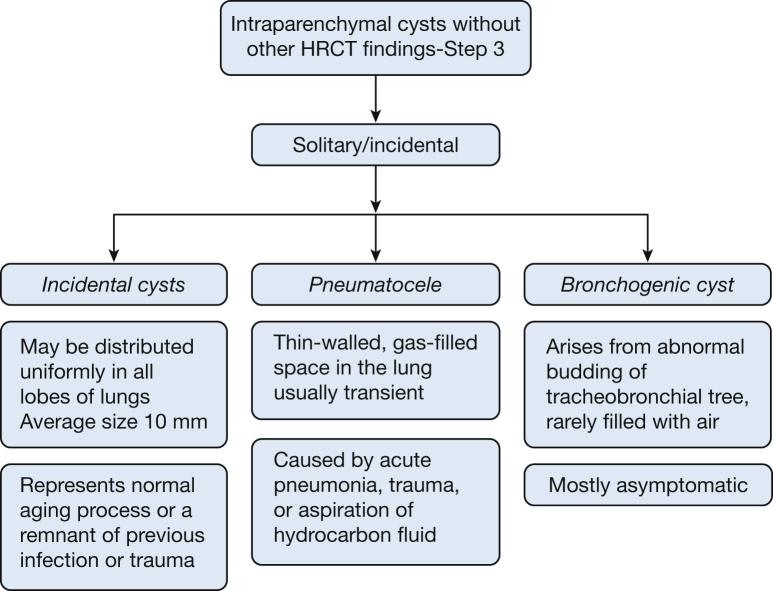
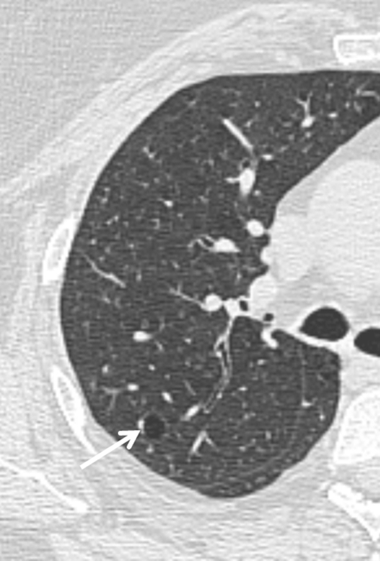
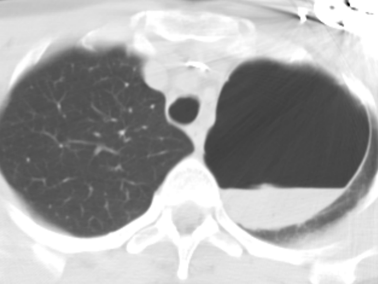
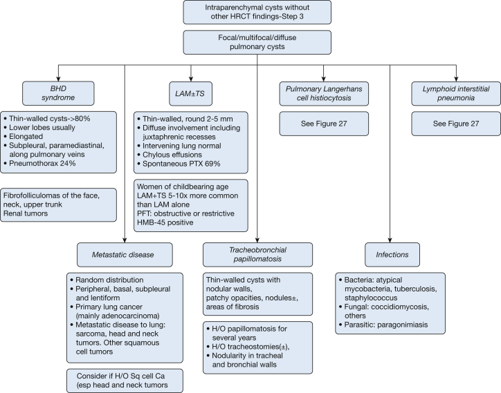
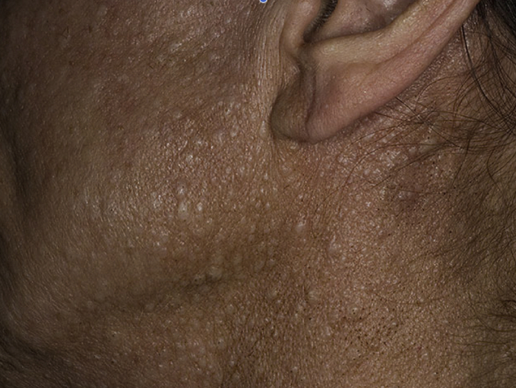
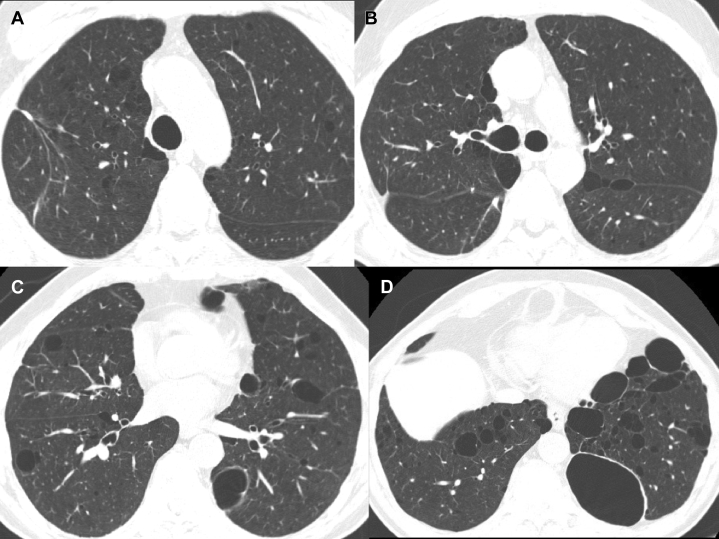
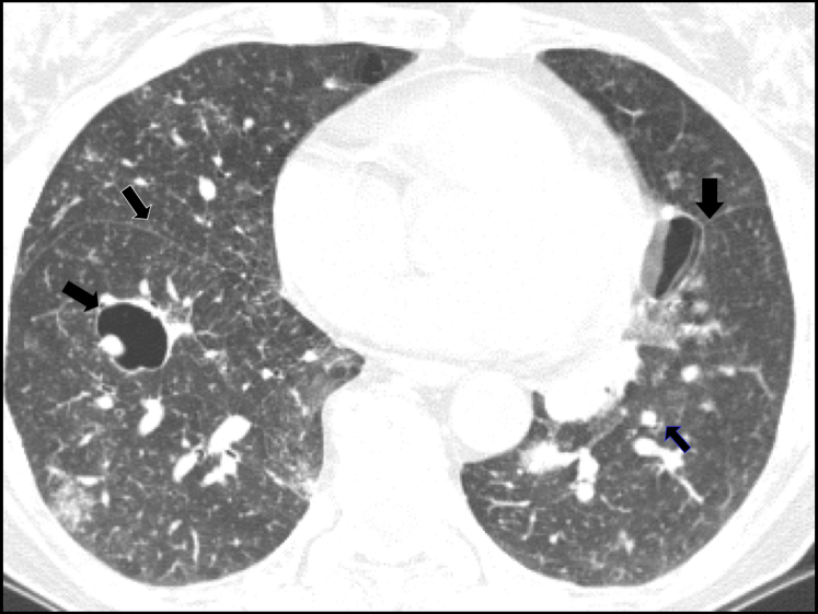
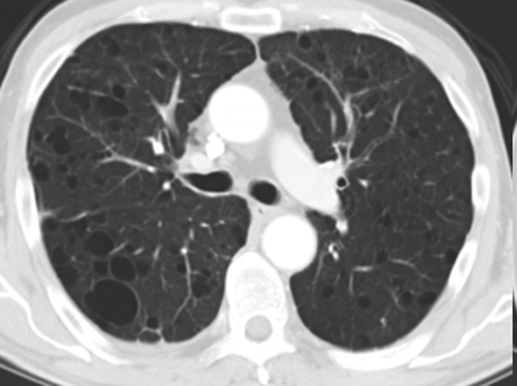
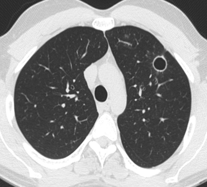
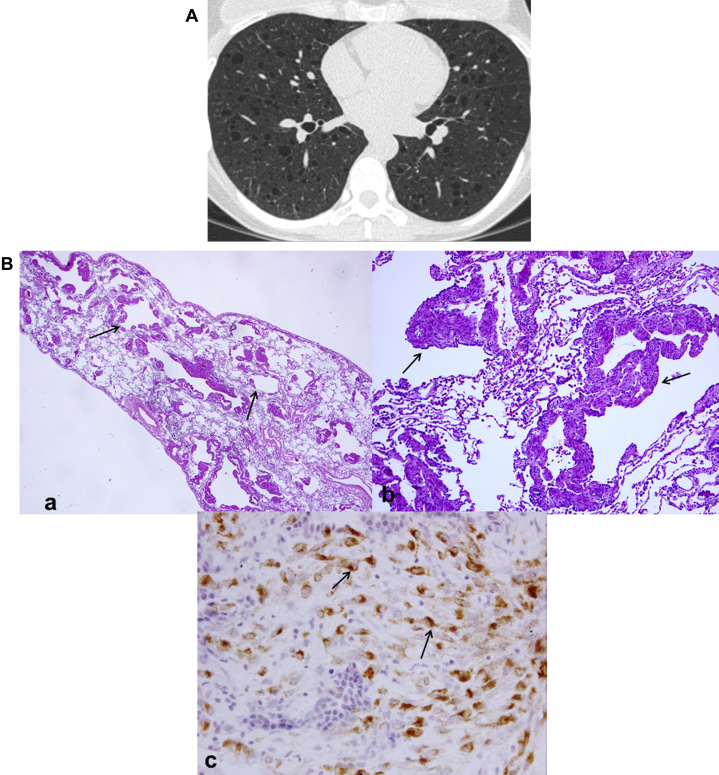
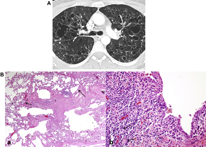
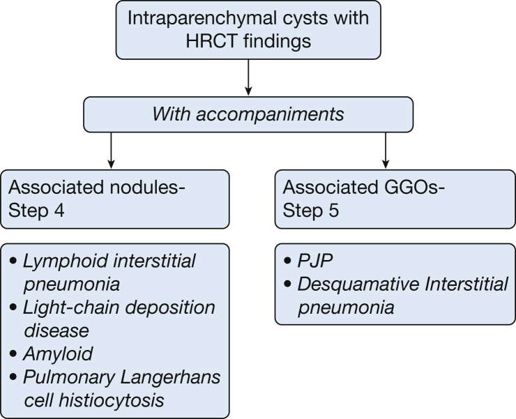
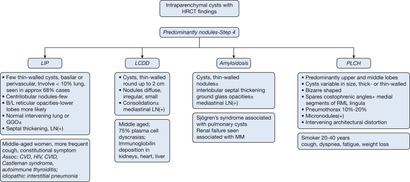
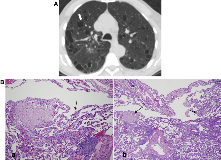
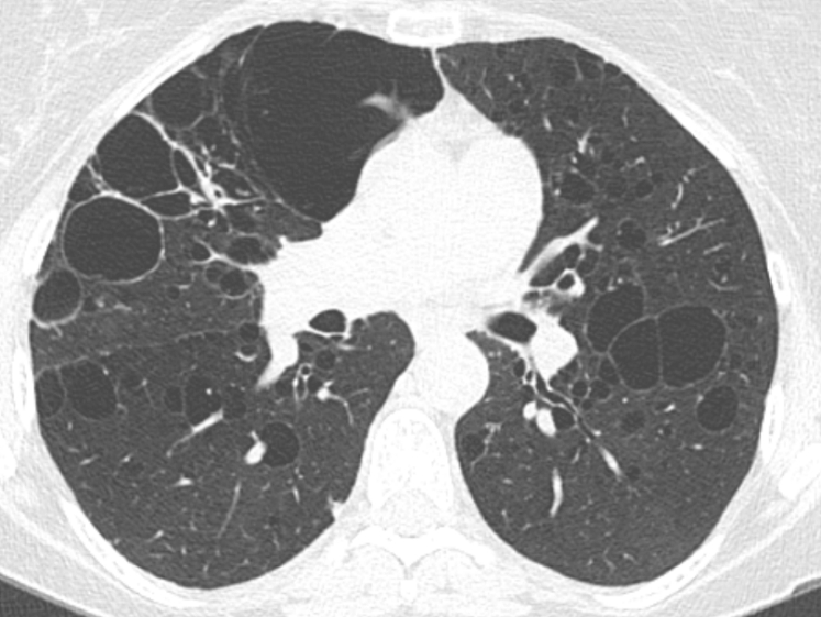
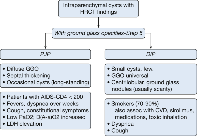
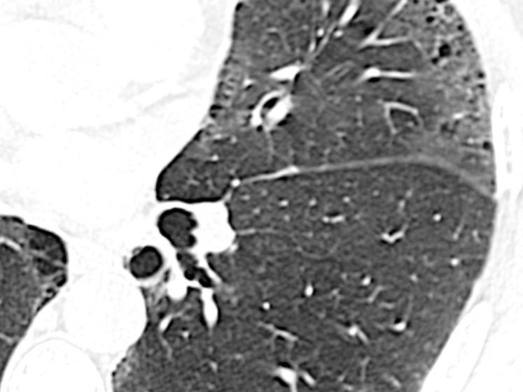
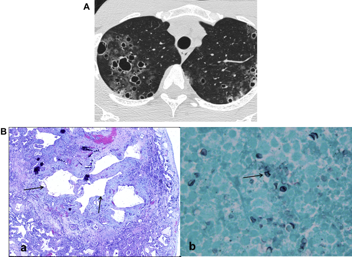
Cysts are commonly seen on CT scans of the lungs, and diagnosis can be challenging. Clinical and radiographic features combined with a multidisciplinary approach may help differentiate among various disease entities, allowing correct diagnosis. It is important to distinguish cysts from cavities because they each have distinct etiologies and associated clinical disorders. Conditions such as emphysema, and cystic bronchiectasis may also mimic cystic disease. A simplified classification of cysts is proposed. Cysts can occur in greater profusion in the subpleural areas, when they typically represent paraseptal emphysema, bullae, or honeycombing. Cysts that are present in the lung parenchyma but away from subpleural areas may be present without any other abnormalities on high-resolution CT scans. These are further categorized into solitary or multifocal/diffuse cysts. Solitary cysts may be incidentally discovered and may be an age related phenomenon or may be a remnant of prior trauma or infection. Multifocal/diffuse cysts can occur with lymphoid interstitial pneumonia, Birt-Hogg-Dubé syndrome, tracheobronchial papillomatosis, or primary and metastatic cancers. Multifocal/diffuse cysts may be associated with nodules (lymphoid interstitial pneumonia, light-chain deposition disease, amyloidosis, and Langerhans cell histiocytosis) or with ground-glass opacities (Pneumocystis jirovecii pneumonia and desquamative interstitial pneumonia). Using the results of the high-resolution CT scans as a starting point, and incorporating the patient's clinical history, physical examination, and laboratory findings, is likely to narrow the differential diagnosis of cystic lesions considerably.
Keywords: cystic lung disease; diffuse lung disease; focal lung luciencies; lung cysts; pulmonary cysts.
Copyright © 2016 American College of Chest Physicians. Published by Elsevier Inc. All rights reserved.
Figures
References
-
- Ryu J.H., Swensen S.J. Cystic and cavitary lung diseases: focal and diffuse. Mayo Clin Proc. 2003;78(6):744–752. - PubMed
-
- Echeveste J., Fernández-Velilla M., Torres M.I., Pardo M., Berrocal T., Martín-Hervás C. Cystic diseases of the lung: high-resolution computed tomography findings. Arch Bronconeumol. 2005 Jan;41(1):42–49. [in Spanish] - PubMed
-
- Devakonda A., Raoof S., Sung A., Travis W.D., Naidich D. Bronchiolar disorders: a clinical-radiological diagnostic algorithm. Chest. 2010;137(4):938–951. - PubMed
-
- Koyama M., Johkoh T., Honda O. Chronic cystic lung disease: diagnostic accuracy of high-resolution CT in 92 patients. AJR Am J Roentgenol. 2003;180(3):827–835. - PubMed
-
- Gupta N., Meraj R., Tanase D. Accuracy of chest high-resolution computed tomography in diagnosing diffuse cystic lung diseases. Eur Respir J. 2015;46(4):1196–1199. - PubMed
Publication types
MeSH terms
Supplementary concepts
Grants and funding
LinkOut - more resources
Full Text Sources
Other Literature Sources
Medical
