

A CCL8 gradient drives breast cancer cell dissemination
- PMID: 27181207
- PMCID: PMC5112152
- DOI: 10.1038/onc.2016.161
A CCL8 gradient drives breast cancer cell dissemination
Abstract
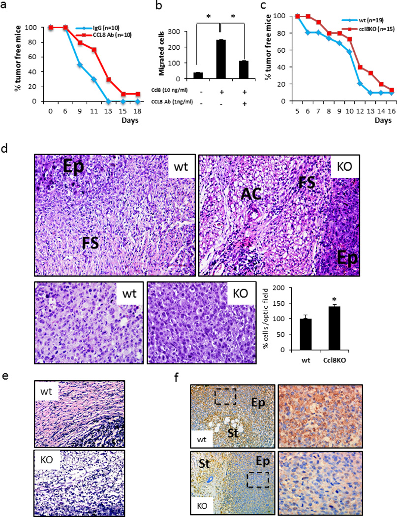
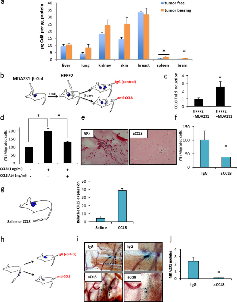
The migration of cancer cells towards gradients of chemoattractive factors represents a potential, yet elusive, mechanism that may contribute to cancer cell dissemination. Here we provide evidence for the maintenance of a gradient of increasing CCL8 concentration between the epithelium, the stroma and the periphery that is instrumental for breast cancer cells' dissemination. In response to signals elicited by the neoplastic epithelium, CCL8 production is enhanced in stromal fibroblasts at the tumor margins and in tissues at which breast cancer cells tend to metastasize such as the lungs and the brain. Manipulation of CCL8 activity influences the histology of the tumors and promotes major steps of the metastatic process such as invasion to adjacent stroma, intravasation and ultimately extravasation and seeding. These findings exemplify how gradients of chemoattractive factors such as CCL8, drive metastasis and suggest that interference with their operation may provide means for breast cancer management.
Conflict of interest statement
The authors declare no conflict of interest.
Figures


Similar articles
-
Tumour but not stromal expression of β3 integrin is essential, and is required early, for spontaneous dissemination of bone-metastatic breast cancer.J Pathol. 2015 Apr;235(5):760-72. doi: 10.1002/path.4490. Epub 2015 Jan 7. J Pathol. 2015. PMID: 25430721
-
Induction of the MCP chemokine cluster cascade in the periphery by cancer cell-derived Ccl3.Cancer Lett. 2017 Mar 28;389:49-58. doi: 10.1016/j.canlet.2016.12.028. Epub 2016 Dec 29. Cancer Lett. 2017. PMID: 28041977 Free PMC article.
-
Essential roles of the interaction between cancer cell-derived chemokine, CCL4, and intra-bone CCR5-expressing fibroblasts in breast cancer bone metastasis.Cancer Lett. 2016 Aug 1;378(1):23-32. doi: 10.1016/j.canlet.2016.05.005. Epub 2016 May 10. Cancer Lett. 2016. PMID: 27177471
-
Hypoxia-inducible factor 1 and breast cancer metastasis.J Zhejiang Univ Sci B. 2015 Jan;16(1):32-43. doi: 10.1631/jzus.B1400221. J Zhejiang Univ Sci B. 2015. PMID: 25559953 Free PMC article. Review.
-
New insights into the pathogenesis of breast cancer metastasis.Breast Dis. 2006-2007;26:13-25. doi: 10.3233/bd-2007-26103. Breast Dis. 2006. PMID: 17473363 Review.
Cited by
-
A Phase 1/2 Trial Combining Avelumab and Trabectedin for Advanced Liposarcoma and Leiomyosarcoma.Clin Cancer Res. 2022 Jun 1;28(11):2306-2312. doi: 10.1158/1078-0432.CCR-22-0240. Clin Cancer Res. 2022. PMID: 35349638 Free PMC article. Clinical Trial.
-
A novel predictive model incorporating immune-related gene signatures for overall survival in melanoma patients.Sci Rep. 2020 Jul 27;10(1):12462. doi: 10.1038/s41598-020-69330-2. Sci Rep. 2020. PMID: 32719391 Free PMC article.
-
CD200 Blockade Modulates Tumor Immune Microenvironment but Fails to Show Efficacy in Inhibiting Tumor Growth in a Murine Model of Melanoma.Front Cell Dev Biol. 2021 Oct 8;9:739816. doi: 10.3389/fcell.2021.739816. eCollection 2021. Front Cell Dev Biol. 2021. PMID: 34692697 Free PMC article.
-
Influence of Fibroblasts on Mammary Gland Development, Breast Cancer Microenvironment Remodeling, and Cancer Cell Dissemination.Cancers (Basel). 2020 Jun 26;12(6):1697. doi: 10.3390/cancers12061697. Cancers (Basel). 2020. PMID: 32604738 Free PMC article. Review.
-
Protein profiling of fine-needle aspirates reveals subtype-associated immune signatures and involvement of chemokines in breast cancer.Mol Oncol. 2019 Feb;13(2):376-391. doi: 10.1002/1878-0261.12410. Epub 2019 Jan 7. Mol Oncol. 2019. PMID: 30451357 Free PMC article.
References
-
- Chatzistamou I, Dioufa N, Trimis G, Sklavounou A, Kittas C, Kiaris H, et al. p21/waf1 and smooth-muscle actin alpha expression in stromal fibroblasts of oral cancers. Cell Oncol (Dordr) 2011;34:483–488. - PubMed
-
- Trimis G, Chatzistamou I, Politi K, Kiaris H, Papavassiliou AG. Expression of p21waf1/Cip1 in stromal fibroblasts of primary breast tumors. Hum Mol Genet. 2008;17:3596–3600. - PubMed
-
- Su S, Liu Q, Chen J, Chen F, He C, Huang D, et al. A positive feedback loop between mesenchymal-like cancer cells and macrophages is essential to breast cancer metastasis. Cancer Cell. 2014;25:605–620. - PubMed
Publication types
MeSH terms
Substances
Grants and funding
LinkOut - more resources
Full Text Sources
Other Literature Sources
Medical
Molecular Biology Databases
