Review
. 2016 May 15;129(10):1951-61.
doi: 10.1242/jcs.179788.

Nuclear migration events throughout development

Affiliations
Review

Nuclear migration events throughout development

Courtney R Bone et al. J Cell Sci. .

Abstract

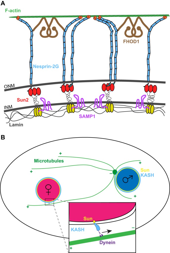
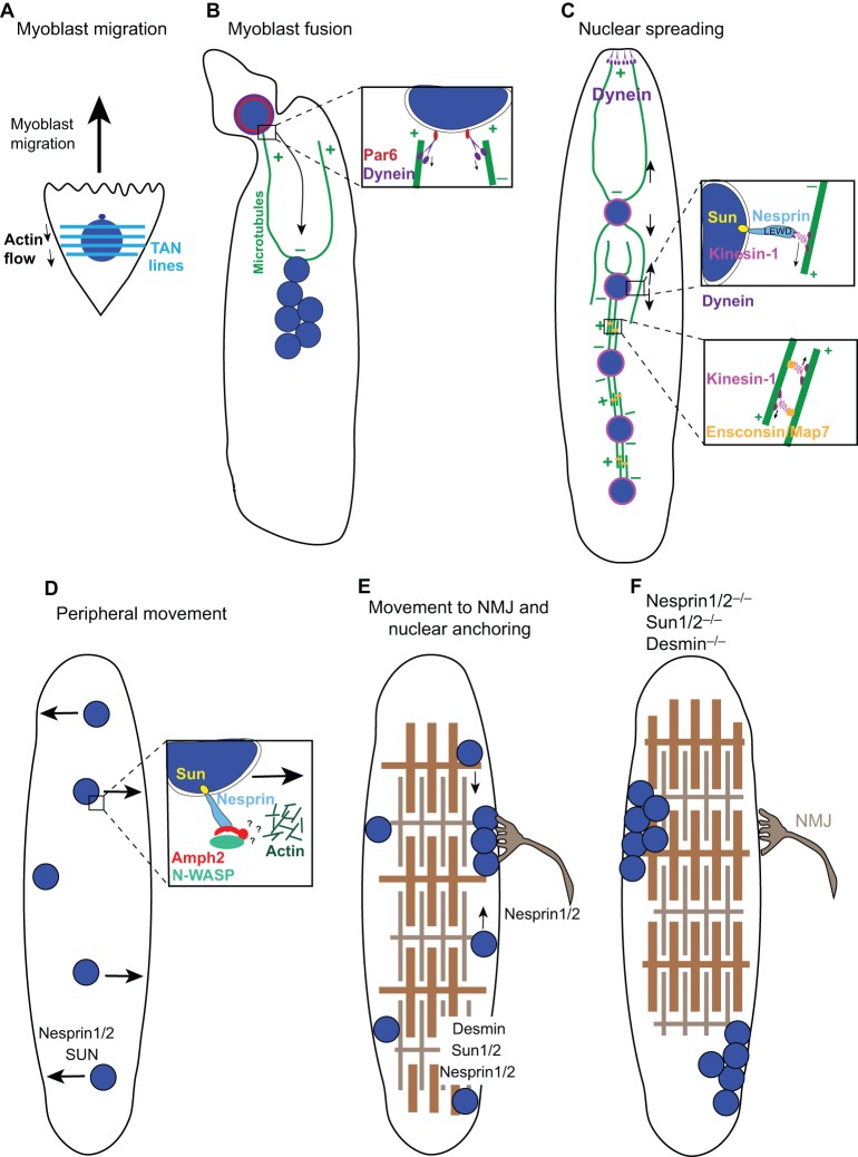
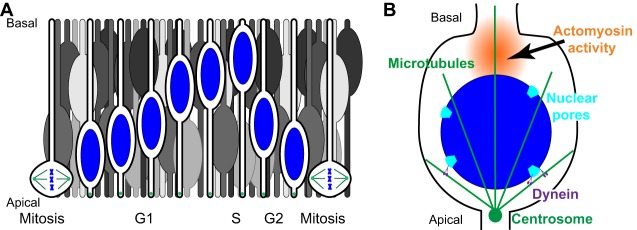
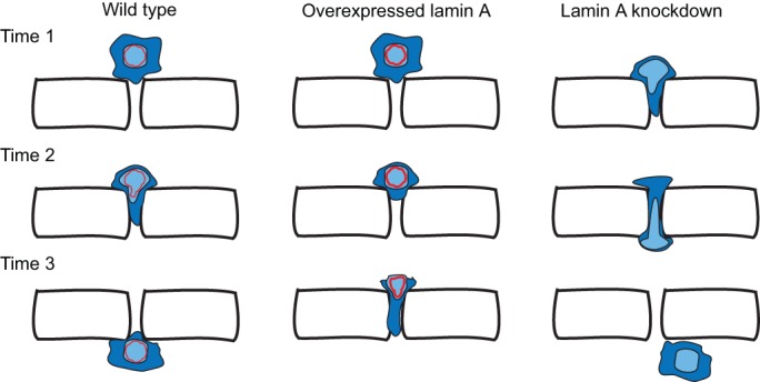
Moving the nucleus to a specific position within the cell is an important event during many cell and developmental processes. Several different molecular mechanisms exist to position nuclei in various cell types. In this Commentary, we review the recent progress made in elucidating mechanisms of nuclear migration in a variety of important developmental models. Genetic approaches to identify mutations that disrupt nuclear migration in yeast, filamentous fungi, Caenorhabditis elegans, Drosophila melanogaster and plants led to the identification of microtubule motors, as well as Sad1p, UNC-84 (SUN) domain and Klarsicht, ANC-1, Syne homology (KASH) domain proteins (LINC complex) that function to connect nuclei to the cytoskeleton. We focus on how these proteins and various mechanisms move nuclei during vertebrate development, including processes related to wound healing of fibroblasts, fertilization, developing myotubes and the developing central nervous system. We also describe how nuclear migration is involved in cells that migrate through constricted spaces. On the basis of these findings, it is becoming increasingly clear that defects in nuclear positioning are associated with human diseases, syndromes and disorders.

Keywords: Development; LINC complex; Nuclear envelope; Nuclear migration.


Conflict of interest statement

Competing interests

The authors declare no competing or financial interests.

Figures

Fig. 1.
Fig. 1.
Examples of nuclear migration mediated by LINC complexes. (A) A transmembrane actin-associated nuclear (TAN) line connects the retrograde-moving actin filaments to the nucleoskeleton. F-actin (green) in the cytoplasm interacts with nesprin-2G (blue), an integral outer nuclear membrane (ONM) protein with a large cytoplasmic domain. This interaction is reinforced by the formin FHOD1 (brown). Nesprin-2G interacts in the peri-nuclear space with the inner nuclear membrane (INM) protein Sun2 (red and yellow). Samp1 (pink) in the inner nuclear membrane is also a component of the TAN line. Finally, the TAN line ends in the nucleoplasm, where the nucleoplasmic domain of Sun2 (yellow) interacts with lamins (gray). (B) Schematic of pronuclear migration showing the capturing of the female pronucleus (pink) by microtubules (green) that have nucleated from a centrosome (green circle) attached to the male pronucleus (blue). This process is mediated by a bridge comprising SUN- and KASH-domain proteins on the nuclear envelope (yellow and blue in the inset), which recruits dynein (purple) to the surface of the female pronucleus in order to move it towards the male pronucleus.
Fig. 2.
Fig. 2.
Nuclear migration events in muscle development. During muscle development, numerous mononucleated myoblasts fuse to form a myotube, which further develops into a myofiber. (A) Myoblast nuclei (blue) are polarized with TAN lines (light blue horizontal lines, see Fig. 1A), which harness actin flow during cell migration. (B) Upon the fusion of a myoblast to the end of a myotube, the new nucleus rapidly migrates towards the minus ends of microtubules (green) at the center of the myotube. This migration is mediated by dynein (purple), which recruited to the nuclear envelope through an interaction with Par6 (red). (C) As the myotube develops, nuclei spread out and become evenly spaced throughout the syncytium. Several mechanisms function to spread these nuclei. First, dynein at the cortex of the ends of myotubes pull nuclei towards the poles. Second, as illustrated in the upper inset, kinesin-1 (pink) is directly recruited to the nuclear envelope through a LEWD amino acid motif within nesprins (light blue) to move nuclei towards distal plus ends of microtubules. Last, this process is mediated by kinesin-1 and the microtubule crosslinker Ensconsin/MAP7 (orange), which slide microtubules apart, thereby pushing nuclei away from each other. (D) Subsequently, nuclei move from the center to the periphery of the myotube as sarcomeres begin to develop. This process is likely to utilize a complex between nesprins, amphiphysin-2 (Amph2, red) and N-WASP (green), which connects to a branched actin network (dark green) to move nuclei to the periphery through unknown mechanisms. (E) A few nuclei move to and anchor at the neuromuscular junction (NMJ, gray), whereas the remaining nuclei remain equally spaced at the periphery of the myofiber. (F) In mutant muscles that either lack both nesprin-1 and nesprin-2, both Sun1 and Sun2, or the intermediate filament desmin, nuclei lose their ability to anchor and become clustered, potentially disrupting the function of the muscle.
Fig. 3.
Fig. 3.
Interkinetic nuclear migration. (A) Illustrated here are nuclei (blue) that undergo interkinetic nuclear migration within a pseudostratified epithelium. Cells with blue nuclei are organized chronologically to show one cell cycle. Other cells (in gray) illustrate the crowded nature of this epithelium. As G2-phase nuclei actively move towards the apical side, they passively push G1-phase nuclei out of the way, resulting in their migration towards the basal surface. (B) Active apical movement during G2 phase; dynein (purple) is recruited to the nuclear surface at nuclear pores (light blue) to move nuclei on microtubules (green). Actomyosin contracts in a zone (orange) behind the nucleus to push the nucleus to the apical surface of the epithelium just prior to mitosis.
Fig. 4.
Fig. 4.
Nuclei limit cell migration through constricted spaces. Schematic of an experiment that uses fabricated constrictions and in which a cell is induced to migrate towards a chemoattractant through a constricted space. (Left) Wild-type cells (blue) migrate through constricted spaces at a given rate that is influenced by the composition and stiffness of the nucleus (light blue) . (Middle) Upon overexpression of lamin A (red), nuclei become stiffer and less deformable, resulting in slower migration of cells through constricted spaces. (Right) By contrast, knockdown of lamin A reduces nuclear stiffness and increases their deformability, allowing cells to squeeze more easily through a constricted space than wild-type cells (right panel).

References

    1. Almonacid M., Ahmed W. W., Bussonnier M., Mailly P., Betz T., Voituriez R., Gov N. S. and Verlhac M.-H. (2015). Active diffusion positions the nucleus in mouse oocytes. Nat. Cell Biol. 17, 470-479. 10.1038/ncb3131 - DOI - PubMed
    1. Attali R., Warwar N., Israel A., Gurt I., McNally E., Puckelwartz M., Glick B., Nevo Y., Ben-Neriah Z. and Melki J. (2009). Mutation of SYNE-1, encoding an essential component of the nuclear lamina, is responsible for autosomal recessive arthrogryposis. Hum. Mol. Genet. 18, 3462-3469. 10.1093/hmg/ddp290 - DOI - PubMed
    1. Baffet A. D., Hu D. J. and Vallee R. B. (2015). Cdk1 activates pre-mitotic nuclear envelope dynein recruitment and apical nuclear migration in neural stem cells. Dev. Cell 33, 703-716. 10.1016/j.devcel.2015.04.022 - DOI - PMC - PubMed
    1. Banerjee I., Zhang J., Moore-Morris T., Pfeiffer E., Buchholz K. S., Liu A., Ouyang K., Stroud M. J., Gerace L., Evans S. M. et al. (2014). Targeted ablation of nesprin 1 and nesprin 2 from murine myocardium results in cardiomyopathy, altered nuclear morphology and inhibition of the biomechanical gene response. PLoS Genet. 10, e1004114 10.1371/journal.pgen.1004114 - DOI - PMC - PubMed
    1. Baye L. M. and Link B. A. (2008). Nuclear migration during retinal development. Brain Res. 1192, 29-36. 10.1016/j.brainres.2007.05.021 - DOI - PMC - PubMed