

Diabetic Macular Edema
- PMID: 27182271
- PMCID: PMC4859054
- DOI: 10.12669/pjms.322.8496
Diabetic Macular Edema
Abstract
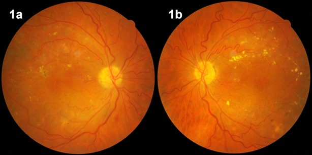
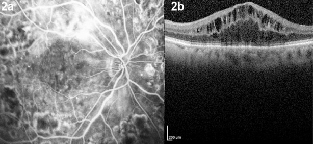
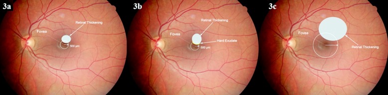
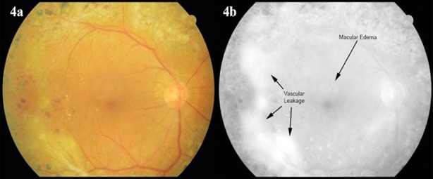
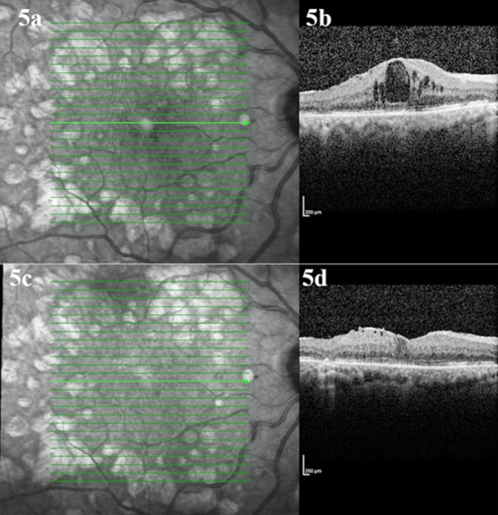
Diabetic macular edema (DME), one the most prevalent causes of visual loss in industrialized countries, may be diagnosed at any stage of diabetic retinopathy. The diagnosis, treatment, and follow up of DME have become straightforward with recent developments in fundus imaging, such as optical coherence tomography. Laser photocoagulation, intravitreal injections, and pars plana vitrectomy surgery are the current treatment modalities; however, the positive effects of currently available intravitreally injected agents are temporary. At this point, further treatment choices are needed for a permanent effect.
Sources of data selection: The articles published between 1985-2015 years on major databases were searched and most appropriate 40 papers were used to write this review article.
Keywords: Bevacizumab; Diabetic macular edema; Fluorescein angiography; Optical coherence tomography; Pars plana vitrectomy; Ranibizumab; Triamcinolone acetonide.
Figures
References
-
- Paulus YM, Gariano RF. Diabetic retinopathy: A growing concern in an aging population. Geriatrics. 2009;64(2):16–20. - PubMed
-
- Bhagat N, Grigorian RA, Tutela A, Zarbin MA. Diabetic macular edema: pathogenesis and treatment. Surv Ophthalmol. 2009;54(1):1–32. doi:10.1016/j.survophthal.2008.10.001. - PubMed
Publication types
LinkOut - more resources
Full Text Sources
Medical
Research Materials
Miscellaneous