Review
. 2016 May;203(1):65-107.
doi: 10.1534/genetics.115.186221.

Mechanism and Regulation of Protein Synthesis in Saccharomyces cerevisiae

Affiliations
Review

Mechanism and Regulation of Protein Synthesis in Saccharomyces cerevisiae

Thomas E Dever et al. Genetics. 2016 May.

Abstract

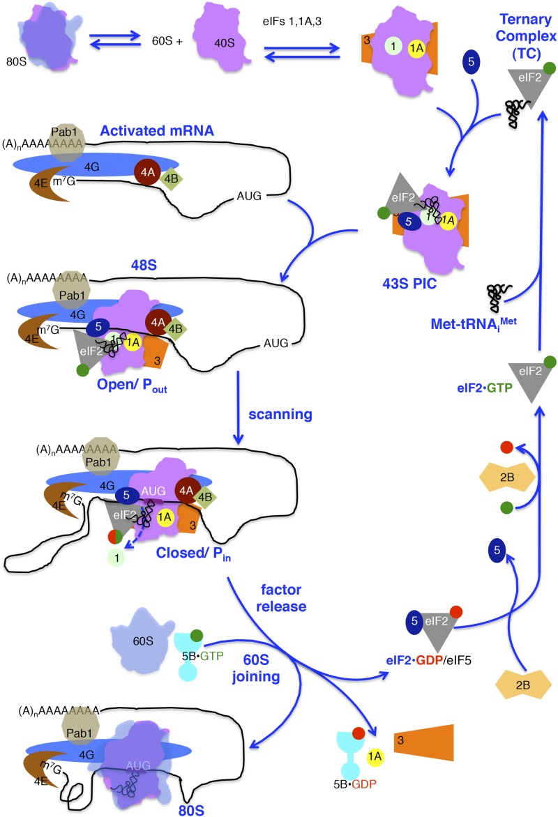
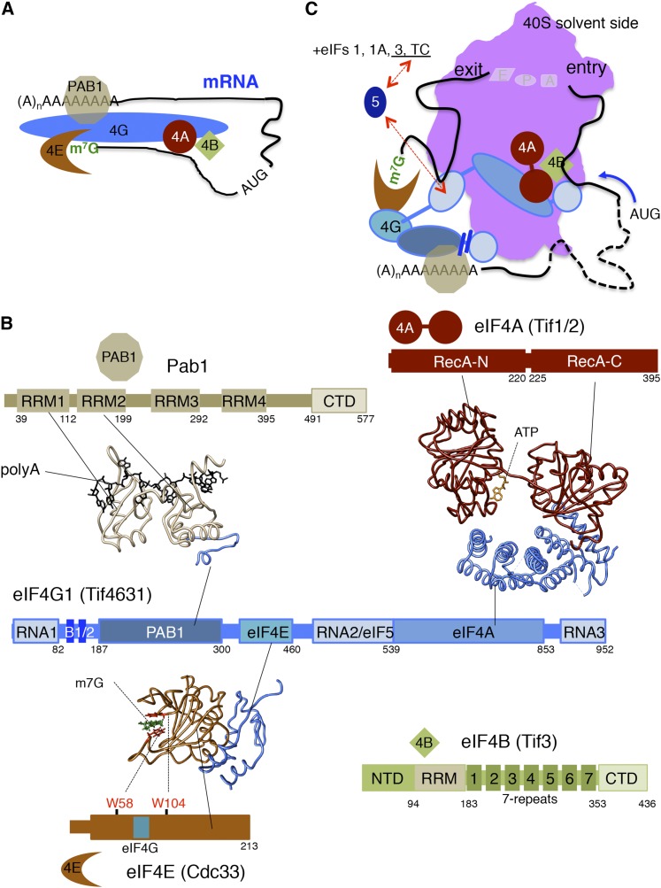
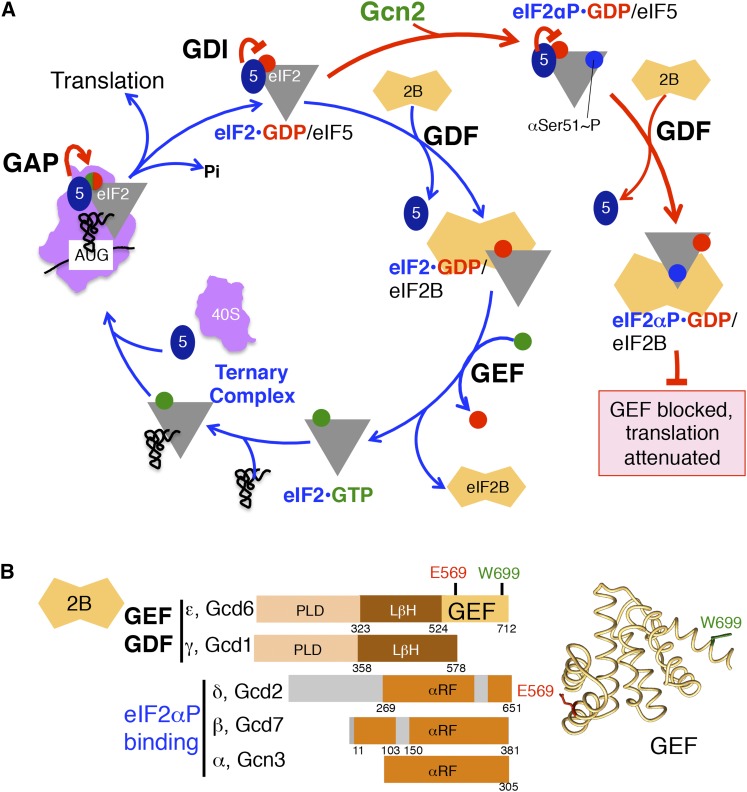
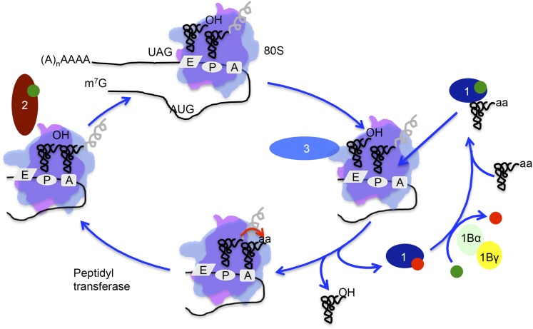
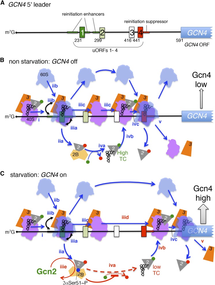
In this review, we provide an overview of protein synthesis in the yeast Saccharomyces cerevisiae The mechanism of protein synthesis is well conserved between yeast and other eukaryotes, and molecular genetic studies in budding yeast have provided critical insights into the fundamental process of translation as well as its regulation. The review focuses on the initiation and elongation phases of protein synthesis with descriptions of the roles of translation initiation and elongation factors that assist the ribosome in binding the messenger RNA (mRNA), selecting the start codon, and synthesizing the polypeptide. We also examine mechanisms of translational control highlighting the mRNA cap-binding proteins and the regulation of GCN4 and CPA1 mRNAs.

Keywords: GCN4; translation elongation; translation initiation.


Figures

Figure 1
Figure 1
Pathway for yeast cytoplasmic translation initiation. Protein synthesis begins with the dissociation of ribosomal subunits and assembly of a 43S PIC. This is shown as consecutive steps in which eukaryotic initiation factors (eIFs) 1, 1A, and 3 bind to the 40S subunit first, followed by the eIF2–GTP (green circle)–Met-tRNAiMet ternary complex (TC) and eIF5. The 43S PIC binds an activated mRNA near the 5′ cap, forming a 48S complex. Activated mRNAs bear eIF4E at the 5′ cap, Pab1 bound to the poly(A) tail, bridged by eIF4G to form a loop along with eIF4A and eIF4B. During scanning, the 43S PIC in an open conformation, where Met-tRNAiMet is not fully base paired within the P site (Pout), moves in a 3′ direction along the 5′ UTR to the AUG codon. Either prior to or upon AUG recognition, GTP bound to TC is hydrolyzed to GDP+Pi (green and red hybrid circle), but Pi is not released until AUG recognition. Start codon selection is accompanied by release of eIF1, Pi loss from eIF2–GDP (red circle), release of eIF2 and eIF5, and reorganization of the 43S PIC to a closed state with Met-tRNAiMet in the Pin conformation and tightly bound to the complex. eIF5B–GTP promotes joining of the 60S subunit to the AUG-bound PIC. GTP hydrolysis and release of eIF5B–GDP and eIF1A forms the 80S complex with Met-tRNAiMet bound in the P site and a vacant A site ready for the elongation phase of protein synthesis. Recycling of eIF2 is accomplished by eIF2B displacing eIF5 from eIF2–GDP and then facilitating nucleotide exchange on eIF2. Met-tRNAiMet binds to eIF2–GTP reforming TC.
Figure 2
Figure 2
tRNAiMet features important for translation initiation. Features that enhance tRNAiMet function in initiation or restrict it from functioning in elongation are highlighted on the tertiary structure of yeast tRNAiMet (pdb 1YFG). Highlighted residues include A1:U72 and C3:G70 base pairs in the acceptor stem, residues A54 and A60 in the T loop, and a 2’-O-ribosyl phosphate modification on residue A64. Three consecutive G:C base pairs in the anticodon loop are important for the accuracy of start site selection. The anticodon 5′-CAU-3′ is depicted in green. Structure was generated using the PyMol Molecular Graphics System (version 1.7.6.6, Schrödinger).
Figure 3
Figure 3
Schematic and structural models of eIF2 and eIF5. (A) Structural model of the eIF2–GTP–Met-tRNAiMet ternary complex bound to an mRNA AUG codon (right) and cartoons depicting the eIF2 α, β, and γ subunit structural domains (left) using the same color schemes. The structural model is adapted from the structure of the yeast 48S complex (pdb 3JAP) with the 40S ribosome and other initiation factors omitted for clarity (Llacer et al. 2015). The eIF2α residue Ser51 (blue), GTP analog (green), Met-tRNAiMet (gray), and mRNA (cyan) with AUG codon (yellow) are indicated. (B) eIF5 domains and activities (left) and structural models (right) for the human GAP domain bearing R15 (pdb 2E9H) and the yeast CTD bearing W391 (pdb 2FUL) (Wei et al. 2006). Structures were drawn using Chimera software (University of California, San Francisco, UCSF).
Figure 4
Figure 4
Schematic and structural models of eIF1, eIF1A, and AUG codon selection. (A) Structural model (right) and schematics (left) of eIF1 (green) and eIF1A (yellow) bound to the 48S PIC (pdb 3JAP) along with Met-tRNAiMet (black) and mRNA (blue, AUG codon in red), but other factors and the ribosome are removed for clarity. Structure was generated using the PyMol Molecular Graphics System (version 1.7.6.6, Schrödinger). (B) Cartoon showing approximate positions of eIFs 1 and 1A with TC and eIF5 in the open scanning conformation (left) with Met-tRNAiMet not fully engaged in the P site (Pout), and factor movements (black arrows) induced by AUG codon recognition (right) and the transition to the closed complex (Pin) signaled by movement of eIF1 that triggers Pi release prior to eIF2–GDP–eIF5 release from the PIC.
Figure 5
Figure 5
Schematic and structural models of eIF3. Schematics depict the eIF3 subunit organization and indicate major structural domains and protein–protein interactions (black arrows) within the eIF3 core complex. Structural models depicting these interactions are shown using Chimera software (UCSF) using pdb coordinates 4U1C (eIF3a/c), 4U1E (eIF3b-CTD/eIF3i/eIF3g-NTD), 4U1F (eIF3b β-propeller domain) (Erzberger et al. 2014), and 2KRB (eIF3b RRM/eIF3j peptide) (Elantak et al. 2010). The cartoon depicting eIF3 binding to the 40S solvent-exposed surface is based on cryo-EM reconstructions (Erzberger et al. 2014; Aylett et al. 2015; Llacer et al. 2015). The same color scheme is used for consistency between images.
Figure 6
Figure 6
Interactions among the m7G cap- and mRNA-binding factors. (A) Cartoon of mRNA recruitment step as in Figure 1. (B) Schematics of eIF4G (middle), Pab1 and eIF4A (top), and eIF4E and eIF4B (bottom). Factor binding domains on eIF4G are labeled, and structural models of the interacting factors are depicted. Structural models of human Pabp-poly(A)–eIF4G (Safaee et al. 2012), yeast eIF4A–eIF4G (Schutz et al. 2008), and yeast eIF4E–eIF4G (Gross et al. 2003) were drawn using Chimera software (UCSF). (C) Model for interactions of eIF4G domains with initiation factors and with the mRNA 5′ UTR on both the mRNA entrance and exit sides of the 40S ribosome to enhance mRNA binding to the ribosome.
Figure 7
Figure 7
Recycling and regulation of eIF2 by eIF2B. (A) Pathway of eIF2 nucleotide cycle and its regulation by eIF2α phosphorylation, adapted from Figure 1. GDI function of eIF5, GDF and GEF activities of eIF2B, and GAP function of eIF5 (5) are described in the text. Phosphorylation of eIF2α on Ser51 by GCN2 is represented by the blue circle; GDP, red circle; and GTP, green circle. (B) Schematics of eIF2B subunits and domain organization (left) and structure of the eIF2Bε GEF domain (right, pdb 1PAQ) (Boesen et al. 2004). Homologous domains are shown in identical color shades. PLD and LβH indicate the pyrophosphorylase-like and the left-handed β-helical domains, respectively (Reid et al. 2012). αRF indicates the α-helical domain followed by a Rossmann-like fold shared by the α-, β-, and δ-subunits. Structural models were drawn using Chimera software (UCSF).
Figure 8
Figure 8
Model of yeast translation elongation. Starting at the top, an elongating ribosome contains a peptidyl-tRNA in the P site and a deacylated tRNA in the E site. eEF1A(1)–GTP (green circle) binds aa-tRNA for delivery to the cognate codon in the A site. The codon–anticodon match in the A site triggers conformational changes in eEF1A, GTP hydrolysis, and release of eEF1A–GDP (red circle), leaving the aa-tRNA in the A site. Guanine nucleotide exchange on eEF1A is catalyzed by the α-subunit of the eEF1Bαγ complex. Following ribosome-catalyzed formation of the peptide bind, the ribosomal translocase eEF2 (2)–GTP stimulates movement of the A-site peptidyl-tRNA to the P site and of the now deacylated tRNA in the P site to the E site. The fungal-specific and essential factor eEF3 (3) interacts with eEF1A and is proposed to assist in the release of the E-site tRNA to allow continued cycles of elongation.
Figure 9
Figure 9
Translational regulation by reinitiation on GCN4 mRNA. (A) The GCN4 5′ leader sequences showing uORFs 1–4 and the start of the GCN4 ORF as filled boxes in their relative positions. The nucleotide positions of each AUG codon are shown relative to the transcription start site. The approximate location of reinitiation enhancer and suppressor sequences is indicated. (B) Reinitiation model in nonstarvation conditions with stepwise depiction of ribosomes and key factor interactions with the GCN4 leader sequence (cartoons as per Figure 1). Blue arrows depict the movement and blue numbered steps (i–v) are explained in the main text. Note: uORF spacing has been altered to accommodate the ribosome cartoons. As depicted, following uORF1 translation high TC levels enable reinitiation at uORF4 leading to ribosome disengagement from the mRNA and GCN4 expression is repressed. (C) Reinitiation model under amino acid starvation conditions. Initial steps through translation of uORF1 (blue numbered i–iiic) are unchanged from nonstarvation conditions. Subsequent steps (red numbered iiid–v) are altered by activation of the eIF2α kinase Gcn2 (step iiie) resulting in low levels of TC. Ribosomes traverse past uORF4 without initiating and then reacquire TC (step ivb). The scanning ribosome (step ivc) recognizes the GCN4 start codon and GCN4 expression is derepressed.

References

    1. Abastado J. P., Miller P. F., Jackson B. M., Hinnebusch A. G., 1991. Suppression of ribosomal reinitiation at upstream open reading frames in amino acid-starved cells forms the basis for GCN4 translational control. Mol. Cell. Biol. 11: 486–496. - PMC - PubMed
    1. Acker M. G., Shin B. S., Dever T. E., Lorsch J. R., 2006. Interaction between eukaryotic initiation factors 1A and 5B is required for efficient ribosomal subunit joining. J. Biol. Chem. 281: 8469–8475. - PubMed
    1. Acker M. G., Shin B. S., Nanda J. S., Saini A. K., Dever T. E., et al. , 2009. Kinetic analysis of late steps of eukaryotic translation initiation. J. Mol. Biol. 385: 491–506. - PMC - PubMed
    1. Advani V. M., Belew A. T., Dinman J. D., 2013. Yeast telomere maintenance is globally controlled by programmed ribosomal frameshifting and the nonsense-mediated mRNA decay pathway. Translation (Austin) 1: e24418. - PMC - PubMed
    1. Aitken C. E., Lorsch J. R., 2012. A mechanistic overview of translation initiation in eukaryotes. Nat. Struct. Mol. Biol. 19: 568–576. - PubMed

Publication types

MeSH terms

Substances

LinkOut - more resources