

Elastin-Derived Peptides Promote Abdominal Aortic Aneurysm Formation by Modulating M1/M2 Macrophage Polarization
- PMID: 27183603
- PMCID: PMC4880455
- DOI: 10.4049/jimmunol.1502454
Elastin-Derived Peptides Promote Abdominal Aortic Aneurysm Formation by Modulating M1/M2 Macrophage Polarization
Abstract
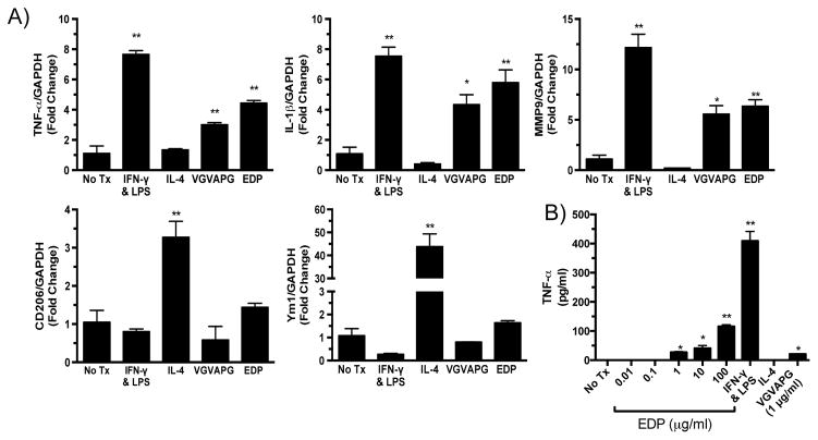
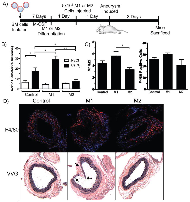
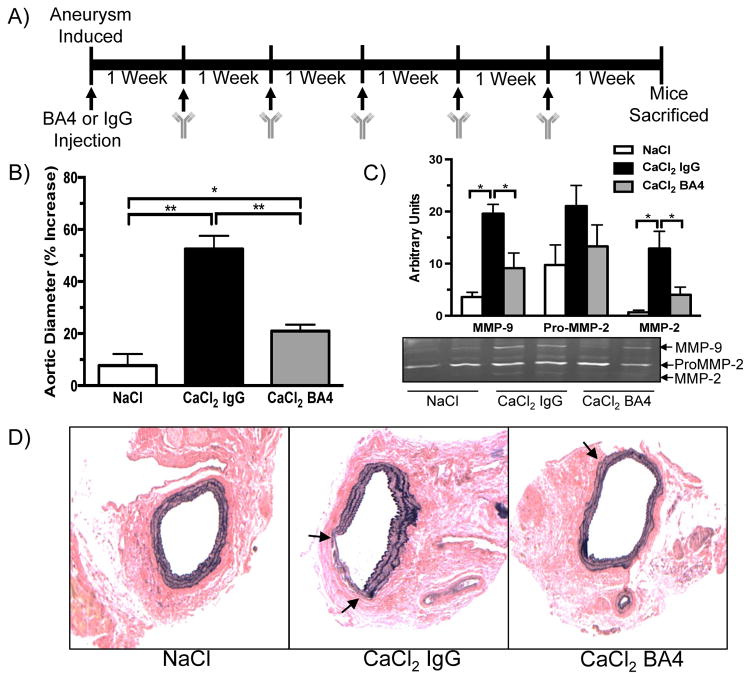
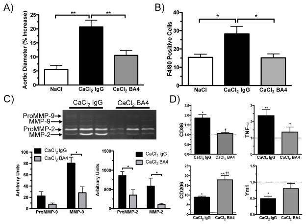
Abdominal aortic aneurysm is a dynamic vascular disease characterized by inflammatory cell invasion and extracellular matrix degradation. Damage to elastin in the extracellular matrix results in release of elastin-derived peptides (EDPs), which are chemotactic for inflammatory cells such as monocytes. Their effect on macrophage polarization is less well known. Proinflammatory M1 macrophages initially are recruited to sites of injury, but, if their effects are prolonged, they can lead to chronic inflammation that prevents normal tissue repair. Conversely, anti-inflammatory M2 macrophages reduce inflammation and aid in wound healing. Thus, a proper M1/M2 ratio is vital for tissue homeostasis. Abdominal aortic aneurysm tissue reveals a high M1/M2 ratio in which proinflammatory cells and their associated markers dominate. In the current study, in vitro treatment of bone marrow-derived macrophages with EDPs induced M1 macrophage polarization. By using C57BL/6 mice, Ab-mediated neutralization of EDPs reduced aortic dilation, matrix metalloproteinase activity, and proinflammatory cytokine expression at early and late time points after aneurysm induction. Furthermore, direct manipulation of the M1/M2 balance altered aortic dilation. Injection of M2-polarized macrophages reduced aortic dilation after aneurysm induction. EDPs promoted a proinflammatory environment in aortic tissue by inducing M1 polarization, and neutralization of EDPs attenuated aortic dilation. The M1/M2 imbalance is vital to aneurysm formation.
Copyright © 2016 by The American Association of Immunologists, Inc.
Figures


Similar articles
-
GDF15 suppresses abdominal aortic aneurysm by upregulating AREG expression to adjust macrophage polarization.Int Immunopharmacol. 2025 Jun 26;159:114899. doi: 10.1016/j.intimp.2025.114899. Epub 2025 May 24. Int Immunopharmacol. 2025. PMID: 40414071
-
IL-1β (Interleukin-1β) and TNF-α (Tumor Necrosis Factor-α) Impact Abdominal Aortic Aneurysm Formation by Differential Effects on Macrophage Polarization.Arterioscler Thromb Vasc Biol. 2018 Feb;38(2):457-463. doi: 10.1161/ATVBAHA.117.310333. Epub 2017 Dec 7. Arterioscler Thromb Vasc Biol. 2018. PMID: 29217508 Free PMC article.
-
D-series resolvins inhibit murine abdominal aortic aneurysm formation and increase M2 macrophage polarization.FASEB J. 2016 Dec;30(12):4192-4201. doi: 10.1096/fj.201600144RR. Epub 2016 Sep 12. FASEB J. 2016. PMID: 27619672 Free PMC article.
-
Macrophage polarization and metabolic reprogramming in abdominal aortic aneurysm.Immun Inflamm Dis. 2024 Nov;12(11):e1268. doi: 10.1002/iid3.1268. Immun Inflamm Dis. 2024. PMID: 39530309 Free PMC article. Review.
-
Targeting Macrophage Polarization in Infectious Diseases: M1/M2 Functional Profiles, Immune Signaling and Microbial Virulence Factors.Immunol Invest. 2024 Oct;53(7):1030-1091. doi: 10.1080/08820139.2024.2367682. Epub 2024 Jun 24. Immunol Invest. 2024. PMID: 38913937 Review.
Cited by
-
Distinct subsets of T cells and macrophages impact venous remodeling during arteriovenous fistula maturation.JVS Vasc Sci. 2020;1:207-218. doi: 10.1016/j.jvssci.2020.07.005. Epub 2020 Sep 1. JVS Vasc Sci. 2020. PMID: 33748787 Free PMC article.
-
The role of phosphoinositide 3-kinases in immune-inflammatory responses: potential therapeutic targets for abdominal aortic aneurysm.Cell Cycle. 2022 Nov;21(22):2339-2364. doi: 10.1080/15384101.2022.2094577. Epub 2022 Jul 6. Cell Cycle. 2022. PMID: 35792922 Free PMC article. Review.
-
Unraveling Elastic Fiber-Derived Signaling in Arterial Aging and Related Arterial Diseases.Biomolecules. 2025 Jan 21;15(2):153. doi: 10.3390/biom15020153. Biomolecules. 2025. PMID: 40001457 Free PMC article. Review.
-
Ex vivo magnetic particle imaging of vascular inflammation in abdominal aortic aneurysm in a murine model.Sci Rep. 2020 Jul 24;10(1):12410. doi: 10.1038/s41598-020-69299-y. Sci Rep. 2020. PMID: 32709967 Free PMC article.
-
Modulation of Immune-Inflammatory Responses in Abdominal Aortic Aneurysm: Emerging Molecular Targets.J Immunol Res. 2018 Jun 3;2018:7213760. doi: 10.1155/2018/7213760. eCollection 2018. J Immunol Res. 2018. PMID: 29967801 Free PMC article. Review.
References
-
- Thom T, Haase N, Rosamond W, et al. Heart disease and stroke statistics--2006 update: a report from the American Heart Association Statistics Committee and Stroke Statistics Subcommittee. Circulation. 2006;113:e85–151. - PubMed
-
- Shimizu K, Mitchell RN, Libby P. Inflammation and cellular immune responses in abdominal aortic aneurysms. Arterioscler Thromb Vasc Biol. 2006;26:987–994. - PubMed
-
- Botnar RM, Wiethoff AJ, Ebersberger U, Lacerda S, Blume U, Warley A, Jansen CH, Onthank DC, Cesati RR, Razavi R, Marber MS, Hamm B, Schaeffter T, Robinson SP, Makowski MR. In vivo assessment of aortic aneurysm wall integrity using elastin-specific molecular magnetic resonance imaging. Circ Cardiovasc Imaging. 2014;7:679–689. - PubMed
Publication types
MeSH terms
Substances
Grants and funding
LinkOut - more resources
Full Text Sources
Other Literature Sources
