Review
. 2016 May 15;196(10):3983-91.
doi: 10.4049/jimmunol.1600251.

Alloimmunity and Tolerance in Corneal Transplantation

Affiliations
Review

Alloimmunity and Tolerance in Corneal Transplantation

Afsaneh Amouzegar et al. J Immunol. .

Abstract

Corneal transplantation is one of the most prevalent and successful forms of solid tissue transplantation. Despite favorable outcomes, immune-mediated graft rejection remains the major cause of corneal allograft failure. Although low-risk graft recipients with uninflamed graft beds enjoy a success rate ∼90%, the rejection rates in inflamed graft beds or high-risk recipients often exceed 50%, despite maximal immune suppression. In this review, we discuss the critical facets of corneal alloimmunity, including immune and angiogenic privilege, mechanisms of allosensitization, cellular and molecular mediators of graft rejection, and allotolerance induction.


Figures

Figure 1
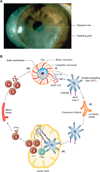
Figure 1. Immunobiology of corneal transplantation
A. Clinical manifestation of corneal graft rejection. Infiltrating monocytes and T cells attack the graft, often involving a rejection line that marches across the inner endothelial layer of the transplant, leaving an opaque and swollen graft behind. B. Schematic representation of corneal alloimmunity (I). Following transplant surgery, upregulation of pro-inflammatory cytokines, adhesion molecules and pro-angiogenic factors results in corneal infiltration of immune cells and formation of new blood and lymphatic vessels. (II). Antigen presenting cells (APCs), which acquire MHC class II and co-stimulatory molecules in the inflammatory environment, egress from the cornea through lymphatic vessels to the draining lymph nodes, where they present alloantigens to naïve T cells (Th0). Newly formed lymphatic vessels may also contribute to resolution of inflammation by mediating clearance of inflammatory cells and debris. (III). Primed T cells undergo clonal expansion and differentiate primarily into IFNγ-secreting CD4+ Th1 cells. Regulatory T cells (Tregs) modulate induction of alloimmune response either through inhibition of T cell activation or suppression of APC stimulatory potential. (IV). Alloreactive Th1 cells, migrate through blood vessels and along a chemokine gradient toward the graft, where they mount a delayed-type hypersensitivity response against the allogeneic tissue, resulting in graft opacification and failure.
Figure 2
Figure 2. Migration of donor graft-derived APCs and activation of the direct pathway of allosensitization
A. Ex vivo staining of corneal grafts with Hoechst vital dye tracks the egress of donor APCs posttransplantation. Stained isografts (BALB/c → BALB/c) demonstrates that ex vivo staining (blue; A1) is mostly restricted to the graft and not the host bed. In contrast, stained allografts (C57BL/6, IAb → BALB/c, IAd) evaluated at 24 h posttransplantation demonstrate that exiting donor cells in the host bed are largely CD45+ (red; A2) and express donor IAb (green; A3) (inlays are respective digitally enlarged portions of host beds). B. The frequencies of IFNγ-producing T cells 2 weeks after transplantation were assessed using ELISPOT. In high risk (HR) graft recipients, a significant higher IFNγ response is generated in directly primed allospecific T cells compared to low risk (LR) recipients and ungrafted controls (Naïve) (**p <0.01), suggesting that the direct alloresponse is the predominant form of allosensitization in the high-risk graft setting. Adopted and modified from Saban, D. R., et al. & Huq, S., et al.
Figure 3
Figure 3. Treg function and their interactions with antigen presenting cells in the draining lymph nodes of corneal allograft acceptors versus allograft rejectors
A. Mean fluorescent intensity (MFI) and Western blot analysis of Foxp3 expression in CD4+CD25+Foxp3+ Tregs from draining lymph nodes of allograft acceptors and rejectors 3 weeks posttransplantation demonstrates significantly higher expression levels of Foxp3 in Tregs from graft acceptors compared to Tregs from grafts undergoing rejection. B. Tregs isolated from the lymph nodes of graft acceptors are significantly more potent in suppressing the proliferation of CD3-stimulated naïve T cells compared to Tregs isolated from lymph nodes of graft rejectors and syngeneic recipients. C. Confocal micrographs of draining lymph nodes show that only Tregs from graft acceptors colocalize with CD11c+ APCs (white arrows, 60×). D. Adoptive transfer of CCR7hi Tregs into corneal allograft recipients significantly improves corneal allograft survival (*p=0.022). Allograft recipients that receive CCR7lo Tregs demonstrate no improvement in allograft survival. Adopted and modified from Chauhan, S. K., et al.,

References

    1. Zirm EK. Eine erfolgreiche totale Keratoplastik (A successful total keratoplasty). 1906. Refractive & corneal surgery. 1989;5:258–261. - PubMed
    1. Dana MR, Qian Y, Hamrah P. Twenty-five-year panorama of corneal immunology: emerging concepts in the immunopathogenesis of microbial keratitis, peripheral ulcerative keratitis, and corneal transplant rejection. Cornea. 2000;19:625–643. - PubMed
    1. Eye banking statistical report. Washington, DC: Eye Bank Association of America (EBAA); 2014.
    1. Anshu A, Price MO, Price FW., Jr Risk of corneal transplant rejection significantly reduced with Descemet's membrane endothelial keratoplasty. Ophthalmology. 2012;119:536–540. - PubMed
    1. van Dooren BT, Mulder PG, Nieuwendaal CP, Beekhuis WH, Melles GR. Endothelial cell density after deep anterior lamellar keratoplasty (Melles technique) American journal of ophthalmology. 2004;137:397–400. - PubMed

LinkOut - more resources