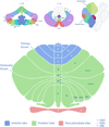
Structure-function relationships in the developing cerebellum: Evidence from early-life cerebellar injury and neurodevelopmental disorders
- PMID: 27184461
- PMCID: PMC5282860
- DOI: 10.1016/j.siny.2016.04.010
Structure-function relationships in the developing cerebellum: Evidence from early-life cerebellar injury and neurodevelopmental disorders
Abstract
The increasing appreciation of the role of the cerebellum in motor and non-motor functions is crucial to understanding the outcomes of acquired cerebellar injury and developmental lesions in high-risk fetal and neonatal populations, children with cerebellar damage (e.g. posterior fossa tumors), and neurodevelopmental disorders (e.g. autism). We review available data regarding the relationship between the topography of cerebellar injury or abnormality and functional outcomes. We report emerging structure-function relationships with specific symptoms: cerebellar regions that interconnect with sensorimotor cortices are associated with motor impairments when damaged; disruption to posterolateral cerebellar regions that form circuits with association cortices impact long-term cognitive outcomes; and midline posterior vermal damage is associated with behavioral dysregulation and an autism-like phenotype. We also explore the impact of age and the potential role for critical periods on cerebellar structure and child function. These findings suggest that the cerebellum plays a critical role in motor, cognitive, and social-behavioral development, possibly via modulatory effects on the developing cerebral cortex.
Keywords: Autism; Cerebellum; Cognition; Developmental diaschisis; Fetal; Preterm.
Copyright © 2016 Elsevier Ltd. All rights reserved.
Conflict of interest statement
statement None declared.
Figures

References
-
- Limperopoulos C, Soul JS, Gauvreau K, et al. Late gestation cerebellar growth is rapid and impeded by premature birth. Pediatrics. 2005;115:688–695. - PubMed
-
- Clouchoux C, Guizard N, Evans AC, du Plessis AJ, Limperopoulos C. Normative fetal brain growth by quantitative in vivo magnetic resonance imaging. Am J Obstet Gynecol. 2012;206:173. e1–8. - PubMed
Publication types
MeSH terms
Grants and funding
LinkOut - more resources
Full Text Sources
Other Literature Sources

