

Malignant-looking thyroid nodules with size reduction: core needle biopsy results
- PMID: 27184652
- PMCID: PMC5040134
- DOI: 10.14366/usg.15082
Malignant-looking thyroid nodules with size reduction: core needle biopsy results
Abstract
Purpose: The aim of this study was to evaluate whether malignant-looking thyroid nodules with size reduction were malignant or not.
Methods: From November 2010 to July 2011, we retrospectively enrolled 16 patients with 16 nodules (11 females and five males; mean age, 55 years) who underwent core needle biopsy (CNB), and whose thyroid nodules had malignant ultrasonographic (US) features, although they showed size reduction (>20% decrease in maximum diameter) during the follow-up period (mean, 37±27 months). The histologic findings of the CNB specimen were reviewed and correlated with the US findings. US studies were analyzed for their internal content, shape, margin, echogenicity, the presence of microcalcification and macrocalcification, inner isoechoic rim, and low-echoic halo.
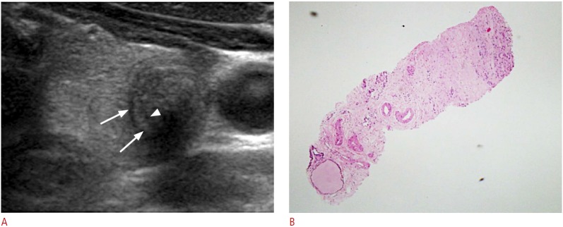
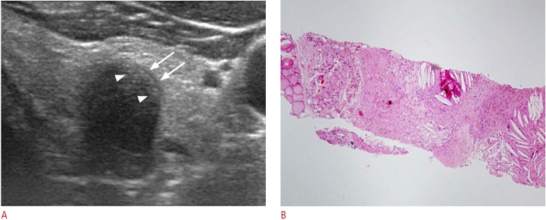
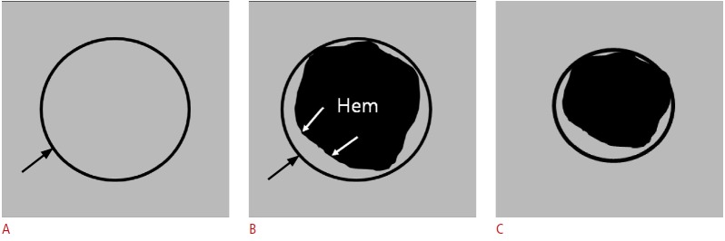
Results: All nodules were confirmed as benign by CNB. Pathologic analysis was available for 12 CNB specimens. US imaging showed central hypoechogenicity or marked hypoechogenicity in all cases and a peripheral isoechoic rim in 15 nodules. US-pathologic correlation showed that the central hypoechoic area was primarily composed of fibrosis (12/12) and hemorrhage (8/12) and that the isoechoic rim was composed of follicular cells.
Conclusion: In our study, the CNB results of all of the malignant-looking thyroid nodules with size reduction were benign and were primarily composed of internal fibrosis and hemorrhage. Understanding these US and pathologic features could prevent repeated fine-needle aspiration or unnecessary diagnostic surgery.
Keywords: Biopsy, fine-needle; Biopsy, large-core needle; Thyroid nodule; Ultrasonography.
Conflict of interest statement
No potential conflict of interest relevant to this article was reported.
Figures
References
-
- American Thyroid Association (ATA) Guidelines Taskforce on Thyroid Nodules and Differentiated Thyroid Cancer. Cooper DS, Doherty GM, Haugen BR, Kloos RT, Lee SL, et al. Revised American Thyroid Association management guidelines for patients with thyroid nodules and differentiated thyroid cancer. Thyroid. 2009;19:1167–1214. - PubMed
-
- Gharib H, Papini E, Paschke R, Duick DS, Valcavi R, Hegedus L, et al. American Association of Clinical Endocrinologists, Associazione Medici Endocrinologi, and EuropeanThyroid Association medical guidelines for clinical practice for the diagnosis and management of thyroid nodules. Endocr Pract. 2010;16 Suppl 1:1–43. - PubMed
-
- Koo JH, Shin JH, Han BK, Ko EY, Kang SS. Cystic thyroid nodules after aspiration mimicking malignancy: sonographic characteristics. J Ultrasound Med. 2010;29:1415–1421. - PubMed
LinkOut - more resources
Full Text Sources
Other Literature Sources
