. 2017 Jan;8(1):44-55.
doi: 10.1111/jdi.12540. Epub 2016 Jul 8.

Mild hyperglycemia triggered islet function recovery in streptozotocin-induced insulin-deficient diabetic rats

Affiliations

Mild hyperglycemia triggered islet function recovery in streptozotocin-induced insulin-deficient diabetic rats

Yu Cheng et al. J Diabetes Investig. 2017 Jan.

Abstract

Aims/introduction: Moderate elevation of glucose level has been shown to effectively promote β-cell replication in various models in vitro and in normal rodents. Here, we aimed to test the effect of moderately elevated glucose on β-cell mass expansion and islet function recovery in diabetic animal models.

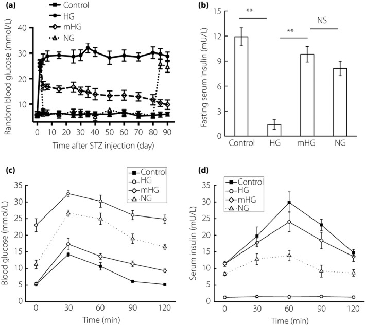
Materials and methods: A single high dose of streptozotocin was given to induce insulin-deficient diabetes in adult male Sprague-Dawley rats. Then, 48 h after streptozotocin injection, newly diabetic rats were randomly divided into three groups: (i) no treatment to maintain hyperglycemia; (ii) daily exogenous long-acting human insulin analog injection that maintained mild hyperglycemia (15 mmol/L < blood glucose < 18 mmol/L); (iii) daily exogenous long-acting human insulin analog injection to restore normoglycemia (blood glucose <8 mmol/L) as a control. Islet function, β-cell regeneration and β-cell replication were monitored during the entire analysis period.

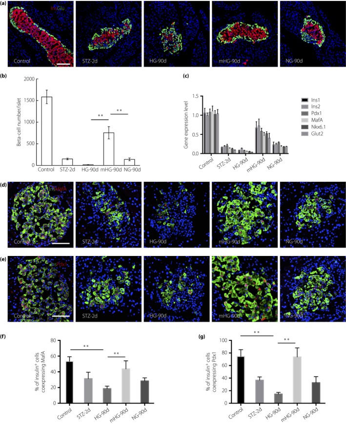
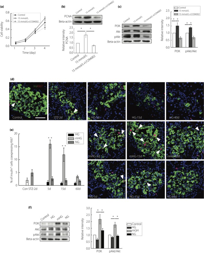
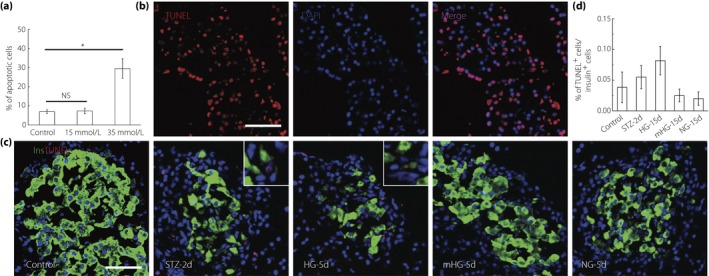
Results: A single high dose of streptozotocin induced massive loss of β-cells, resulting in irreversible hyperglycemia. Mild hyperglycemia markedly promoted β-cell proliferation, leading to robust β-cell regeneration. Importantly, rats that maintained mild hyperglycemia showed nearly normal glucose-stimulated insulin secretion, glucose disposal and random blood glucose levels, suggesting almost full restoration of the islet function. Normalization of blood glucose levels profoundly blunted β-cell replication, regeneration and islet function recovery observed in mild hyperglycemia.

Conclusions: Our research provides a feasible approach to stimulate in situ β-cell regeneration in diabetic rats, offering new perspectives for diabetes therapy.

Keywords: Islet function recovery; Mild hyperglycemia; β-Cell proliferation.


Figures

Figure 1
Figure 1
Chronic mild hyperglycemia incubation reversed diabetes in single high‐dose streptozotocin (STZ)‐treated rats. Eight‐week‐old male SD rats were treated with a single high dose of STZ (65 mg/kg), and 48 h post‐STZ injection newly diabetic rats (blood glucose >25 mmol/L) were randomly treated with: no treatment (HG; n = 6), daily exogenous long‐acting human insulin analog injection that restored normoglycemia (NG; blood glucose <8 mmol/L, n = 8), or maintained mild hyperglycemia (mHG; 15 mmol/L < blood glucose < 18 mmol/L, n = 15). Six normal rats were used as normal controls. Insulin withdrawal was executed within 30 days in the mHG group and at day 80 in the NG group. (a) Random blood glucose levels were monitored for 90 days. (b) Fasting serum insulin levels were detected at the end of observation. (c,d) Blood glucose levels and insulin levels during intraperitoneal glucose tolerance tests at the end of observation. The results are presented as the mean ± standard deviation. **P < 0.01. NS, not significant.
Figure 2
Figure 2
Mild hyperglycemia resulted in robust β‐cell regeneration. (a) Representative islets stained with antibodies against insulin (red) and glucagon (green) of normal rats (Control; n = 6), streptozotocin (STZ)‐treated rats at day 2 (STZ‐2d; n = 6) and rats at day 90 after no treatment (HG‐90d; n = 6), mild hyperglycemia incubation (mGH‐90d; n = 8) and euglycemia incubation (NG‐90d; n = 8). Nuclei were labeled with 40′,6‐diamidino‐2‐phenylindole. Scale bars, 100 μm. (b) Quantification of β‐cell number. The results are presented as the mean ± standard deviation. **P < 0.01. (c) Expression of β‐cell enriched genes in isolated islets (pooled from five rats). Gene expression levels were shown as fold change to the levels of normal rats. The results are presented as the mean ± standard deviation. (d) Photomicrographs of islets double stained with anti‐insulin (green) and anti‐v‐maf musculopeoneurotic fiberosarcoma oncogene homologue A (MafA; red) antibodies of Control, STZ‐2d and HG‐90d, mGH‐90d and NG‐90d. Nuclei were labeled with 40′,6‐diamidino‐2‐phenylindole. Scale bars, 50 μm. (e) Photomicrographs of islets double stained with anti‐insulin (green) and anti‐Pdx1 (red) antibodies. Nuclei were labeled with 40′,6‐diamidino‐2‐phenylindole. Scale bars, 50 μm. (f) The percentage of insulin+ cells co‐expressing MafA (n = 5) was quantified. The results are presented as the mean ± standard deviation. **P < 0.01. (g) The percentage of insulin+ cells co‐expressing Pdx1 (n = 5) was quantified. The results are presented as the mean ± standard deviation. **P < 0.01.
Figure 3
Figure 3
Mild hyperglycemia enhanced β‐cell replication. (a) INS1 cells were treated with 11.1 mmol/L glucose, 15 mmol/L glucose or a combination of 15 mmol/L glucose and a PI3K inhibitor (Ly294002, 10 mol/L). Cell viability of these cells was detected at indicated time‐points. Values are mean ± standard deviation of three individual experiments. (b) Western blot detection of proliferating cell nuclear antigen (PCNA) in INS1 cells exposed to 11.1 mmol/L glucose, 15 mmol/L glucose or a combination of 15 mmol/L glucose and 10 mol/L Ly294002, with densitometry analysis. (c) Western blot detection of PI3K, Akt and pAkt in INS1 cells exposed to 11.1 mmol/L glucose, 15 mmol/L glucose or a combination of 15 mmol/l glucose and 10 mol/L Ly294002, with densitometry analysis. (d) Representative islets stained with antibodies against insulin (green) and Ki67 (red) of normal rats (Control; n = 5), streptozotocin (STZ)‐treated rats at day 2 (STZ‐2d; n = 5), and rats at day 5, day 15 and day 60 with no treatment (HG‐5d, HG‐15d, HG‐60d; n = 4), mild hyperglycemia incubation (mHG‐5d, mHG‐15d, mHG‐60d; n = 4) and euglycemia incubation (NG‐5d, NG‐15d, NG‐60d; n = 4). The arrows: insulin+ cells showing Ki67 expression. Nuclei were labeled with 40′,6‐diamidino‐2‐phenylindole. Scale bars, 50 μm. (e) Quantification of insulin+ cells co‐expressing Ki67 was shown. The results are presented as the mean ± standard deviation. (f) Western blot detection of PI3K, Akt and pAkt in isolated islets of normal rats, and rats at day 15 with no treatment (HG; n = 3), hyperglycemia incubation (mHG; n = 3) and euglycemia incubation (NG; n = 3), with densitometry analysis. The results of western blot are presented as the mean ± standard deviation from three independent experiments. *P < 0.05, **P < 0.01.
Figure 4
Figure 4
Mild hyperglycemia did not induce β‐cell apoptosis. (a) Apoptosis quantification in INS1 cells after 72‐h culture in 11.1 mmol/L, 15 mmol/L and 35 mmol/L glucose. Values are mean ± standard deviation (n = 6) of three individual experiments. *P < 0.05. (b) Pancreas sections were incubated with DNase I recombinant for 10 min to induce deoxyribonucleic acid strand breaks before terminal dexynucleotidyl transferase‐mediated dUTP nick end labeling labeling (red) as positive control. Nuclei were labeled with 40′,6‐diamidino‐2‐phenylindole. Scale bars, 50 μm. (c) Representative islets labeling with insulin (green) and terminal dexynucleotidyl transferase‐mediated dUTP nick end labeling (red) of normal rats (Control; n = 6), streptozotocin (STZ)‐treated rats at day 2 (STZ‐2d; n = 6), and rats at day 5 with no treatment (HG‐5d; n = 6), mild hyperglycemia incubation (mHG‐5d; n = 6) and euglycemia incubation (NG‐5d; n = 6). Nuclei were labeled with 40′,6‐diamidino‐2‐phenylindole. Scale bars, 50 μm. (d) Quantification of apoptosis. The results are presented as the mean ± standard deviation.
Figure 5
Figure 5
Chronic mild hyperglycemia slightly enhanced α‐cell replication with no excess glucagon secretion. (a) Representative islets stained with antibodies against glucagon (green) and Ki67 (red) of normal rats (Control; n = 5), streptozotocin (STZ)‐treated rats at day 2 (STZ‐2d; n = 5), and rats at day 5, day 15 and day 60 with no treatment (HG‐5d, HG‐15d, HG‐60d; n = 4), mild hyperglycemia incubation (mHG‐5d, mHG‐15d, mHG‐60d; n = 4) and euglycemia incubation (NG‐5d, NG‐15d, NG‐60d; n = 4). The arrows: glucagon+ cells showing Ki67 expression. Nuclei were labeled with 40′,6‐diamidino‐2‐phenylindole. Scale bars, 50 μm. (b) Quantification of glucagon+ cells co‐expressing Ki67. The results are presented as the mean ± standard deviation. *P < 0.05, **P < 0.01. Quantification of (c) α‐cell number was shown in and (d) serum glucagon level. The results are presented as the mean ± standard deviation. *P < 0.05, **P < 0.01.

References

    1. Atkinson MA, Bluestone JA, Eisenbarth GS, et al How does type 1 diabetes develop?: the notion of homicide or beta‐cell suicide revisited. Diabetes 2011; 60: 1370–1379. - PMC - PubMed
    1. Yi P, Park JS, Melton DA. Betatrophin: a hormone that controls pancreatic beta cell proliferation. Cell 2013; 153: 747–758. - PMC - PubMed
    1. Porat S, Weinberg‐Corem N, Tornovsky‐Babaey S, et al Control of pancreatic beta cell regeneration by glucose metabolism. Cell Metab 2011; 13: 440–449. - PMC - PubMed
    1. Hugl SR, White MF, Rhodes CJ. Insulin‐like growth factor I (IGF‐I)‐stimulated pancreatic β‐cell growth is glucose‐dependent: synergistic activation of insulin receptor substrate‐mediated signal transduction pathways by glucose and IGF‐I in INS‐1 cells. J Biol Chem 1998; 273: 17771–17779. - PubMed
    1. Assmann A, Ueki K, Winnay JN, et al Glucose effects on beta‐cell growth and survival require activation of insulin receptors and insulin receptor substrate 2. Mol Cell Biol 2009; 29: 3219–3228. - PMC - PubMed

MeSH terms