. 2016 Jun 15;5(6):669-77.
doi: 10.1242/bio.015552.

MicroRNA-93 promotes the malignant phenotypes of human glioma cells and induces their chemoresistance to temozolomide

Affiliations

MicroRNA-93 promotes the malignant phenotypes of human glioma cells and induces their chemoresistance to temozolomide

Rui Chen et al. Biol Open. .

Abstract

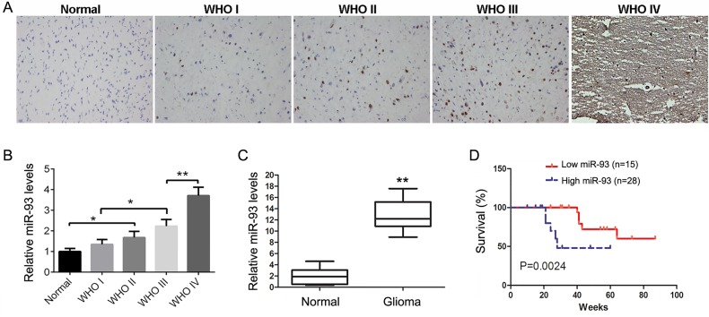
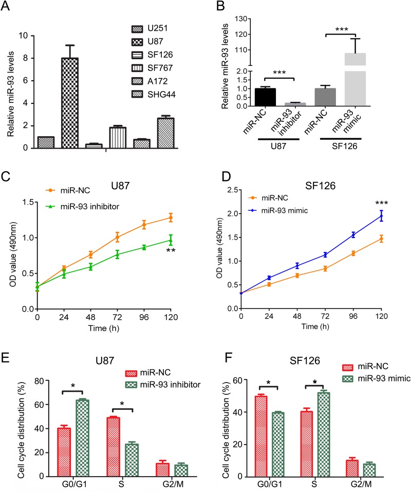
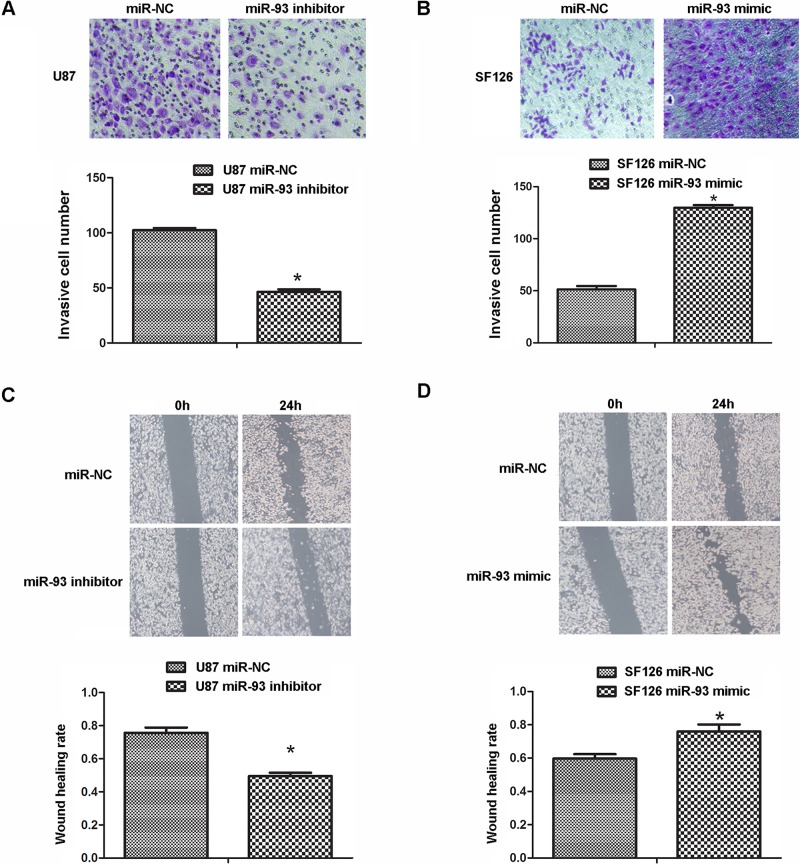
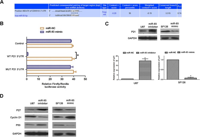
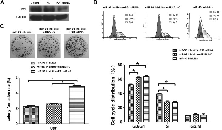
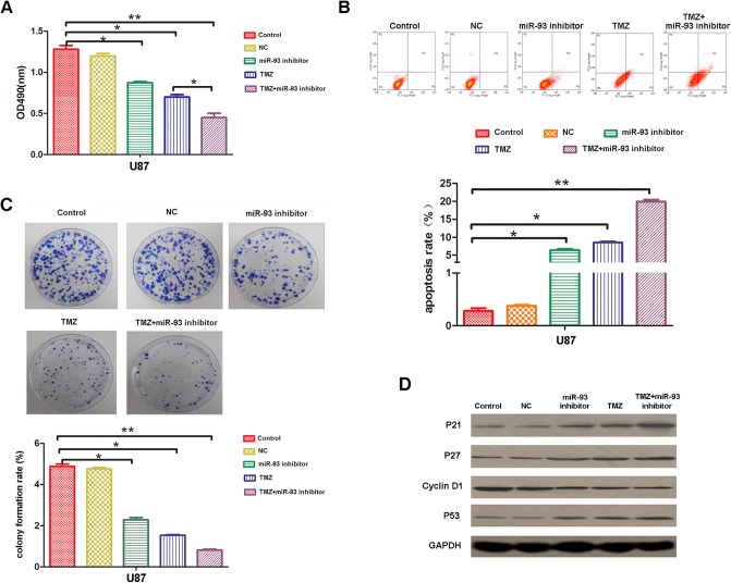
MicroRNAs (miRNAs), a class of small non-coding RNAs, can induce mRNA degradation or repress translation by binding to the 3'-untranslated region (UTR) of its target mRNA. Recently, some specific miRNAs, e.g. miR-93, have been found to be involved in pathological processes by targeting some oncogenes or tumor suppressors in glioma. However, the regulatory mechanism of miR-93 in the biological behaviors and chemoresistance of glioma cells remains unclear. In the present study, in situ hybridization and real-time RT-PCR data indicated that miR-93 was significantly upregulated in glioma patients (n=43) compared with normal brain tissues (n=8). Moreover, the upregulated miR-93 level was significantly associated with the advanced malignancy. We also found that upregulation of miR-93 promoted the proliferation, migration and invasion of glioma cells, and that miR-93 was involved in the regulation of cell cycle progression by mediating the protein levels of P21, P27, P53 and Cyclin D1. P21 was further identified as a direct target of miR-93. Knockdown of P21 attenuated the suppressive effects of miR-93 inhibition on cell cycle progression and colony formation. In addition, inhibition of miR-93 enhanced the chemosensitization of glioma cells to temozolomide (TMZ). Based on these above data, our study demonstrates that miR-93, upregulated in glioma, promotes the proliferation, cell cycle progression, migration and invasion of human glioma cells and suppresses their chemosensitivity to TMZ. Therefore, miR-93 may become a promising diagnostic marker and therapeutic target for glioma.

Keywords: Cell cycle; Glioma; Invasion; MicroRNA; Proliferation; Temozolomide.


Conflict of interest statement

Competing interests

The authors declare no competing or financial interests.

Figures

Fig. 1.
Fig. 1.
The expression of miR-93 in glioma. (A) Representative images of in situ hybridization staining in glioma tissues. Magnification, 200×. (B) Relative score of miR-93 expression in normal brain tissues and in different grade glioma tissues, indicating that the miR-93 level was gradually upregulated as the advanced malignancy of glioma. Data represented as mean±s.d; *P<0.05, **P<0.01. (C) Real-time RT-PCR data showed that miR-93 was significantly upregulated in glioma tissues compared to normal brain tissues. **P<0.01. (D) Kaplan–Meier curves showed worse overall survival rates for glioma patients with high miR-93 expression (n=28) compared to patients with low miR-93 expression (n=15) (P=0.0024).
Fig. 2.
Fig. 2.
Downregulation of miR-93 inhibits cell proliferation and arrests cell cycle in U87 and SF126 cells. (A,B) Real-time RT-PCR was performed to analyze the miR-93 levels in several glioma cell lines including U87, U251, SF126, SF767, A172 and SHG44 (A), and in U87 and SF126 cells transfected with miR-93 inhibitor or mimic, respectively (B). Cells transfected with scramble miRNA (miR-NC) were used as control. (C,D) MTT assay was performed to determine the cell proliferation in U87 cells (C) and SF126 cells (D) after miR-93 inhibitor or mimic transfection. (E,F) Cell cycle analysis was performed to examine the cell cycle distribution in U87 cells (E) and SF126 cells (F) after miR-93 inhibitor or mimic transfection. Data represented as mean±s.d; *P<0.05, **P<0.01, ***P<0.001 vs miR-NC.
Fig. 3.
Fig. 3.
Downregulation of miR-93 inhibits cell invasion in U87 and SF126 cells. U87 and SF126 cells were transfected with miR-93 inhibitor or mimic, respectively. Cells transfected with scramble miRNA (miR-NC) were used as control. (A,B) Transwell assay was conducted to examine the cell invasion in U87 cells (A) and SF126 cells (B). (C,D) Wound healing assay was performed to determine the cell migration in U87 cells (C) and SF126 cells (D). Data represented as mean±s.d; *P<0.05 vs miR-NC.
Fig. 4.
Fig. 4.
MiR-93 directly targets p21. (A) Targetscan software data indicated that CDKN1A (P21) was a potential target of miR-93. (B) We constructed the wild-type (WT) P21 3′UTR containing the putative binding sequences (GCACUUU) of miR-93 and the mutant type (MUT) P21 3′UTR with the binding sequences were changed into ‘AAAAAAA’. We then inserted them into the pMIR- REPORT miRNA Expression Reporter vector, generating WT P21-3′UTR plasmid and MUT P21-3′UTR plasmid, respectively. HEK293 cells were co-transfected with WT P21-3′UTR or MUT P21-3′UTR plasmid, and miR-NC or miR-93 mimic, respectively. After co-transfection for 48 h, dual-luciferase reporter assay was performed to examine the luciferase activity in each group. *P<0.05. (C,D) U87 and SF126 cells were transfected with miR-93 inhibitor or mimic, respectively. Cells transfected with scramble miRNA (miR-NC) were used as control. Western blot was conducted to examine the protein expression of P21 (C), P27, P53 and Cyclin D1 (D) in each group. GAPDH was used as an internal reference. Data represented as mean±s.d; *P<0.05.
Fig. 5.
Fig. 5.
Knockdown of p21 reverses miR-93 inhibitor mediated inhibition of cell proliferation. (A) U87 cells were transfected with non-specific siRNA (NC) or P21 siRNA, respectively. Western blot was conducted to examine the protein expression of P21. (B) Cell cycle analysis was performed to examine the cell cycle distribution in each group, and quantification. (C) Colony formation assay was performed to examine the colony formation capacity in each group, and formation quantified. Data represented as mean±s.d; *P<0.05.
Fig. 6.
Fig. 6.
Knockdown of miR-93 enhances the chemosensitivity of U87 cells to TMZ. U87 cells were treated with miR-93 inhibitor or TMZ alone, or co-treated with both. Cells without any treatment or treated with miR-NC were used as mock or negative control. (A) MTT assay was used to measure the cell proliferation in each group. (B) Cell apoptosis assay was used to determine the cell apoptosis in each group. (C) Colony formation assay was used to evaluate colony formation capacity in each group. (D) Western blot was conducted to examine the protein expression of P21, P27, P53 and Cyclin D1 in each group. GAPDH was used as an internal reference. Data represented as mean±s.d; *P<0.05, **P<0.01, ***P<0.001.

Similar articles

Cited by

References

    1. Agarwal R. (2000). Cell signaling and regulators of cell cycle as molecular targets for prostate cancer prevention by dietary agents. Biochem. Pharmacol. 60, 1051-1059. 10.1016/S0006-2952(00)00385-3 - DOI - PubMed
    1. An L., Liu Y., Wu A. and Guan Y. (2013). microRNA-124 inhibits migration and invasion by down-regulating ROCK1 in glioma. PLoS ONE 8, e69478 10.1371/journal.pone.0069478 - DOI - PMC - PubMed
    1. Areeb Z., Stylli S. S., Koldej R., Ritchie D. S., Siegal T., Morokoff A. P., Kaye A. H. and Luwor R. B. (2015). MicroRNA as potential biomarkers in Glioblastoma. J. Neurooncol. 125, 237-248. 10.1007/s11060-015-1912-0 - DOI - PubMed
    1. Auffinger B., Thaci B., Ahmed A., Ulasov I. and Lesniak M. S. (2013). MicroRNA targeting as a therapeutic strategy against glioma. Curr. Mol. Med. 13, 535-542. 10.2174/1566524011313040006 - DOI - PubMed
    1. Buscemi G., Ricci C., Zannini L., Fontanella E., Plevani P. and Delia D. (2014). Bimodal regulation of p21(waf1) protein as function of DNA damage levels. Cell Cycle 13, 2901-2912. 10.4161/15384101.2014.946852 - DOI - PMC - PubMed