. 2016 Jul 21;84(8):2162-2174.
doi: 10.1128/IAI.00190-16. Print 2016 Aug.

Crude Preparations of Helicobacter pylori Outer Membrane Vesicles Induce Upregulation of Heme Oxygenase-1 via Activating Akt-Nrf2 and mTOR-IκB Kinase-NF-κB Pathways in Dendritic Cells

Affiliations

Crude Preparations of Helicobacter pylori Outer Membrane Vesicles Induce Upregulation of Heme Oxygenase-1 via Activating Akt-Nrf2 and mTOR-IκB Kinase-NF-κB Pathways in Dendritic Cells

Su Hyuk Ko et al. Infect Immun. .

Abstract

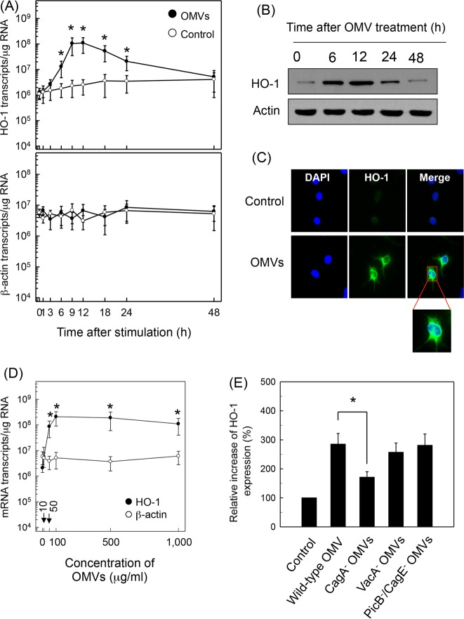
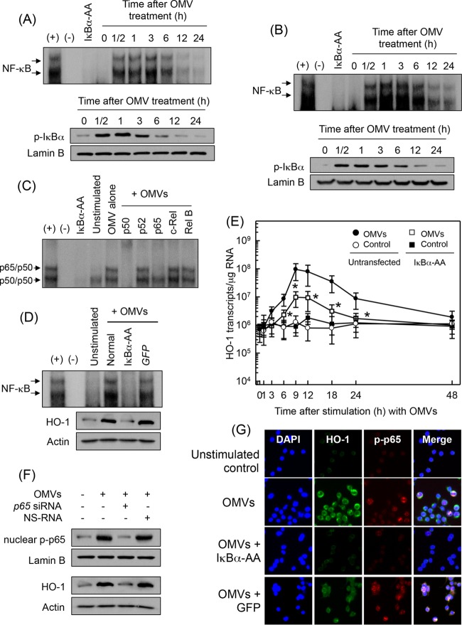
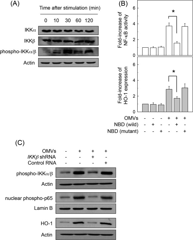
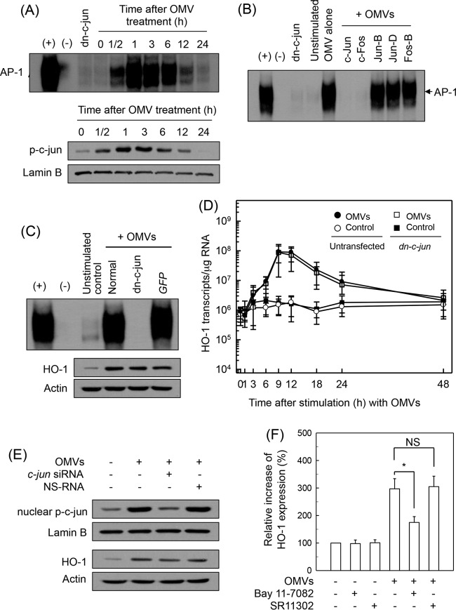
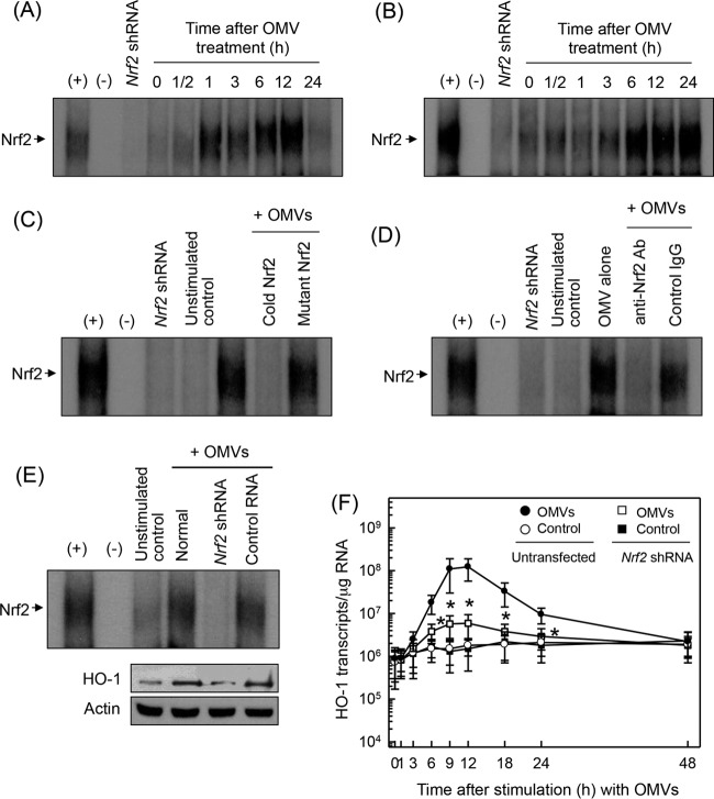
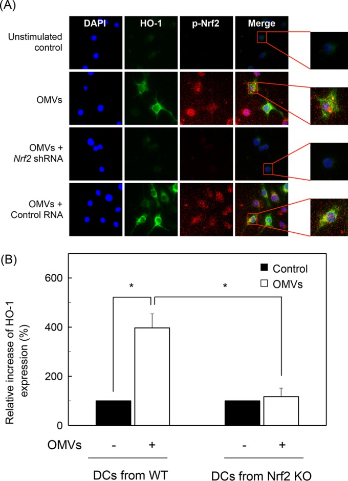
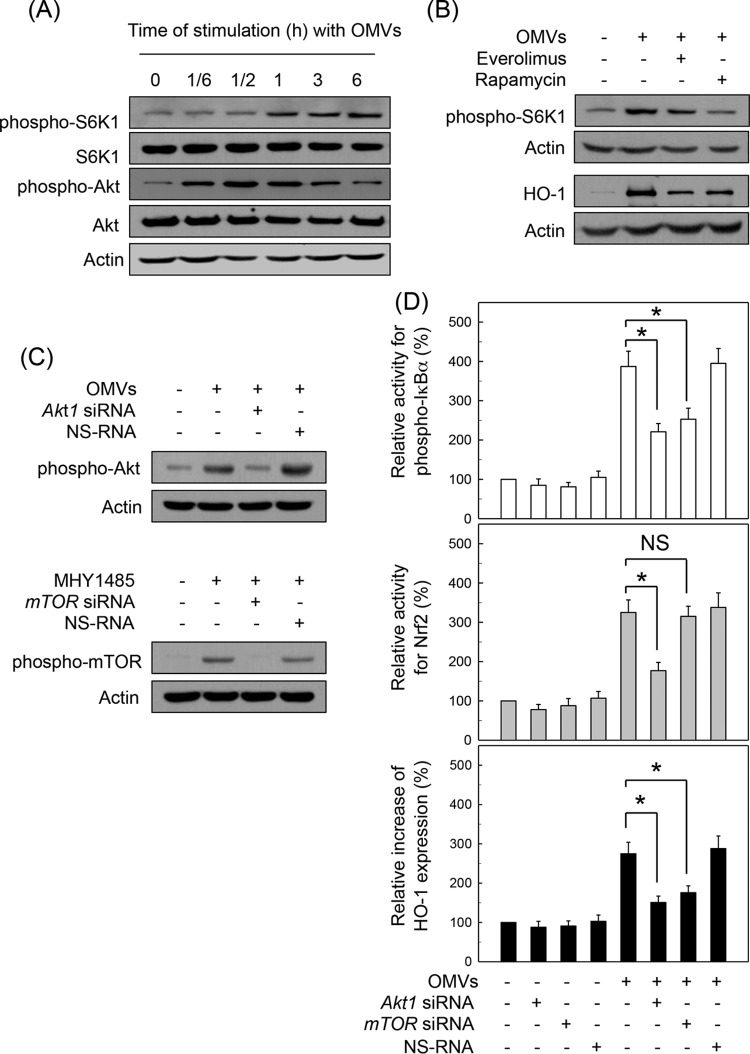
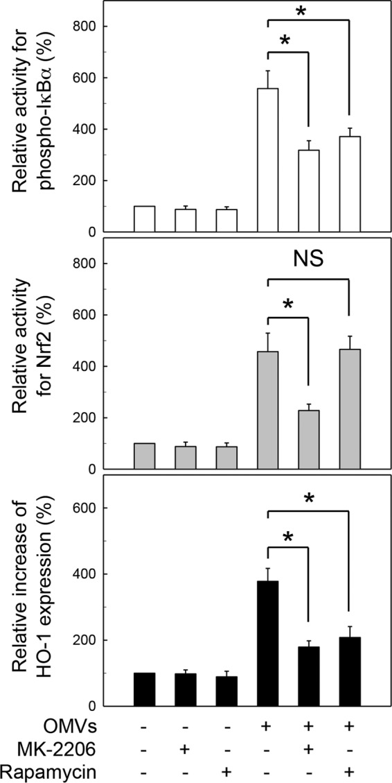
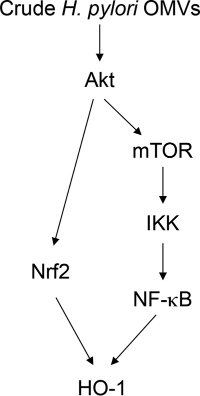
Helicobacter pylori sheds outer membrane vesicles (OMVs) that contain many surface elements of bacteria. Dendritic cells (DCs) play a major role in directing the nature of adaptive immune responses against H. pylori, and heme oxygenase-1 (HO-1) has been implicated in regulating function of DCs. In addition, HO-1 is important for adaptive immunity and the stress response. Although H. pylori-derived OMVs may contribute to the pathogenesis of H. pylori infection, responses of DCs to OMVs have not been elucidated. In the present study, we investigated the role of H. pylori-derived crude OMVs in modulating the expression of HO-1 in DCs. Exposure of DCs to crude H. pylori OMVs upregulated HO-1 expression. Crude OMVs obtained from a cagA-negative isogenic mutant strain induced less HO-1 expression than OMVs obtained from a wild-type strain. Crude H. pylori OMVs activated signals of transcription factors such as NF-κB, AP-1, and Nrf2. Suppression of NF-κB or Nrf2 resulted in significant attenuation of crude OMV-induced HO-1 expression. Crude OMVs increased the phosphorylation of Akt and downstream target molecules of mammalian target of rapamycin (mTOR), such as S6 kinase 1 (S6K1). Suppression of Akt resulted in inhibition of crude OMV-induced Nrf2-dependent HO-1 expression. Furthermore, suppression of mTOR was associated with inhibition of IκB kinase (IKK), NF-κB, and HO-1 expression in crude OMV-exposed DCs. These results suggest that H. pylori-derived OMVs regulate HO-1 expression through two different pathways in DCs, Akt-Nrf2 and mTOR-IKK-NF-κB signaling. Following this induction, increased HO-1 expression in DCs may modulate inflammatory responses in H. pylori infection.


Figures

FIG 1
FIG 1
Upregulation of HO-1 in DCs treated with crude OMVs. (A) Time courses of HO-1 mRNA expression in murine BM-derived DCs after treatment with crude OMVs. DCs were treated with crude OMVs (50 μg/ml) for the indicated periods of time. Levels of HO-1 and β-actin mRNAs were analyzed by quantitative RT-PCR using each standard RNA. Values are expressed as means ± SD (n = 5). *, P < 0.05, for results compared to those with untreated control (0 h). (B) DCs were treated with OMVs (50 μg/ml) for the indicated period of time. Protein levels of HO-1 and actin were determined using immunoblot analysis. These results are a representative of three independent experiments. (C) DCs were incubated with or without crude OMVs (50 μg/ml) for 12 h. Cells were stained with anti-HO-1 (green) and 4′,6′-diamidino-2-phenylindole (DAPI; blue, nucleus) and were visualized with fluorescence microscopy (magnification, ×400). Results are representative of three independent experiments. (D) DCs were treated with indicated concentrations of crude OMVs for 9 h. Expression of HO-1 (filled circles) and β-actin (open circles) mRNAs was analyzed by quantitative RT-PCR using a standard RNA for each. Values are expressed as means ± SD (n = 5). *, P < 0.05, for results compared with those of the untreated control. (E) Murine BM-derived DCs were stimulated with crude OMVs (50 μg/ml) obtained from wild-type H. pylori or the indicated isogenic mutants for 12 h. Protein expression of HO-1 was measured using ELISA kits. Data are expressed as mean fold induction ± SEM (%) relative to that of the untreated controls (n = 5). *, P < 0.05.
FIG 2
FIG 2
Effects of NF-κB suppression on HO-1 expression in DCs treated with crude OMVs. (A and B) Activation of NF-κB in DCs stimulated with crude OMVs. BM-derived DCs (A) and DC2.4 cells (B) were treated with crude OMVs (50 μg/ml) for the indicated times. NF-κB DNA binding activity was assessed by EMSA. Immunoblot results for concurrent phospho-IκBα and lamin B in nuclear extracts under the same conditions are provided beneath the EMSA panels. Results are representative of more than three independent experiments. (C) Supershift assays using nuclear extracts from BM-derived DCs treated with crude OMVs (50 μg/ml) for 1 h were performed using Abs against p50, p52, p65, c-Rel, and Rel B. +, positive control; −, no extracts; IκBα-AA, nuclear extracts obtained from lentivirus containing IκBα superrepressor-transfected DC2.4 cells stimulated with crude OMVs (50 μg/ml) for 1 h. (D) DC2.4 cells were transfected with either lentivirus containing IκBα-superrepressor (IκBα-AA) or control virus (GFP). Transfected cells were stimulated with crude OMVs (50 μg/ml) for 1 h. NF-κB binding activity was assayed by EMSA (top panel). Transfected or untransfected cells were treated with crude OMVs (50 μg/ml) for 12 h. Expression of HO-1 and actin proteins was analyzed by immunoblotting (bottom panels). Results are representative of more than three independent experiments. (E) Transfected DC2.4 cells were treated with crude OMVs (50 μg/ml) for the indicated periods of time. Levels of HO-1 mRNA were analyzed by quantitative RT-PCR using a standard RNA. Values are expressed as means ± SD (n = 5). β-Actin mRNA levels in each group remained relatively constant throughout the same periods (∼ 106 transcripts/μg total RNA). *, P < 0.05, for results compared with those in untransfected cells treated with crude OMVs. (F) DC2.4 cells were transfected with NF-κB p65-specific siRNA or a nonsilencing siRNA (NS-RNA) as a control for 48 h, after which cells were combined with crude OMVs (50 μg/ml) for 1 h. Nuclear extracts were analyzed by immunoblotting with the indicated Abs (top panels). Transfected cells were stimulated with crude OMVs (50 μg/ml) for 12 h. Expression of HO-1 and actin proteins was analyzed by immunoblotting (bottom panels). Results shown are representative of more than three independent experiments. (G) DC2.4 cells were transfected with IκBα-AA or control virus (GFP). Transfected cells were stimulated with crude OMVs (50 μg/ml) for 6 h, and immunofluorescence microscopy was performed. Cells were stained with anti-HO-1 Ab (green), anti-phospho-p65 Ab (red), and 4′,6′-diamidino-2-phenylindole (DAPI; blue, nucleus). Data are representative of at least five experiments.
FIG 3
FIG 3
IKK activation is associated with HO-1 expression in DCs treated with crude OMVs. (A) BM-derived DCs were treated with crude OMVs (50 μg/ml) for the indicated times. Protein expression of IKKα, IKKβ, phospho-IKKα/β, and actin was assessed by immunoblot analysis. Results are representative of three independent experiments. (B) BM-derived DCs were preincubated with NBD peptide (200 μM) for 1 h, after which crude OMVs (50 μg/ml) were added for an additional 1 h (NF-κB) or 12 h (HO-1). NF-κB activity and HO-1 expression were measured by ELISA kits. Data are expressed as mean fold induction ± SEM relative to that of untreated controls (n = 5). *, P < 0.05. (C) DC2.4 cells were transfected with lentiviral vectors containing an IKKβ shRNA or control shRNA. Transfected cells were stimulated with crude OMVs (50 μg/ml) for 1 h (phospho-IKKα/β and nuclear phospho-65) or 12 h (HO-1). Expression of each protein was analyzed by immunoblotting. Results are representative of more than three independent experiments.
FIG 4
FIG 4
Effects of AP-1 suppression on HO-1 expression in DCs treated with crude OMVs. (A) Activation of AP-1 in DCs stimulated with crude OMVs. BM-derived DCs were treated with OMVs (50 μg/ml) for the indicated times. AP-1 DNA binding activity was assessed by EMSA. Immunoblot results for concurrent phospho-c-Jun and lamin B in nuclear extracts under the same conditions are provided beneath the EMSA panel. Results are representative of more than three independent experiments. (B) Supershift assays were performed using Abs against c-Jun, c-Fos, JunB, JunD, and FosB in BM-derived DCs stimulated with crude OMVs (50 μg/ml) for 1 h. +, positive control; −, no extracts; dn-c-jun, nuclear extracts obtained from with lentivirus-dn–c-jun-transfected DC2.4 cells stimulated with crude OMVs (50 μg/ml) for 1 h. (C) DC2.4 cells were transfected with either lentivirus containing dominant negative c-jun plasmid (dn–c-jun) or control virus (GFP). Transfected cells were stimulated with crude OMVs (50 μg/ml) for 1 h. AP-1 binding activity was assayed by EMSA. Transfected or untransfected cells were treated with OMVs (50 μg/ml) for 12 h (top panel). Expression of HO-1 and actin proteins was analyzed by immunoblotting (bottom panels). Results are representative of more than three independent experiments. (D) Transfected DC2.4 cells were treated with crude OMVs (50 μg/ml) for the indicated periods of time. Levels of HO-1 mRNA were analyzed by quantitative RT-PCR using a standard RNA. Values are expressed as means ± SD (n = 5). β-Actin mRNA levels in each group remained relatively constant throughout the same periods (∼ 106 transcripts/μg total RNA). *, P < 0.05, for results compared with those in untransfected cells treated with crude OMVs. (E) DC2.4 cells were transfected with AP-1 c-jun-specific siRNA or nonsilencing siRNA (NS-RNA) as a control for 48 h, after which cells were combined with crude OMVs (50 μg/ml) for 1 h. Nuclear extracts were analyzed by immunoblotting with the indicated Abs (top panels). Transfected cells were stimulated with crude OMVs (50 μg/ml) for 12 h. Expression of HO-1 and actin proteins was analyzed by immunoblotting (bottom panels). Results shown are representative of more than three independent experiments. (F) Murine BM-derived DCs were preincubated with NF-κB inhibitor Bay 11-7082 (50 μM) or AP-1 inhibitor SR11302 (10 μM) for 30 min, followed by stimulation with crude OMVs (50 μg/ml) for an additional 12 h. Expression levels of HO-1 were measured by ELISA (means ± SEM; n = 5). *, P < 0.05, for results compared with those with OMV alone. NS, statistically nonsignificant.
FIG 5
FIG 5
Activation of Nrf2 in DCs stimulated with crude OMVs. (A and B) Murine BM-derived DCs (A) and DC2.4 cells (B) were treated with crude OMVs (50 μg/ml) for the indicated periods of time. Nrf2 DNA binding activity was assessed by EMSA. Results are representative of more than three independent experiments. (C) Competition assays for Nrf2 signals. Murine BM-derived DCs were treated with crude OMVs (50 μg/ml) for 6 h, and nuclear extracts were then prepared. (D) Nuclear extracts were obtained from murine BM-derived DCs treated with crude OMVs (50 μg/ml) for 6 h. Supershift assays using nuclear extracts were performed using anti-Nrf2 Ab and IgG isotype control Ab. Results are representative of more than three independent experiments. +, positive control; −, no extracts; Nrf2 shRNA, nuclear extracts obtained with Nrf2 shRNA-transfected DC2.4 cells stimulated with crude OMVs (50 μg/ml) for 6 h. (E) DC2.4 cells were transfected with Nrf2-specific shRNA or a control RNA. Transfected cells were combined with crude OMVs (50 μg/ml) for 6 h. Nrf2 binding activity was assayed by EMSA (top panels). Transfected cells were treated with crude OMVs (50 μg/ml) for 12 h. Expression of HO-1 and actin proteins was analyzed by immunoblotting (bottom panels). Results are representative of more than three independent experiments. (F) Transfected cells were treated with crude OMVs (50 μg/ml) for the indicated periods of time. Levels of HO-1 mRNA were analyzed by quantitative RT-PCR using standard RNA. Values are expressed as means ± SD (n = 5). β-Actin mRNA levels in each group remained relatively constant throughout the same periods (∼ 106 transcripts/μg total RNA). *, P < 0.05, for results compared with those with untransfected cells treated with OMV.
FIG 6
FIG 6
Effects of Nrf2 suppression on HO-1 expression in DCs stimulated with crude OMVs. (A) Nrf2 translocation and HO-1 expression in crude OMV-exposed DCs. DC2.4 cells were transfected with Nrf2-specific shRNA or a control RNA. Transfected cells were treated with crude OMVs (50 μg/ml) for 6 h, and immunofluorescence microscopy was performed. Each group of cells was stained with anti-HO-1 Ab (green), anti-Nrf2 Ab (red), and 4′,6′-diamidino-2-phenylindole (DAPI; blue, nucleus). Data are representative of at least five experiments. (B) HO-1 expression in DCs derived from wild-type and Nrf2−/− knockout mice. DCs derived from wild-type (WT) or Nrf2−/− knockout (KO) mice were stimulated with crude OMVs (50 μg/ml) for 12 h. Expression of HO-1 proteins in each panel was measured by ELISA (means ± SEM; n = 5). *, P < 0.01.
FIG 7
FIG 7
Akt/mTOR pathway is involved in HO-1 expression induced by H. pylori OMV stimulation. (A) Murine BM-derived DCs were stimulated with crude OMVs (50 μg/ml) for the indicated time periods. Expression levels of phospho-S6K1, S6K1, phospho-Akt, Akt, and actin were measured by immunoblot analysis. Results are representative of more than three independent experiments. (B) DCs were pretreated with everolimus (1 μM) or rapamycin (100 nM) for 30 min, after which cells were stimulated with crude OMVs (50 μg/ml) for another 1 h (phospho-S6K1) or 12 h (HO-1). Protein levels of phospho-S6K1, HO-1, and actin in total cell extracts were determined using immunoblot analysis. Results are representative of more than five independent experiments. (C) DC2.4 cells were transfected with Akt1- or mTOR-specific silencing siRNA (siRNA) or nonsilencing control siRNA (NS RNA) for 48 h. Transfected cells were stimulated with crude OMVs (50 μg/ml) or the mTOR inducer MHY1485 (5 μM) for another 1 h, after which expression of phospho-Akt and phospho-mTOR was analyzed by immunoblotting. Results shown are representative of three independent experiments. (D) Transfected cells were either left untreated or stimulated with crude OMVs (50 μg/ml) for another 1 h (phospho-IκBα), 6 h (Nrf2), or 12 h (HO-1). Each ELISA kit measured activities of phospho-IκBα and Nrf2, as well as HO-1 expression. Data are expressed as mean fold induction ± SEM (%) relative to that in untreated controls (n = 5). *, P < 0.05.
FIG 8
FIG 8
Relationship between suppression of Akt-mTOR activity and HO-1 expression in human PBMC-derived DCs stimulated with crude H. pylori OMVs. Human PBMC-derived DCs were pretreated with MK-2206 (100 nM) or rapamycin (100 nM) for 30 min, after which cells were stimulated with crude OMVs (50 μg/ml) for another 1 h (phospho-IκBα), 6 h (Nrf2), or 12 h (HO-1). Activities of phospho-IκBα and Nrf2 and expression of HO-1 were measured by each ELISA kit. Data are expressed as mean fold induction ± SEM (%) relative to that in untreated controls (n = 5). *, P < 0.01; NS, not significant.
FIG 9
FIG 9
Schematic summary indicating how signal transduction by crude H. pylori OMVs induces HO-1 expression in DCs.

Similar articles

Cited by

References

    1. Necchi V, Manca R, Ricci V, Solcia E. 2009. Evidence for transepithelial dendritic cells in human H. pylori active gastritis. Helicobacter 14:208–222. doi:10.1111/j.1523-5378.2009.00679.x. - DOI - PubMed
    1. Algood HM, Gallo-Romero J, Wilson KT, Peek RM Jr, Cover TL. 2007. Host response to Helicobacter pylori infection before initiation of the adaptive immune response. FEMS Immunol Med Microbiol 51:577–586. doi:10.1111/j.1574-695X.2007.00338.x. - DOI - PubMed
    1. Oghumu S, Satoskar A. 2014. The emerging role of dendritic cells in the host immune response against Helicobacter pylori. Front Microbiol 5:560. doi:10.3389/fmicb.2014.00560. - DOI - PMC - PubMed
    1. Shiu J, Blanchard TG. 2013. Dendritic cell function in the host response to Helicobacter pylori infection of the gastric mucosa. Pathog Dis 67:46–53. doi:10.1111/2049-632X.12014. - DOI - PubMed
    1. Ko SH, Jeon JI, Kim YJ, Yoon HJ, Kim H, Kim N, Kim JS, Kim JM. 2015. Helicobacter pylori outer membrane vesicle proteins induce human eosinophil degranulation via a β2 Integrin CD11/CD18- and ICAM-1-dependent mechanism. Mediators Inflamm 2015:301716. doi:10.1155/2015/301716. - DOI - PMC - PubMed

LinkOut - more resources