. 2016 May 23;213(4):435-50.
doi: 10.1083/jcb.201501120. Epub 2016 May 16.

Asterless is required for centriole length control and sperm development

Affiliations

Asterless is required for centriole length control and sperm development

Brian J Galletta et al. J Cell Biol. .

Abstract

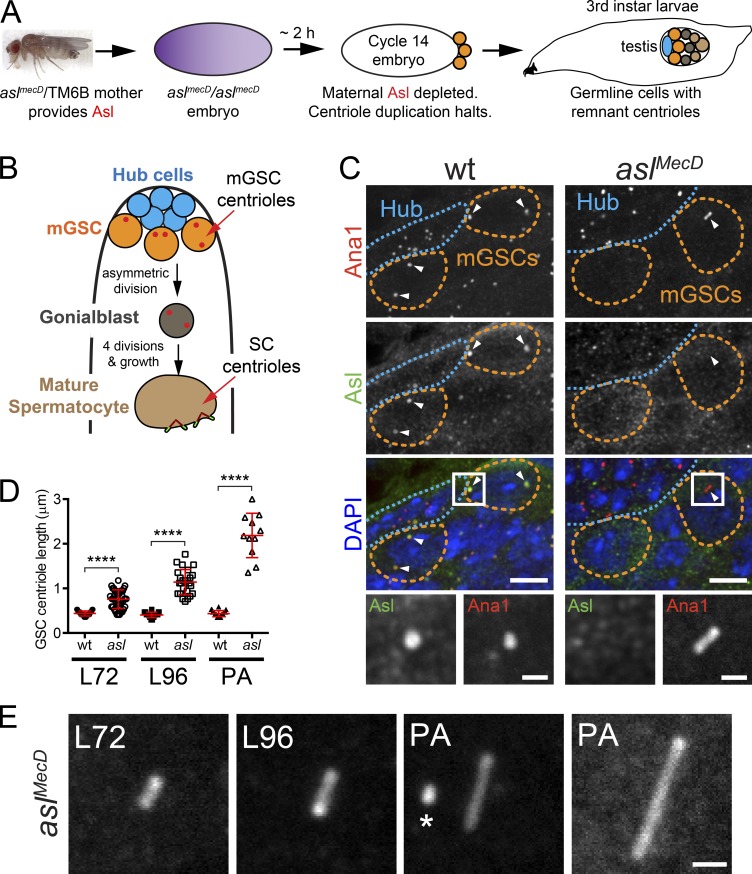
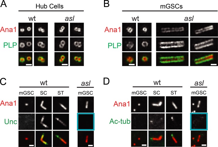
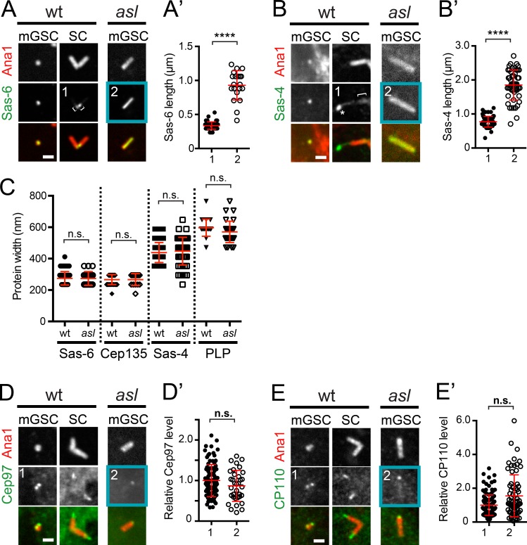
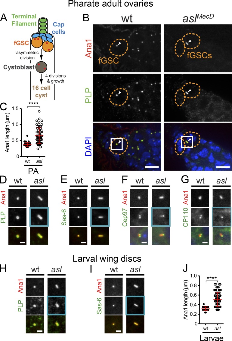
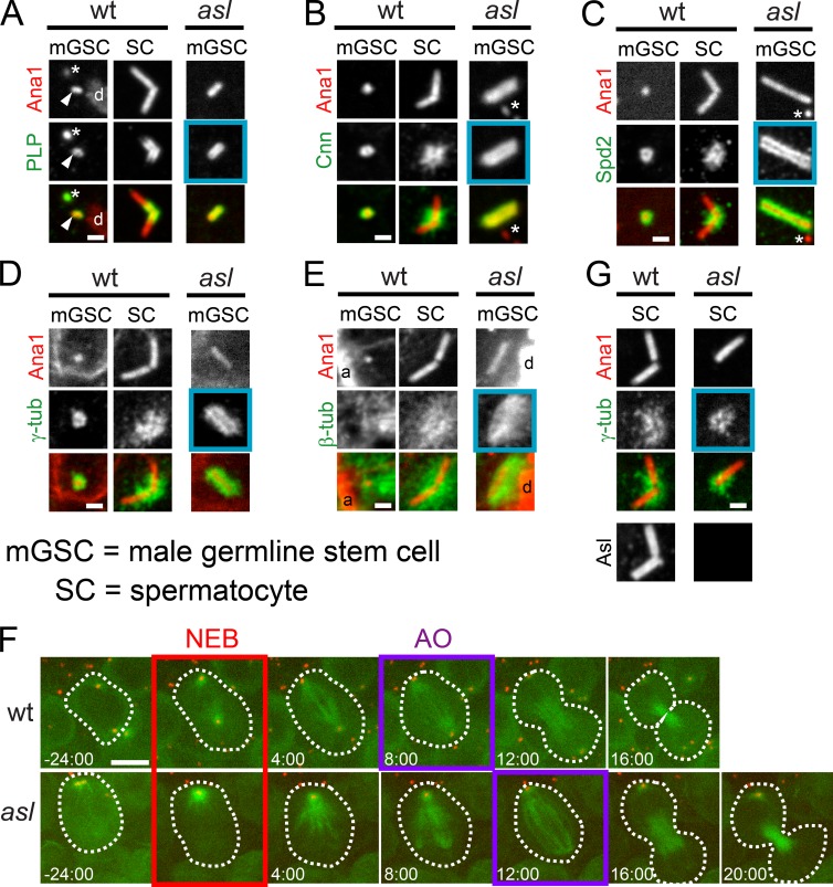
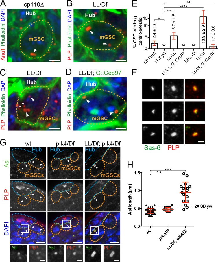
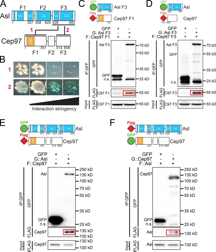
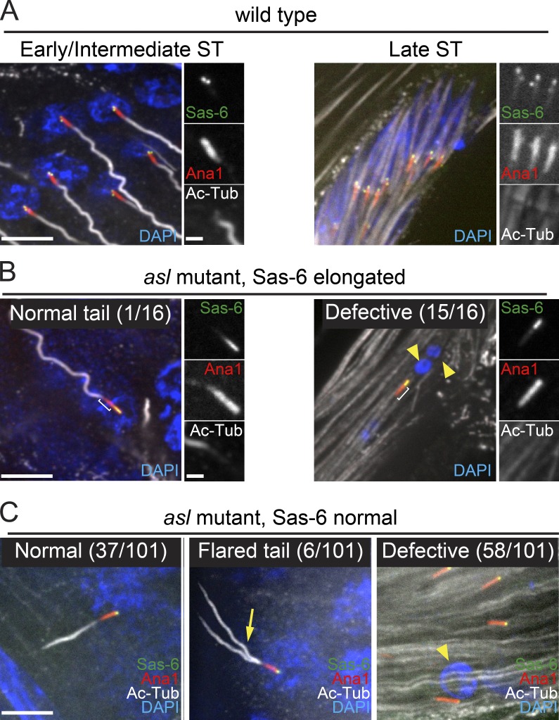
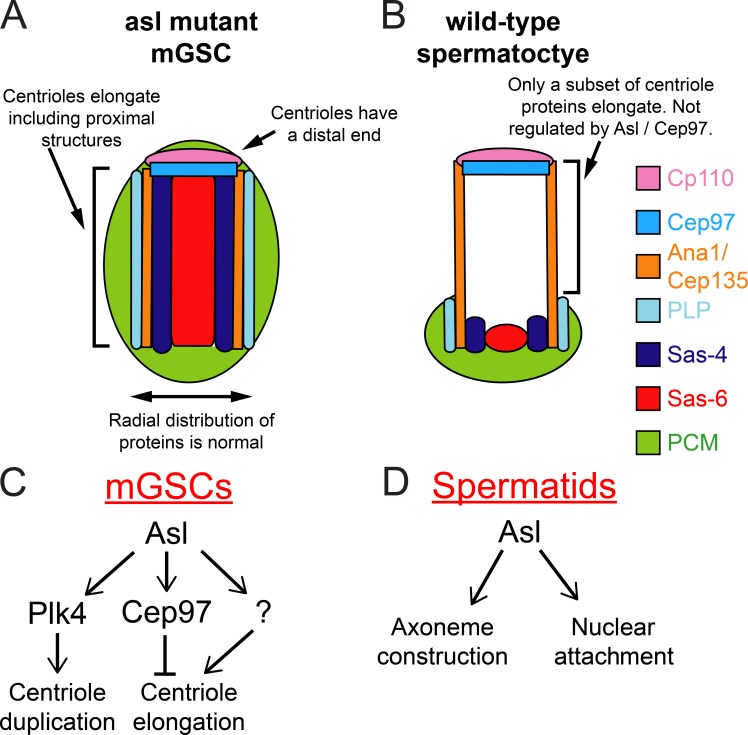
Centrioles are the foundation of two organelles, centrosomes and cilia. Centriole numbers and functions are tightly controlled, and mutations in centriole proteins are linked to a variety of diseases, including microcephaly. Loss of the centriole protein Asterless (Asl), the Drosophila melanogaster orthologue of Cep152, prevents centriole duplication, which has limited the study of its nonduplication functions. Here, we identify populations of cells with Asl-free centrioles in developing Drosophila tissues, allowing us to assess its duplication-independent function. We show a role for Asl in controlling centriole length in germline and somatic tissue, functioning via the centriole protein Cep97. We also find that Asl is not essential for pericentriolar material recruitment or centrosome function in organizing mitotic spindles. Lastly, we show that Asl is required for proper basal body function and spermatid axoneme formation. Insights into the role of Asl/Cep152 beyond centriole duplication could help shed light on how Cep152 mutations lead to the development of microcephaly.


Figures

Figure 1.
Figure 1.
Asl is required for proper centriole length control. (A) asl embryos receive maternal Asl protein and mRNA (purple) used for the first 2 h of development. When this protein runs out, centriole duplication halts. Centrioles present at this point are reliably inherited by the developing testes (remnant centrioles). (B) Cell types in the Drosophila testis (centrioles, red). (C) Remnant centrioles (Ana1::tdTomato, red) in larval asl mGSCs do not have Asl (green) and are longer than in wt. Arrowheads, mGSC centrioles. Boxed region enlarged. DAPI, blue. (D) Centriole length (Ana1::tdTomato length) in mGSCs in wt (aslmecD/TM6B) and asl mutants in larvae 72 h after egg laying (L72), larvae 96 h after egg laying (L96), or PAs. Mean ± SD, red. Comparison: ordinary one-way analysis of variance followed by Tukey’s multiple comparisons test. ****, P < 0.0001. (E) Representative centrioles in mGSCs at indicated stages. The longest centriole observed is on the right. Asterisk, centriole in adjacent hub cell. Bars: (C) 5 µm; (insets) 1 µm; (E) 1 µm.
Figure 2.
Figure 2.
Loss of Asl does not trigger premature centriole to basal body transition in mGSCs. (A and B) SIM of hub (A) and mGSC (B) centrioles in wt (aslmecD/TM6B; left) and asl (right) larval testes. Ana1::tdTomato, red; PLP, green. Two examples of each. (C and D) Confocal images of wt and asl centrioles. (C) Unc (green) was not on asl (blue box) or wt mGSC centrioles (Ana1, red) but was on wt SC and ST centrioles. (D) Ac-tub was on centrioles in 75% of SC and 100% of ST. Ac-tub was not on centrioles in wt or asl (blue box) mGSCs. Bars, 0.5 µm.
Figure 3.
Figure 3.
Asl regulates the position of centriole proteins. (A–E) Centrioles from interphase wt (aslmecD/TM6B) and asl mGSCs or wt mature SCs. (A) GFP::Sas-6 (green) and Ana1::tdTomato (red). Sas-6 extends the length of the centriole in asl mGSCs but is only proximal in wt SC centrioles (brackets). (A′) Length of Sas-6 signal in wt SCs (1) and asl mGSCs (2). (B) Sas-4::GFP (green) and Ana1::tdTomato (red). In SCs, Sas-4::GFP localizes proximally (bracket), where endogenous protein localizes (Gopalakrishnan et al., 2011; Fu and Glover, 2012). Additional localization beyond Ana1 (asterisk) is a consequence of overexpression. In asl mGSCs, Sas-4 is uniformly along the entire length of Ana1. (B′) Lengths of the Sas-4 signal in wt SCs (1, only the proximal signal) and asl mGSCs (2). (C) The radial distribution (diameter) of centriole proteins is unaltered in asl mGSCs. (D) GFP::Cep97 (green) and Ana1::tdTomato (red). (D′) Intensity of GFP::Cep97, normalized to the mean intensity of Cep97, at centrioles in wt (1) and asl (2) mGSCs. Cep97 levels are unaffected in asl. (E) CP110::GFP (green) and Ana1::tdTomato (red). (E′) Intensity of CP110::GFP, normalized as in D, in wt (1) and asl (2) mGSCs. CP110 levels are unaffected in asl. Bars, 1 µm. Mean ± SD, red. Comparison by unpaired t tests, with Welch’s corrections when appropriate. ****, P < 0.0001; n.s., not significant.
Figure 4.
Figure 4.
Asl controls centriole length in female germline and somatic cells. (A–G) Remnant centrioles in wt (aslmecD/TM6B) and asl fGSCs from PAs. (A) Cell types in Drosophila ovaries (centrioles, red). (B) Remnant centrioles (Ana1::tdTomato, red; PLP, green) in asl fGSCs are longer than in wt. Arrowheads, centrioles in fGSCs. DAPI, blue. Boxes are enlarged in D. (C) Measurements of centriole length (Ana1::tdTomato), in fGSCs. (D) Boxed region from B. PLP is along the length of asl centrioles. (E) GFP::Sas-6 (green) extends the length of asl centrioles. (F and G) GFP::Cep97 (F, green) and CP110::GFP (G, green) localize to the proximal tip of asl fGSC centrioles. (H–J) Centrioles in wt (aslmecD/TM6B) and asl larval wing discs. (H) Remnant asl centrioles (Ana1::tdTomato, red) are longer than wt. PLP (red) is along their entire length. (I) GFP::Sas-6 (green) extends the entire length of asl centrioles. (J) Centriole length (Ana1::tdTomato) in wing discs. Mean ± SD, red. Comparison: unpaired t tests, with Welch’s correction. ****, P < 0.0001. Bars: (B) 5 µm; (D–I) 1 µm.
Figure 5.
Figure 5.
Asl is not required for PCM recruitment during mitosis. (A–E) Centrioles (Ana1::tdTomato, red) from mitotic wt (aslmecD/TM6B) and asl mGSCs or wt mature SCs. Asterisk represents centrioles in adjacent hub cells. (A) PLP (green) localizes to centrioles in wt mGSCs, wt SCs, and asl mGSCs (blue box, n = 7/7). mGSC centriole, arrowhead. DNA, d (phosphohistone, not depicted). (B) Cnn (green) localizes to centrioles in wt mGSCs, wt SCs, and asl mGSCs (blue box, n = 3/3). (C) Spd2 (green) localizes to centrioles in wt mGSCs, wt SCs, and asl mGSCs (blue box, n = 4/4). (D) γ-Tubulin (green) localizes to centrioles in wt mGSCs, wt SCs, and asl mGSCs (blue box, n = 10/10). (E) MTs (green) organized around centrioles in wt mGSCs, wt SCs, and asl mGSCs (blue box, n = 8/8). DNA, d (phosphohistone); actin, a (phalloidin). (F) Frames from Videos 1 and 2 of mGSCs in wt (top row) and asl (bottom row) expressing GFP::α-tubulin (MTs, green) and Ana1::tdTomato (red). Time (min) relative to nuclear envelope breakdown (NEB; red box). Anaphase onset (AO; purple box). (G) γ-Tubulin (green) localizes to centrioles in meiotic asl SCs (blue box, n = 15/15). Asl is absent from centrioles in asl (bottom right). Bars: (A–E and G) 1 µm; (F) 5 µm.
Figure 6.
Figure 6.
Cep97 controls mGSC centriole length. (A–D) mGSCs in adult testes of indicated genotypes. Ana1::tdTomato, red (in A); PLP, red (in B–D); phalloidin, green; DAPI, blue. Arrowheads represent centrioles in mGSCs. Asterisks represent centrioles from the hub. (A) An mGSC from a cp110Δ testis with a long centriole. The other centriole is not visible in this projection. (B) cep97LL/Df(2L)A1 (LL/Df) mGSC with a long apical centriole. (C) LL/Df mGSC with a long nonapical centriole. (D) An LL/Df mGSC expressing GFP::Cep97 has normal-length centrioles. (E) Percentage of mGSCs with long centrioles per testis ± SEM for indicated genotypes. Testes counted include cp110Δ, 36; LL/CyO, 17; LL/LL, 34; LL/CyO; GFP::Cep97, 23; LL/Df, 32; and LL/Df; GFP::Cep97, 27. Comparison: unpaired t tests, with Welch’s correction. ****, P < 0.0001; ***, P < 0.001; *, P ≤ 0.05; n.s., not significant. (F) GFP::Sas-6 (green) and PLP (red) extend along the length of cep97 centrioles. Procentriole can position at one end (middle row) or the middle (bottom row) of the elongated centriole. (G) Remnant centrioles (Asl, green; PLP, red) in cep97;plk4 double-mutant adult mGSCs are longer than in wt (yw) or plk4. mGSCs, orange line; DAPI, blue; arrowheads, centrioles in mGSCs. Boxed region enlarged (bottom). (H) Centriole (Ana1::tdtomato) length (red line, mean ± SD) in mGSCs in adults of the indicated genotypes. Dashed line is the mean of wt plus two SDs. Almost all centrioles in mGSCs are long in cep97;plk4. Comparison: ordinary one-way analysis of variance followed by Tukey’s multiple comparisons test. ****, P < 0.0001; n.s., not significant. Bars: (A–D) 2 µm; (F and G, bottom row) 1 µm; (G) 5 µm.
Figure 7.
Figure 7.
Asterless and Cep97 interact. (A) Schematics of the subfragments (F1–F3) of Asl and Cep97 used for interaction assays. Red numbers refer to B. Numbers are amino acids. Lines, Y2H interactions; CC, coiled coil; LRR, leucine-rich repeat. (B) Asl F3 interacts with Cep97 F1 (1) and Cep97 F3 (2) by Y2H screening. The first column shows the presence of Y2H plasmids. The remaining columns show interaction under conditions of increasing stringency (see Materials and methods). Red numbers refer to A. (C) GFP::Asl F3 coIPs Flag::Cep97-F1 from S2 cells. (D) GFP::Asl F3 coIPs Flag::Cep97-F3 from S2 cells. (E) GFP::Asl full-length coIPs Flag::Cep97 full length from S2 cells. (F) GFP::Cep97 full-length coIPs Flag::Asl full length from S2 cells.
Figure 8.
Figure 8.
Cep97 functions downstream of Asl in mGSC centriole length control. (A) Centriole targeted Cep97 construct. LRR, leucine-rich repeat; PACT, centriole targeting domain of PLP. (B) A representative centriole in asl mGSCs expressing Cep97PACT. PLP, blue; Cep97PACT, green; Ana1::tdTomato, red. Linescans (dashed line) along the long axis of the centriole, normalized to peak intensity. (C) A representative centriole in asl mutant mGSCs. PLP, blue; Ana1::tdTomato, red. Linescans as in B. (D) Length of Ana1::tdTomato in asl mutant mGSC centrioles at indicated developmental time from individuals with or without Cep97PACT expression. Additional examples are shown in Fig. S4. Error bars are ±SD. Comparison: unpaired t tests with Welch’s corrections when appropriate. N, centrioles measured. Bars, 1 µm.
Figure 9.
Figure 9.
Asl is required for proper function of sperm basal bodies. (A–C) Sas-6 (green) on BBs (Ana1::tdTomato, red) in early/intermediate (left) or late (right) STs. Axonemes, Ac-tub (gray); DNA, DAPI (blue). (A) wt (asl/TM6) STs. In early/intermediate STs (left), basal bodies are docked at the nucleus and form axonemes. The nuclei of late STs are elongated and clustered. (B) Remnant centrioles in asl mutant STs with “elongated” region of Sas-6 (n = 16). ST at all stages were examined. Bracket, distal extension of Ana1 beyond Sas-6. (left) The only remnant centriole (early/intermediate ST) with elongated Sas-6 exhibiting normal nuclear attachment and axoneme nucleation. (right) Representative remnant ST centriole not attached to a nucleus or forming an axoneme. The nuclei of late stage asl STs are fragmented and dispersed (yellow arrowheads). (C) Remnant centrioles in asl mutants with a “normal” distribution of Sas-6 (restricted to the very proximal end; n = 101). (left) Representative remnant centriole in an early/intermediate ST exhibiting normal nuclear attachment and axoneme formation. (middle) Representative remnant centriole in an early/intermediate ST with a disrupted, flared axoneme (arrow). (right) Representative remnant centrioles in late STs not attached to nuclei or nucleating axonemes. Yellow arrowhead indicates nucleus that has not undergone reshaping. Additional examples are shown in Fig. S5. Bars: 5 µm; (insets) 1 µm. Defective, no axoneme or no nuclear attachment or both.
Figure 10.
Figure 10.
Model for Asl function in centriole organization and length control. (A and B) Not all long centrioles are alike. Centrioles in asl mutant mGSCs (A) are organized in a manner distinct from the giant centrioles normally found in SCs (B). This indicates that these two structures, although of similar scale, arise via distinct mechanisms. (C and D) Expanded roles for Asterless at the centriole. In the male germline, Asl plays a critical role in regulating centrosome duplication via Plk4, and controlling centriole length upstream of, and likely directly through, Cep97. (D) In developing STs, Asl is required for normal nuclear attachment and axoneme formation.

Similar articles

Cited by

References

    1. Angus K.L., and Griffiths G.M.. 2013. Cell polarisation and the immunological synapse. Curr. Opin. Cell Biol. 25:85–91. 10.1016/j.ceb.2012.08.013 - DOI - PMC - PubMed
    1. Baker J.D., Adhikarakunnathu S., and Kernan M.J.. 2004. Mechanosensory-defective, male-sterile unc mutants identify a novel basal body protein required for ciliogenesis in Drosophila. Development. 131:3411–3422. 10.1242/dev.01229 - DOI - PubMed
    1. Balestra F.R., Strnad P., Flückiger I., and Gönczy P.. 2013. Discovering regulators of centriole biogenesis through siRNA-based functional genomics in human cells. Dev. Cell. 25:555–571. (published erratum appears in Dev. Cell. 2013. 26:220)10.1016/j.devcel.2013.05.016 - DOI - PubMed
    1. Bennett M.A., Shern J.F., and Kahn R.A.. 2004. Reverse two-hybrid techniques in the yeast Saccharomyces cerevisiae. Methods Mol. Biol. 261:313–326. 10.1385/1-59259-762-9:313 - DOI - PubMed
    1. Bettencourt-Dias M., Rodrigues-Martins A., Carpenter L., Riparbelli M., Lehmann L., Gatt M.K., Carmo N., Balloux F., Callaini G., and Glover D.M.. 2005. SAK/PLK4 is required for centriole duplication and flagella development. Curr. Biol. 15:2199–2207. 10.1016/j.cub.2005.11.042 - DOI - PubMed

LinkOut - more resources