. 2016 May 30;213(6):887-96.
doi: 10.1084/jem.20151720. Epub 2016 May 16.

Bridging channel dendritic cells induce immunity to transfused red blood cells

Affiliations

Bridging channel dendritic cells induce immunity to transfused red blood cells

Samuele Calabro et al. J Exp Med. .

Abstract

Red blood cell (RBC) transfusion is a life-saving therapeutic tool. However, a major complication in transfusion recipients is the generation of antibodies against non-ABO alloantigens on donor RBCs, potentially resulting in hemolysis and renal failure. Long-lived antibody responses typically require CD4(+) T cell help and, in murine transfusion models, alloimmunization requires a spleen. Yet, it is not known how RBC-derived antigens are presented to naive T cells in the spleen. We sought to answer whether splenic dendritic cells (DCs) were essential for T cell priming to RBC alloantigens. Transient deletion of conventional DCs at the time of transfusion or splenic DC preactivation before RBC transfusion abrogated T and B cell responses to allogeneic RBCs, even though transfused RBCs persisted in the circulation for weeks. Although all splenic DCs phagocytosed RBCs and activated RBC-specific CD4(+) T cells in vitro, only bridging channel 33D1(+) DCs were required for alloimmunization in vivo. In contrast, deletion of XCR1(+)CD8(+) DCs did not alter the immune response to RBCs. Our work suggests that blocking the function of one DC subset during a narrow window of time during RBC transfusion could potentially prevent the detrimental immune response that occurs in patients who require lifelong RBC transfusion support.


Figures

Figure 1.
Figure 1.
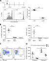
RBC alloimmunization requires MHC II antigen presentation. (A) RBC alloimmunization model: predicted HOD antigen on RBC membrane (left), in vivo RBC alloimmunization model (middle), and the time course of alloantibody induction in serum (right); n = 10 mice. Representative of two independent experiments. (B) Using an antibody (anti-Fy3) specific for the transgenic Duffy alloantigen on the surface of the HOD RBCs, we tracked the persistence of transfused RBCs. n = 5 mice/group. Representative of three independent experiments. (C) Absence of alloantibodies to transfused HOD RBCs in MHC II (CIIta) KO mice as measured by ELISA for HEL-specific IgG1 in day 21 sera. *, P < 0.02 or (D) WT mice depleted of CD4+ T cells or MHC II KO mice as measured by flow cytometric cross match of day 21 posttransfusion sera. *, P < 0.02; **, P < 0.01. Isotype mice were injected with an isotype-matched antibody that did not deplete CD4+ T cells. Dashed line indicates antibody level in naive mice. n = 5–10 mice/group. Representative of three independent experiments.
Figure 2.
Figure 2.
The presentation of RBC antigens to CD4+ T cells requires conventional DCs. (A) Cell populations in the spleen that phagocytose GFP+ RBCs 30 min after transfusion as compared with untransfused (naive) mice. n = 2–3 mice/group. Representative of four independent experiments. (B) Fluorescent image of a spleen from a mouse transfused with GFP-expressing RBCs 6 h prior. Bar, 100 µm. RP, red pulp; WP, white pulp. Representative image from one of three mice. (C) The same populations as in A were sorted from spleens of mice transfused with HOD RBCs 6–8 h prior and used to stimulate OT-II CD4+ T cells in vitro for 3 d. Proliferation measured by CFSE dilution of stimulated (open histogram) versus unstimulated (shaded histogram) T cells. (D) Percentage of DCs in the spleen in CD11c-DTr mice injected i.p. with DT on day 0. Gated from TCRβB220 cells. (E) WT and CD11c-DTr mice were treated with DT and received 106 CFSE-labeled CD45.1+ CD4+ OT-II cells. 48 h after T cell transfer, mice were transfused with HOD RBCs and proliferation in the spleen was measured by CFSE dilution (left). Anti-RBC antibody in the sera of mice 21 d after HOD RBC transfusion (naive mice did not receive HOD RBCs; right). n = 2–9 mice/group. Representative of three independent experiments. **, P < 0.002. (F) Zbtb46-DTR BM chimeras were left untreated or treated with DT and transferred with CFSE-labeled OT-II cells. 3 d after DT treatment, mice were transfused with HOD RBCs and proliferation in the spleen was measured by CFSE dilution 3 d later (left). Anti-RBC antibody levels in the sera of Zbtb46-DTr chimeric mice at different time points after HOD RBC transfusion (right). ***, P = 0.0002; ****, P < 0.0001. (G) Representative flow cytometry plots of class-switched germinal center B cells (B220+ GL7+ IgG1+) 8 d after HOD RBC transfusion in Zbtb46-DTr BM chimeric mice treated or not treated with DT. n = 2–10 mice/group. Representative of three independent experiments.
Figure 3.
Figure 3.
Bridging channel 33D1+ DCs are required for alloimmunization. (A) Representative flow cytometry plot of DC subset gating (pregated on B220TCRβ singlets) in a naive spleen. Cartoon represents these two populations along with commonly associated markers that roughly define the same populations. (B, top) GFP+ RBCs were transfused into WT mice and 33D1+ and XCR1+ splenic DCs were analyzed by flow cytometry 30 min later. Representative images from one of three mice/group in three independent experiments. (bottom) Ex vivo T cell proliferation of CFSE+ OT-II T cells induced by 33D1+ or XCR1+ splenic DCs isolated from WT mice transfused with HOD RBCs 8 h prior. (C and D) Anti-RBC antibodies in the sera of mice lacking XCR1+ DCs (BATF3 KO; C) or 33D1+ DCs (IRF4fl/flxCD11cCre; D) 21 d after transfusion of HOD RBCs. ***, P < 0.0006. n = 3–7 mice/group. Representative of two to three independent experiments.
Figure 4.
Figure 4.
Disruption of DC function during a narrow window of time prevents alloimmunization. (A) 106 CFSE-labeled CD45.1+ CD4+ OT-II cells were adoptively transferred into WT mice treated with (+LPS) or without (−LPS) 10ug of intravenous LPS. 24 h later, mice were transfused with HOD RBCs and, 3 d after transfusion, OT-II cell proliferation in the spleen was determined by CFSE dilution. Cumulative plot showing the percentage of proliferating T cells in each mouse per group. ***, P < 0.0001, n = 4 mice/group. Representative of three independent experiments. (B) WT mice were pretreated i.v. with 10 µg LPS or not as indicated (day 0) and were transfused with HOD RBCs 24 h later, as indicated. 3 wk after transfusion, anti-HOD alloantibody response was analyzed by flow cytometry (left) or ELISA (right). **, P = 0.0027; ***, P < 0.0001; n = 4–5 mice/group. Representative of three independent experiments. (C) WT mice were pretreated i.v. with (+LPS) or without (−LPS) 10 µg LPS 24 h before transfusion of GFP+ RBCs. 30 min later, splenic DCs were analyzed by flow cytometry. n = 2 mice/group; representative of two independent experiments. (D) WT mice were pretreated (square) or not (circle) with 10 µg of LPS i.v. and, 24 h later, transfused with HOD RBCs (Challenge 1). 3 wk later, sera was collected, and then the same mice were rechallenged with HOD RBCs (Challenge 2). 3 wk after the second transfusion, sera was again collected; anti-HOD antibodies were measured in all sets of sera by cross matching. **, P = 0.0027. Dashed line indicates level of alloantibodies detected in a naive mouse. n = 3–4 mice/group. Representative of two independent experiments.

References

    1. Bachem A., Hartung E., Güttler S., Mora A., Zhou X., Hegemann A., Plantinga M., Mazzini E., Stoitzner P., Gurka S., et al. . 2012. Expression of XCR1 Characterizes the Batf3-dependent lineage of dendritic cells capable of antigen cross-presentation. Front. Immunol. 3:214 10.3389/fimmu.2012.00214 - DOI - PMC - PubMed
    1. Boctor F.N., Ali N.M., Mohandas K., and Uehlinger J.. 2003. Absence of D- alloimmunization in AIDS patients receiving D-mismatched RBCs. Transfusion. 43:173–176. 10.1046/j.1537-2995.2003.00289.x - DOI - PubMed
    1. Borges da Silva H., Fonseca R., Cassado A.A., Machado de Salles É., de Menezes M.N., Langhorne J., Perez K.R., Cuccovia I.M., Ryffel B., Barreto V.M., et al. . 2015. In vivo approaches reveal a key role for DCs in CD4+ T cell activation and parasite clearance during the acute phase of experimental blood-stage malaria. PLoS Pathog. 11:e1004598 10.1371/journal.ppat.1004598 - DOI - PMC - PubMed
    1. Casas J., Friedman D.F., Jackson T., Vege S., Westhoff C.M., and Chou S.T.. 2015. Changing practice: red blood cell typing by molecular methods for patients with sickle cell disease. Transfusion. 55:1388–1393. 10.1111/trf.12987 - DOI - PMC - PubMed
    1. Chiaroni J., Dettori I., Ferrera V., Legrand D., Touinssi M., Mercier P., de Micco P., and Reviron D.. 2006. HLA-DRB1 polymorphism is associated with Kell immunisation. Br. J. Haematol. 132:374–378. 10.1111/j.1365-2141.2005.05868.x - DOI - PubMed

Publication types