. 2016 Jul;57(7):1204-18.
doi: 10.1194/jlr.M067108. Epub 2016 May 16.

Endothelial HO-1 induction by model TG-rich lipoproteins is regulated through a NOX4-Nrf2 pathway

Affiliations

Endothelial HO-1 induction by model TG-rich lipoproteins is regulated through a NOX4-Nrf2 pathway

Sally H Latham Birt et al. J Lipid Res. 2016 Jul.

Abstract

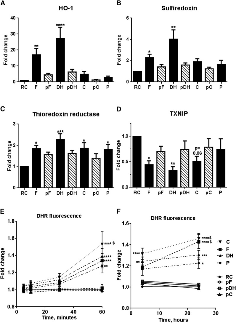
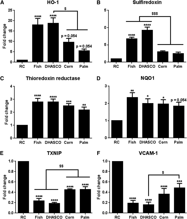
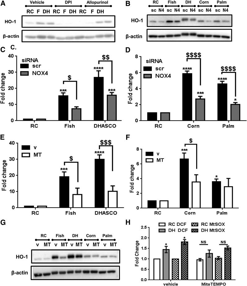
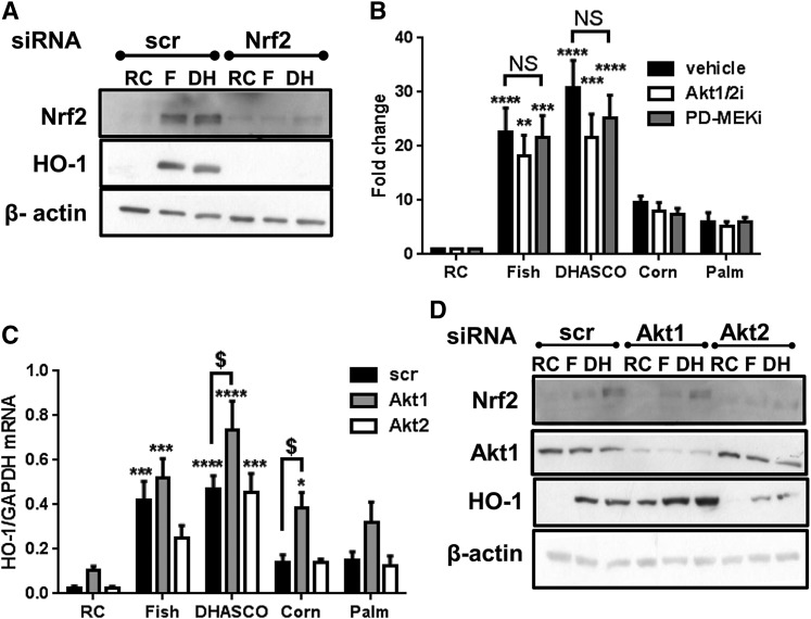
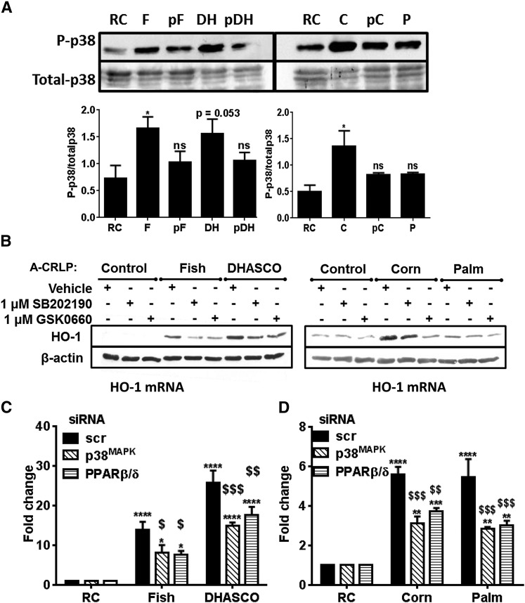
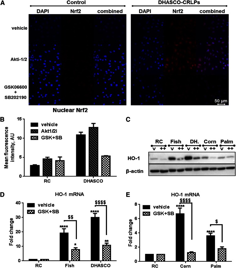
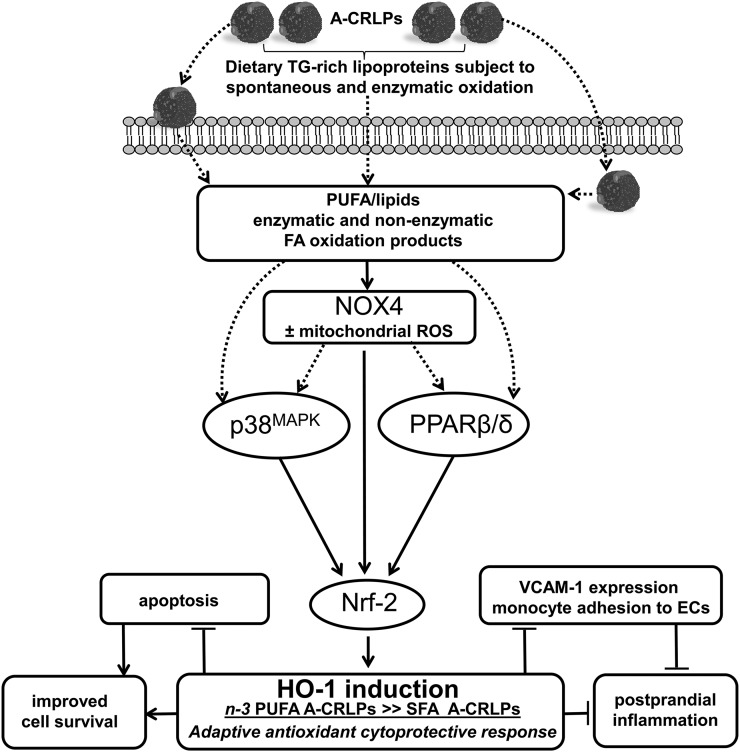
Circulating levels of chylomicron remnants (CMRs) increase postprandially and their composition directly reflects dietary lipid intake. These TG-rich lipoproteins likely contribute to the development of endothelial dysfunction, albeit via unknown mechanisms. Here, we investigated how the FA composition of CMRs influences their actions on human aortic endothelial cells (HAECs) by comparing the effects of model CMRs-artificial TG-rich CMR-like particles (A-CRLPs)-containing TGs extracted from fish, DHA-rich algal, corn, or palm oils. HAECs responded with distinct transcriptional programs according to A-CRLP TG content and oxidation status, with genes involved in antioxidant defense and cytoprotection most prominently affected by n-3 PUFA-containing A-CRLPs. These particles were significantly more efficacious inducers of heme oxygenase-1 (HO-1) than n-6 PUFA corn or saturated FA-rich palm CRLPs. Mechanistically, HO-1 induction by all CRLPs requires NADPH oxidase 4, with PUFA-containing particles additionally dependent upon mitochondrial reactive oxygen species. Activation of both p38 MAPK and PPARβ/δ culminates in increased nuclear factor erythroid 2-related factor 2 (Nrf2) expression/nuclear translocation and HO-1 induction. These studies define new molecular pathways coupling endothelial cell activation by model CMRs with adaptive regulation of Nrf2-dependent HO-1 expression and may represent key mechanisms through which dietary FAs differentially impact progression of endothelial dysfunction.

Keywords: artificial chylomicron remnant-like particles; cell signaling; endothelial cells; fatty acid; heme oxygenase-1; lipids/oxidation; nuclear factor erythroid 2-related factor 2; omega-3 fatty acids; reduced nicotinamide adenine dinucleotide phosphate oxidase 4; triglyceride.


Figures

Fig. 1.
Fig. 1.
A-CRLPs modulate cytoprotective gene expression and elevate ROS production dependent upon oxidative state. A–D: HAECs were serum starved for 5 h and then incubated with A-CRLPs (280 μM TG) for 4 h. HO-1, SRX, TXR, and TXNIP mRNAs were quantified by qPCR and normalized to GAPDH (mean fold change versus RC ± SEM; n = 4–5. Black bars: remnant control (RC), fish (F), DHASCO® (DH), corn (C), and palm (P) A-CRLPs. Striped bars: probucol-fish (pF), probucol-DHASCO® (pDH), and probucol-corn (pC) A-CRLPs. One-way ANOVA with Dunnett’s multiple comparisons test, compared with RC: *P < 0.05, **P < 0.01, ****P < 0.0001. E, F: DHR was used to measure ROS/RNS production, presented as mean fold change relative to RC ± SEM (n = 6 for A-CRLPs, n = 3 for probucol-A-CRLPs with each experiment performed in technical quintuplet). Two-way ANOVA, RC versus A-CRLPs indicated: *P < 0.05, **P < 0.01, ***P < 0.001, ****P < 0.0001. For palm A-CRLPs versus other compositions, $P < 0.05.
Fig. 2.
Fig. 2.
Differential effects of A-CRLPs with different FA compositions on gene expression in aortic ECs. HAECs were incubated with A-CRLPs (280 μM TG) for 16 h and mRNAs for the genes indicated in (A–F) measured by qPCR and normalized to GAPDH. Data are mean fold change from RC (±SEM; n = 5). One-way ANOVA with Tukey’s multiple comparisons test versus RC: *P < 0.05, **P < 0.01, ***P < 0.001, ****P < 0.0001 and compared with group(s) indicated: $P < 0.05, $$P < 0.01, $$$P < 0.001.
Fig. 3.
Fig. 3.
A-CRLP-driven HO-1 expression is controlled by NOX4 and mitochondrial ROS. A: After serum starvation, HAECs were pretreated with DMSO (vehicle), 5 μM diphenyleneiodonium, or 200 μM allopurinol for 1 h and then incubated with remnant control (RC), fish (F), or DHASCO® (DH) A-CRLPs (280 μM TG) in the continued presence or absence of inhibitor for 4 h. Representative HO-1 and β-actin blots from three independent experiments are shown. B–D: HAECs transfected with scrambled (scr/sc) siRNA or NOX4 (N4) siRNA were serum-starved then incubated with A-CRLPs for 4 h. B: Representative Western blots from three independent experiments. C, D: qPCR for HO-1 mRNA normalized to GAPDH presented as fold change of RC, mean ± SEM (n = 3). Two-way ANOVA with Bonferroni’s multiple comparisons test, scr versus NOX4 siRNA; $P < 0.05, $$P < 0.01, $$$$P < 0.0001. RC versus A-CRLPs: *P < 0.05, ***P < 0.001, ****P < 0.0001. E–G: Serum-starved HAECs were pretreated with DMSO (v) or 25 μM MitoTEMPO (MT) for 1 h then incubated with A-CRLPs for 4 h in the presence of DMSO or MT. E, F: qPCR for HO-1 mRNA normalized to GAPDH presented as fold change relative to RC, mean ± SEM, n = 3. Two-way ANOVA with Bonferroni’s multiple comparisons test, v versus MT; $P < 0.05, $$$P < 0.001. RC versus A-CRLP indicated; *P < 0.05, ***P < 0.001, ****P < 0.0001. G: Representative Western blots. H: HUVECs were pretreated with DMSO (vehicle) or 25 μM MitoTEMPO then incubated with RC or DHASCO® (DH) A-CRLPs in the presence of vehicle/MitoTEMPO for 2 h prior to loading with the ROS probe, 2′7′-DCF-DA (DCF), or MitoSOX (MtSOX) as indicated. Data are mean fold change relative to RC ± SEM (n = 3 with each experiment performed in technical quintuplet). Two-way ANOVA with Bonferroni’s multiple comparisons test, RC versus DHASCO®; *P < 0.05.
Fig. 4.
Fig. 4.
HO-1 induction by A-CRLPs is Nrf2 dependent but independent of Akt and ERK. A: HAECs transfected with 20 nM noncoding (scr) or Nrf2 siRNA were incubated with remnant control (RC), fish (F), or DHASCO® (DH) A-CRLPs (280 μM TG) for 7 h. Representative blots from three independent experiments are shown. B: Serum-starved HAECs were preincubated with DMSO (vehicle), 1 μM Akt1/2i, or 1 μM PD184352 (PD-MEKi) for 1 h then incubated with A-CRLPs for 4 h in the continued presence or absence of inhibitor. HO-1 mRNA expression normalized to GAPDH is presented as fold change relative to RC (mean ± SEM, n = 3). Two-way ANOVA with Bonferroni’s multiple comparisons test, vehicle versus inhibitors not significant (NS). RC versus A-CRLP indicated: **P < 0.01, ***P < 0.001, ****P < 0.0001. C, D: HAECs transfected with 20 nM noncoding (scr), Akt1, or Akt2 siRNA were serum starved then incubated with A-CRLPs for 4 h. C: HO-1 mRNA normalized to GAPDH (fold change relative to RC; mean ± SEM, n = 4). Two-way ANOVA with Bonferroni’s multiple comparisons test, scr versus Akt1 siRNA: $P < 0.05. RC versus CRLP: *P < 0.05, ***P < 0.001, ****P < 0.0001. D: Representative Western blots from four independent experiments are shown for Nrf2, Akt1, HO-1, and β-actin.
Fig. 5.
Fig. 5.
A-CRLP-induced HO-1 expression is regulated by p38MAPK and PPARβ/δ. A: Serum-starved HAECs were incubated with remnant control (RC), fish (F), DHASCO® (DH), probucol-DHASCO® (pDH), corn (C), probucol-corn (pC), or palm (P) A-CRLPs at 280 μM TG for 10 min. Representative Western blots and densitometry analysis are shown for phospho- and total p38MAPK (n = 3–4) *P < 0.05 versus RC. B: Serum-starved HAECs were pretreated with vehicle, 1 μM SB202190, or 1 μM GSK0660 for 1 h prior to a 4 h A-CRLP incubation in the absence/presence of inhibitor. Representative blots are shown (n = 3). C, D: HAECs were transfected with 20 nM noncoding (scr), MAPK14 (p38MAPKα), or PPARβ/δ siRNA and then incubated with A-CRLPs for 4 h. HO-1 mRNA normalized to GAPDH is given as fold change relative to RC (mean ± SEM, n = 3–4). Two-way ANOVA with Bonferroni’s multiple comparisons test, scr versus p38MAPKα and scr versus PPARβ/δ as indicated: $P < 0.05, $$P < 0.01, $$$P < 0.001. RC versus A-CRLP: **P < 0.01, ***P < 0.001, ****P < 0.0001.
Fig. 6.
Fig. 6.
A-CRLPs increase Nrf2 expression and nuclear translocation via p38MAPK and PPARβ/δ. A, B: ECs were serum depleted for 1 h, pretreated (1 h) with vehicle, 1 μM Akt1/2i, or both GSK0660 and SB202190 (each 1 μM) then incubated with remnant control (Control) or DHASCO® A-CRLPs (280 μM TG) in the continued absence/presence of inhibitor(s) for 4 h. Cells were immunostained for Nrf2 (red) and nuclei labeled with DAPI (blue). No staining was evident in secondary antibody-only controls (not shown). Representative images are in (A) and combined mean nuclear Nrf2 fluorescence intensities from three separate images per condition are shown in (B) (mean ± SEM). C–E: Serum-starved HAECs were preincubated with vehicle or both 1 μM GSK0660 and 1 μM SB202190 (GSK+SB) for 1 h then incubated with A-CRLPs in the presence of vehicle or inhibitors for 4 h. C: Representative HO-1 and β-actin blots: vehicle (v), GSK+SB (++). D, E: HO-1 mRNA expression normalized to GAPDH. Data are fold change relative to remnant control (RC) (mean ± SEM, n = 3). Two-way ANOVA with Bonferroni’s multiple comparisons test: vehicle versus GSK+SB: $P < 0.05, $$P < 0.01, $$$$P < 0.0001. RC versus A-CRLP: *P < 0.05, **P < 0.01, ****P < 0.0001.
Fig. 7.
Fig. 7.
Summary of results: proposed mechanisms mediating the effects of A-CRLPs on HAECs. Artificial model TRLs resembling CMRs (A-CRLPs) upregulate several antioxidant and cytoprotective genes in HAECs with the most efficacious effects evident with A-CRLPs containing TG extracted from n-3 PUFA-containing oils. A-CRLPs regulate HO-1 induction in HAECs through a NOX4-driven pathway involving downstream activation of p38MAPK and parallel activation of the nuclear receptor superfamily member, PPARβ/δ. This results in increased Nrf2 expression and nuclear translocation, culminating in transcriptional upregulation of HO-1 and of other protective/antioxidant genes (e.g., SRX and TXR). We also showed that A-CRLP-driven HO-1 induction in HAECs is accompanied by reduced expression of the pro-inflammatory adhesion molecule, VCAM-1. The lipid species mediating activation of this redox-dependent pathway are yet to be clarified, but it is reasonable to speculate that oxidized FA/lipids acting directly on ECs following metabolism of TG-rich A-CRLPs at the cell surface and/or enzymatic products of FA/lipid oxidation generated intracellularly following particle uptake play key roles. Because HO-1 has known anti-inflammatory and cytoprotective functions, TRLs carrying n-3 PUFAs may act to suppress inflammatory changes in the endothelium and maintain cell survival in the postprandial phase, thus moderating the postprandial inflammation associated with fat consumption.

Similar articles

Cited by

References

    1. Libby P., Ridker P. M., and Hansson G. K.. 2011. Progress and challenges in translating the biology of atherosclerosis. Nature. 473: 317–325. - PubMed
    1. Karpe F., Steiner G., Uffelman K., Olivecrona T., and Hamsten A.. 1994. Postprandial lipoproteins and progression of coronary atherosclerosis. Atherosclerosis. 106: 83–97. - PubMed
    1. Botham K. M., and Wheeler-Jones C. P.. 2013. Postprandial lipoproteins and the molecular regulation of vascular homeostasis. Prog. Lipid Res. 52: 446–464. - PubMed
    1. Bravo E., Napolitano M., and Botham K. M.. 2010. Postprandial lipid metabolism: the missing link between life-style habits and the increasing incidence of metabolic diseases in western countries? Open Transl. Med. J. 2: 1–13.
    1. Nakano T., Nakajima K., Niimi M., Fujita M. Q., Nakajima Y., Takeichi S., Kinoshita M., Matsushima T., Teramoto T., and Tanaka A.. 2008. Detection of apolipoproteins B-48 and B-100 carrying particles in lipoprotein fractions extracted from human aortic atherosclerotic plaques in sudden cardiac death cases. Clin. Chim. Acta. 390: 38–43. - PubMed

Publication types

MeSH terms