. 2016 Jul 1;129(13):2501-13.
doi: 10.1242/jcs.186338. Epub 2016 May 16.

CEP295 interacts with microtubules and is required for centriole elongation

Affiliations

CEP295 interacts with microtubules and is required for centriole elongation

Ching-Wen Chang et al. J Cell Sci. .

Abstract

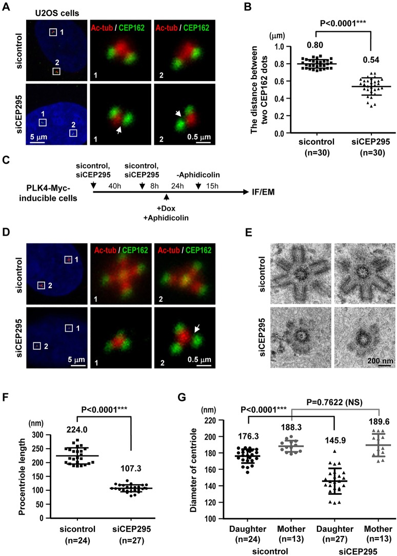
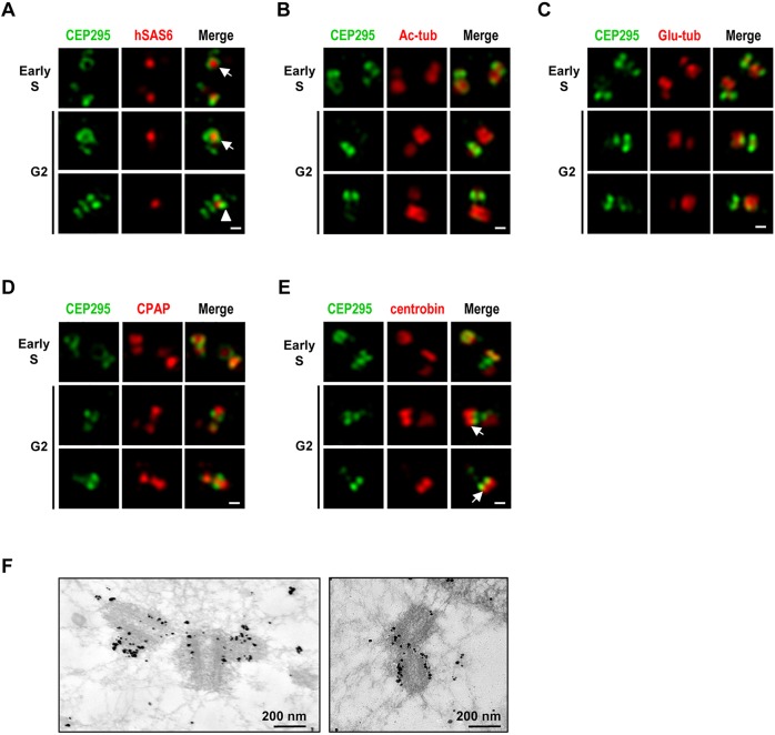
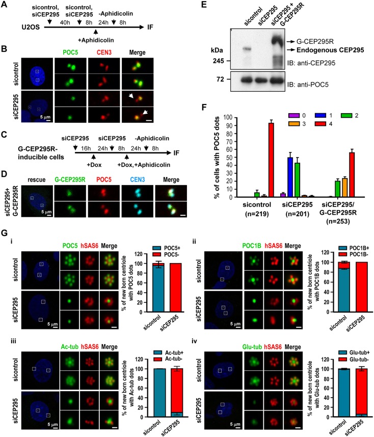
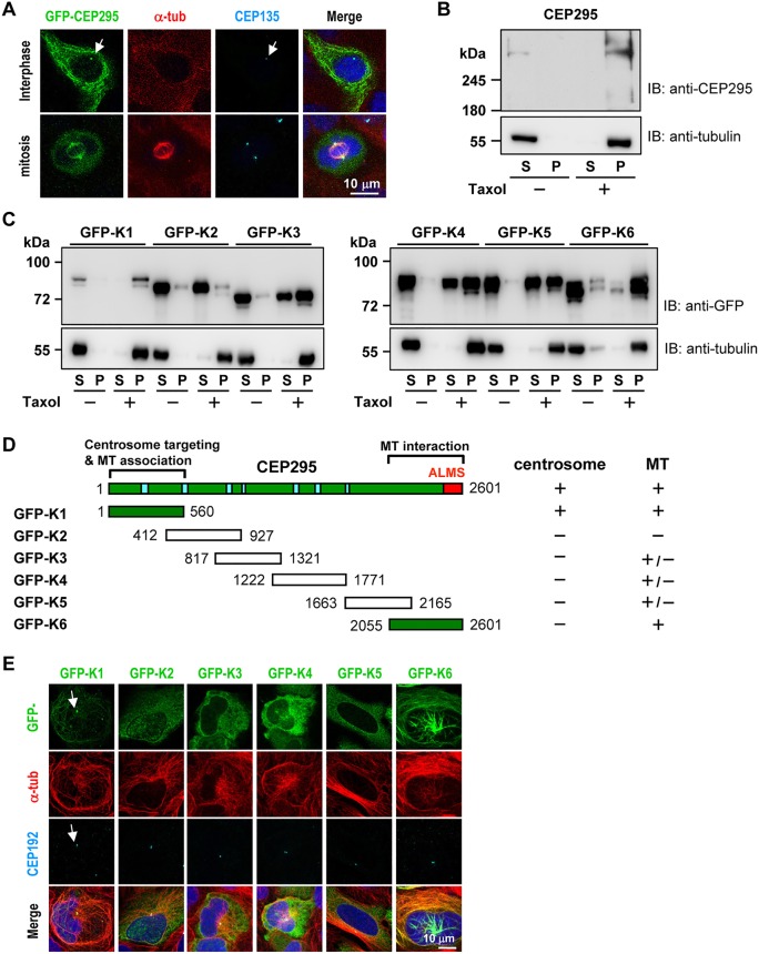
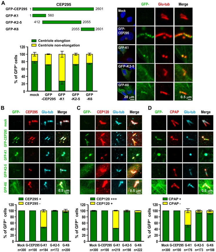
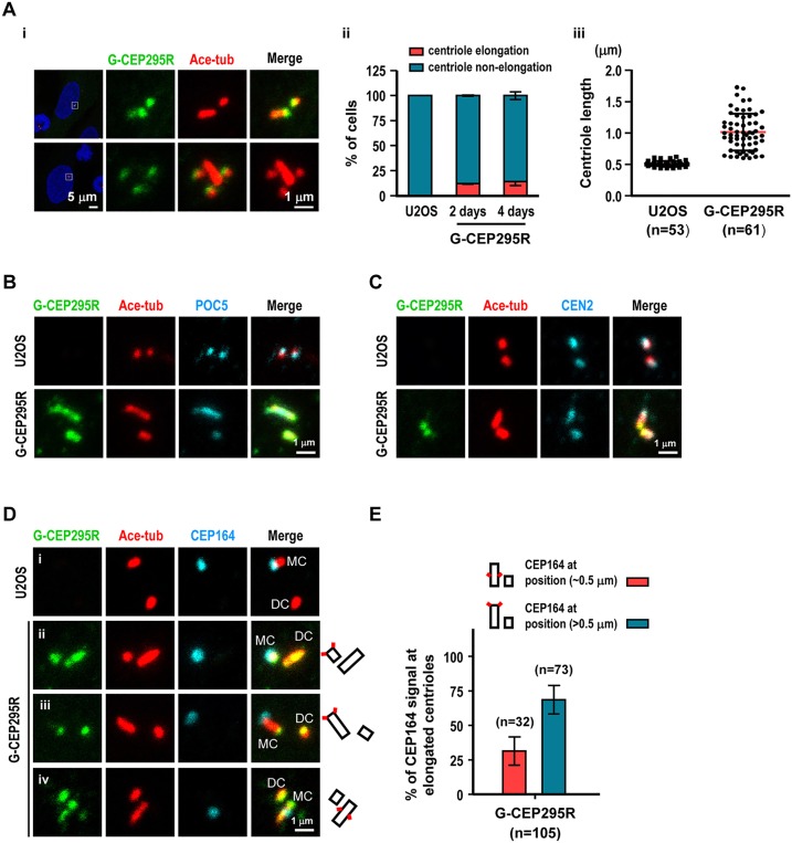
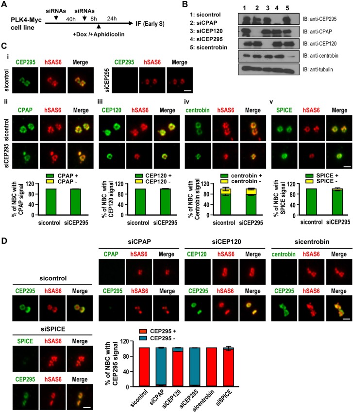
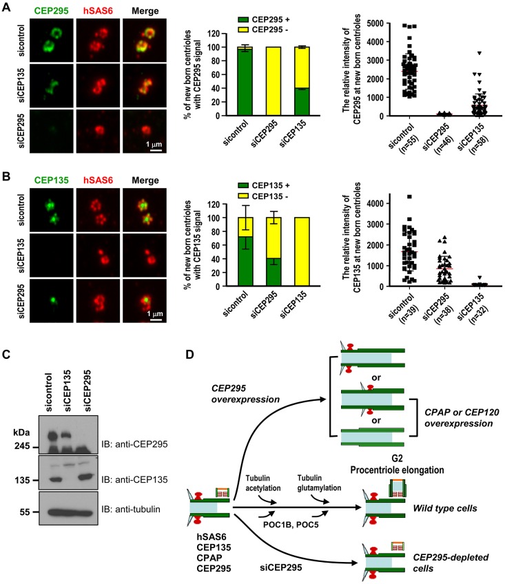
Centriole duplication is a tightly ordered process during which procentrioles are assembled in G1-S and elongate during S and G2. Here, we show that human CEP295 (Drosophila Ana1) is not essential for initial cartwheel assembly, but is required to build distal half centrioles during S and G2. Using super-resolution and immunogold electron microscopy, we demonstrate that CEP295 is recruited to the proximal end of procentrioles in early S phase, when it is also localized at the centriolar microtubule wall that surrounds the human SAS6 cartwheel hub. Interestingly, depletion of CEP295 not only inhibits the recruitments of POC5 and POC1B to the distal half centrioles in G2, resulting in shorter centrioles, it also blocks the post-translational modification of centriolar microtubules (e.g. acetylation and glutamylation). Importantly, our results indicate that CEP295 directly interacts with microtubules, and that excess CEP295 could induce the assembly of overly long centrioles. Furthermore, exogenous expression of the N-terminal domain of CEP295 exerts a dominant-negative effect on centriole elongation. Collectively, these findings suggest that CEP295 is essential for building the distal half centrioles and for post-translational modification of centriolar microtubules.

Keywords: Acetylation; Centriole assembly; Centriole duplication; Centrosome; Polyglutamylation; Procentriole formation.


Conflict of interest statement

Competing interests

The authors declare no competing or financial interests.

Figures

Fig. 1.
Fig. 1.
Centriole elongation is impaired in the absence of CEP295. (A) U2OS cells were transfected with control or CEP295-targeting siRNAs for 4 days. On day 3, aphidicolin was added to arrest cells at early S phase. After 24 h, the cells were released in fresh medium for another 8 h to enrich for G2-phase cells. The treated cells were fixed and stained with antibodies against CEP162 (green) and acetylated tubulin (Ac-tub, red). DNA was counterstained with DAPI. (B) Elongation of centrioles was calculated by measuring the distances between the centers of the two dots (representing staining for CEP162) within each pair of orthogonally arranged G2 centrioles. Error bars indicate the s.d. (standard deviation); ***P<0.0001 (two-tailed unpaired t-test). (C–G) PLK4-inducible cells were treated with sicontrol or siCEP295 as described in (C). IF, immunofluorescence; EM, electron microscopy; Dox, doxycycline. Cells were analyzed by immunofluorescence microscopy using the indicated antibodies (D), or by electron microscopy (E). Quantitative analysis of the centriole lengths (F) or the diameters (G) of daughter and mother centrioles acquired from electron microscopy images of sicontrol- and siCEP295-treated PLK4-inducible cells. Error bars represent the mean±s.d.; NS, not significant (two-tailed unpaired t-test). Arrows indicate reduced intensity of Ac-tub at the nascent centrioles.
Fig. 2.
Fig. 2.
Super-resolution (3D-SIM) analysis of the subcellular localizations of CEP295 and other centriolar proteins during centriole biogenesis. U2OS cells were synchronized by aphidicolin for 24 h (to arrest cells at early S phase) and the synchronized cells were released in fresh medium for another 8 h (to allow progression to G2 phase). The cells were then fixed and dual stained with antibodies against CEP295 (A–E) and hSAS6 (A), Ac-tub (B), glutamylated tubulin (Glu-tub) (C), CPAP (D) or centrobin (E). Super-resolution images were acquired using a Zeiss ELYRA system. (A) Arrows indicate that CEP295 embraces hSAS6 at the proximal end of the procentriole, arrowhead indicates two-dot shape with hSAS6 in the center. (E) Arrows indicate localization of centrobin distal to CEP295 on growing centrioles. Scale bars: 0.2 μm. (F) Immunogold electron microscopy analysis of CEP295 localization.
Fig. 3.
Fig. 3.
The G2 centrioles in CEP295-depleted cells fail to recruit POC5 or POC1B and do not show post-translational modification of centriolar microtubules. (A,B) U2OS cells were treated with sicontrol or siCEP295 as shown in (A) and analyzed by immunofluorescence microscopy (B). Arrows in B indicate inhibited targeting of POC5 to nascent centrioles. (C–F) Rescue experiment. G-CEP295R-inducible cells were treated as described in (C) and analyzed by confocal fluorescence microscopy (D) or immunoblot analysis (E). (F) Histograms illustrating the percentages of cells exhibiting POC5 signals. Error bars represent the mean±s.e.m. from four independent experiments. The total number n as indicated. (G) PLK4–myc-inducible cells were treated with siCEP295 as described in Fig. 1C, analyzed by immunofluorescence staining with antibodies against: (i) POC5 and hSAS6; (ii) POC1B and hSAS6; (iii) hSAS6 and Ac-tub, and (iv) hSAS6 and Glu-tub, and expression in new-born centrioles quantified. Error bars represent the mean±s.e.m. n=3 independent experiments each scoring 100 cells. Scale bars: 0.5 μm in the enlarged figures.
Fig. 4.
Fig. 4.
Mapping the functional domains of CEP295. (A) CEP295 is associated with interphase and mitotic microtubules. HEK 293T cells were transiently transfected with a vector encoding full-length GFP–CEP295 and analyzed by confocal fluorescence microscopy. Arrow indicates exogenously expressed GFP–CEP295 colocalized with CEP135 at the centrosomes. (B) Endogenous CEP295 co-sediments with microtubules in the presence of Taxol. HEK 293T cells were lysed in lysis buffer in the presence or absence of Taxol. The cell lysates were centrifuged, separated into supernatant (S) and pellet (P) fractions, and analyzed by immunoblotting. (C,D) Co-sedimentation assays (C) with six different fragments of CEP295, GFP−K1 to GFP–K6 (D), were used to map the regions of CEP295 that interact with microtubules. (D) Summary of the functional domains of CEP295: the centrosome-targeting region (centrosome), the microtubule-interacting/associating regions (MT), the ALMS motif (red), and the coil-coiled regions (blue). (E) HEK 293T cells were transfected with vectors encoding various GFP-tagged CEP295 truncation mutants. At 24 h post-transfection, the cells were fixed and stained for α-tubulin and CEP192 (a centrosome marker). Arrow indicates colocalization of N-terminal fragment, GFP–K1, with centrosome marker CEP192.
Fig. 5.
Fig. 5.
The N-terminal K1 fragment of CEP295 (GFP–K1) blocks CEP120-induced centriole elongation and suppresses the centriolar localization of CEP295, CPAP and CEP120. (A) Various GFP–CEP295 truncation mutants were transiently expressed in CEP120–myc-inducible cells. At 24 h after doxycycline induction, the cells were analyzed by confocal fluorescence microscopy, and the percentages of cells with elongated (>0.5 μm) or non-elongated centrioles were calculated. Error bars represent the mean±s.e.m. from three independent experiments (n=100 cells/experiment). (B–D) GFP–K1 suppresses the centriolar localization of CEP295, CEP120 and CPAP. Cells treated as described above were fixed and stained with antibodies against CEP295 (B), CEP120 (C), CPAP (D) and polyglutamylated-tubulin (Glu-tub; A–D), and percentages of GFP-positive cells quantified. Error bars represent the mean±s.e.m. from three independent experiments. The total sample size (n) as indicated.
Fig. 6.
Fig. 6.
CEP295 overexpression induces extra-long microtubule-based filaments with low efficiency. (A) G-CEP295R-inducible cells were treated with doxycycline for 2 days (i-iii) or 4 days (ii), and analyzed by confocal fluorescence microscopy using indicated antibodies. Percentages of cells with elongated centrioles and centriole length were quantified. Centriole length labeled by acetylated tubulin (Ace-tub) >0.5 µm was counted as elongated centriole. Error bars in ii represent the mean±s.e.m. from three independent experiments (n=100 cells/experiment). Error bars in iii show the mean±s.d. (B–D) U2OS and G-CEP295R-inducible cells were immunostained with antibodies against Ace-tub and other centriolar proteins including POC5 (B), centrin2 (CEN2; C), and CEP164 (a mother distal appendage marker; D). Schematics on right of D indicate centrioles (black rectangles) and positions of CEP164 (red squares). MC, mother centriole; DC, daughter centriole. (E) The quantitation of percentage of CEP164 signal at elongated centrioles in G-CEP295R-inducible cells. Error bars represent the mean±s.e.m. from two independent experiments. The total number n as indicated.
Fig. 7.
Fig. 7.
CEP295 acts downstream of CPAP. (A) Schematic of the protocol used to analyze the recruitment of centriolar proteins in siRNA-treated PLK4–myc-inducible cells at early S stage. (B) Immunoblot analysis of cell lysates from siRNA-treated cells. (C) Depletion of CEP295 (i-v) did not affect the localizations of CPAP (ii), CEP120 (iii), centrobin (iv) or SPICE (v) to new-born centrioles (NBC). PLK4–myc-inducible cells were treated with sicontrol or siCEP295 as described in A, analyzed by immunofluorescence microscopy, and the results quantified. (D) Depletion of CPAP interferes with the targeting of CEP295 to new-born centrioles (NBC). PLK4–myc-inducible cells were treated with siRNAs against CPAP, CEP120, centrobin or SPICE as described in (A) and analyzed by immunofluorescence microscopy. Scale bars: 0.5 μm. Error bars in C,D represent the mean±s.e.m. n=3 independent experiments each scoring 100 cells.
Fig. 8.
Fig. 8.
The localization of CEP295 and CEP135 on new-born centrioles is mutually affected. (A–C) PLK4–myc-inducible cells were treated with siCEP295 or siCEP135 as described in Fig. 7A and analyzed by confocal immunofluorescence microscopy (A,B) and immunoblotting (C) using indicated antibodies. Percentages of new-born centrioles expressing CEP135 or CEP295 and the relative intensities of CEP135 or CEP295 expression were quantified. Error bars represent the mean±s.e.m. n=3 independent experiments each scoring 100 cells or mean±s.d. from indicated numbers (n). (D) Model showing how CEP295 is proposed to function in the centriole biogenesis of vertebrate cells. Cyan triangles indicate distal appendages; red mushroom shapes indicate subdistal appendages; brown fence shapes indicate cartwheel structure; orange line indicates the distal end of procentrioles; light blue rectangles indicates the internal lumen of centrioles. For simplicity, we did not include CEP63, CEP152, PLK4 or STIL, which were previously reported to be essential for the early initiation of centriole biogenesis. For detail, please see Discussion.

References

    1. Arquint C., Sonnen K. F., Stierhof Y.-D. and Nigg E. A. (2012). Cell-cycle-regulated expression of STIL controls centriole number in human cells. J. Cell Sci. 125, 1342-1352. 10.1242/jcs.099887 - DOI - PubMed
    1. Azimzadeh J. and Marshall W. F. (2010). Building the centriole. Curr. Biol. 20, R816-R825. 10.1016/j.cub.2010.08.010 - DOI - PMC - PubMed
    1. Azimzadeh J., Hergert P., Delouvée A., Euteneuer U., Formstecher E., Khodjakov A. and Bornens M. (2009). hPOC5 is a centrin-binding protein required for assembly of full-length centrioles. J. Cell Biol. 185, 101-114. 10.1083/jcb.200808082 - DOI - PMC - PubMed
    1. Blachon S., Cai X., Roberts K. A., Yang K., Polyanovsky A., Church A. and Avidor-Reiss T. (2009). A proximal centriole-like structure is present in Drosophila spermatids and can serve as a model to study centriole duplication. Genetics 182, 133-144. 10.1534/genetics.109.101709 - DOI - PMC - PubMed
    1. Bobinnec Y., Moudjou M., Fouquet J. P., Desbruyères E., Eddé B. and Bornens M. (1998). Glutamylation of centriole and cytoplasmic tubulin in proliferating non-neuronal cells. Cell Motil. Cytoskeleton 39, 223-232. 10.1002/(SICI)1097-0169(1998)39:3<223::AID-CM5>3.0.CO;2-5 - DOI - PubMed

MeSH terms