. 2016 May 31;113(22):6254-8.
doi: 10.1073/pnas.1606027113. Epub 2016 May 16.

BPTF transduces MITF-driven prosurvival signals in melanoma cells

Affiliations

BPTF transduces MITF-driven prosurvival signals in melanoma cells

Altaf A Dar et al. Proc Natl Acad Sci U S A. .

Abstract

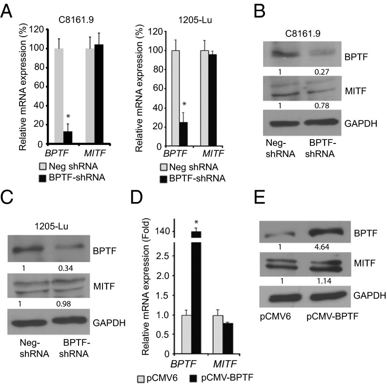
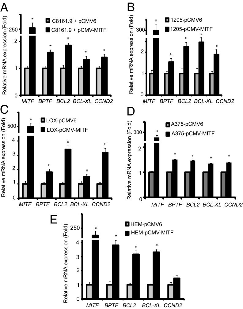
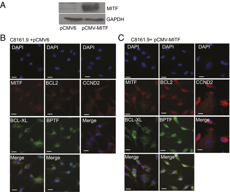
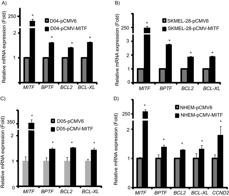
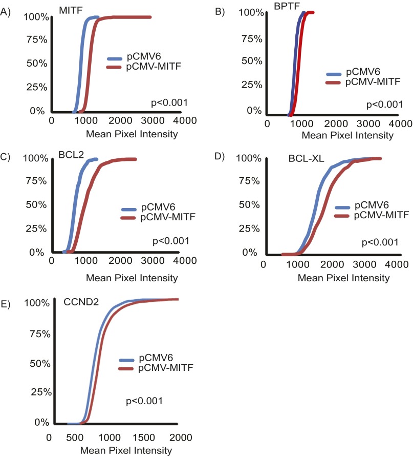
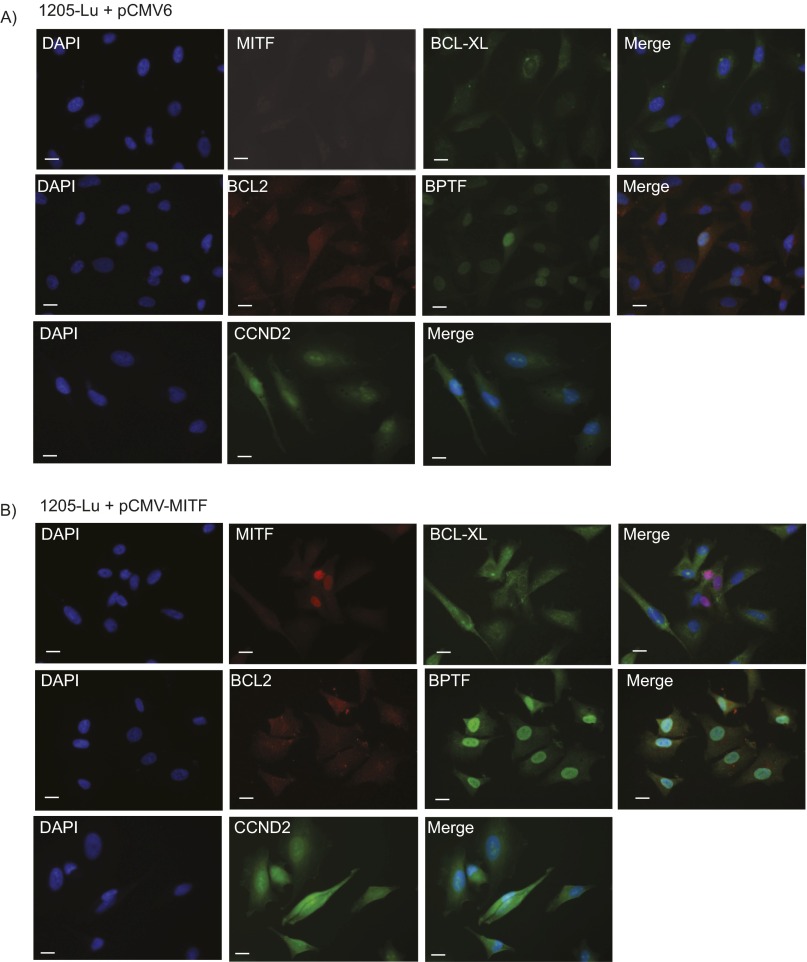
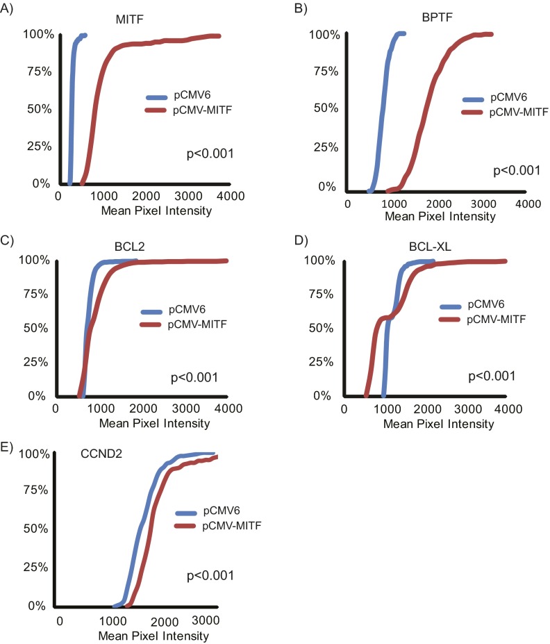
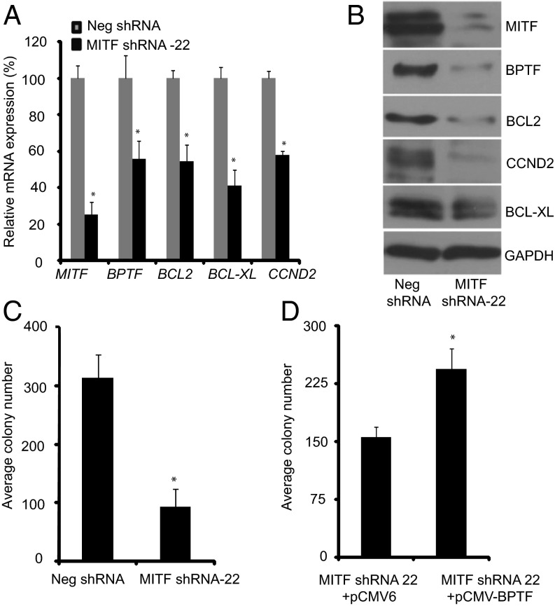
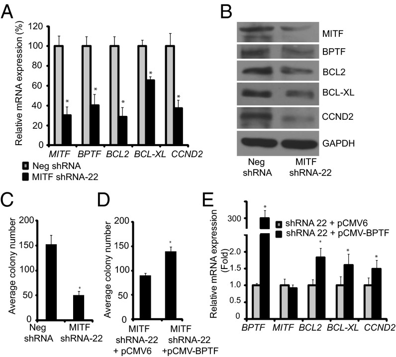
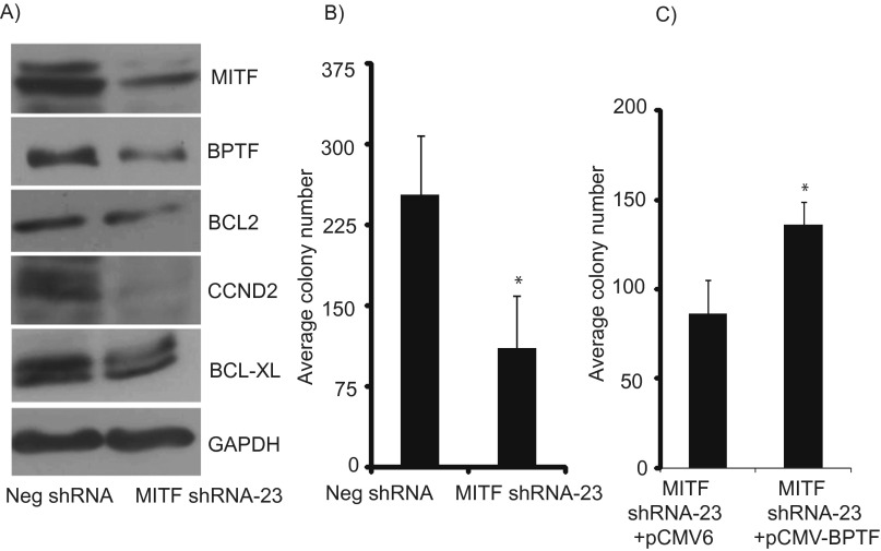
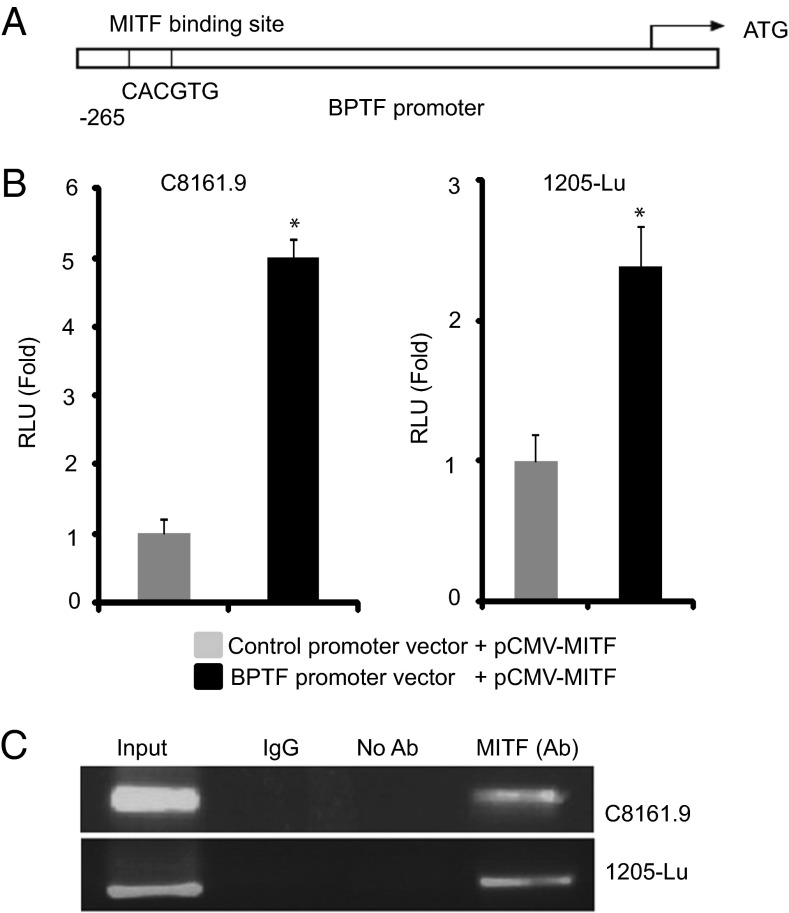
Microphthalmia-associated transcription factor (MITF) plays a critical and complex role in melanocyte transformation. Although several downstream targets of MITF action have been identified, the precise mechanisms by which MITF promotes melanocytic tumor progression are incompletely understood. Recent studies identified an oncogenic role for the bromodomain plant homeodomain finger transcription factor (BPTF) gene in melanoma progression, in part through activation of BCL2, a canonical target of MITF signaling. Analysis of the BPTF promoter identified a putative MITF-binding site, suggesting that MITF may regulate BPTF expression. Overexpression of MITF resulted in up-regulation of BPTF in a panel of melanoma and melanocyte cell lines. shRNA-mediated down-regulation of MITF in melanoma cells was accompanied by down-regulation of BPTF and BPTF-regulated genes (including BCL2) and resulted in reduced proliferative capacity of melanoma cells. The suppression of cell growth mediated by MITF silencing was rescued by overexpression of BPTF cDNA. Binding of MITF to the BPTF promoter was demonstrated using ChIP analysis. MITF overexpression resulted in direct transcriptional activation of BPTF, as evidenced by increased luciferase activity driven by the BPTF promoter. These results indicate that BPTF transduces key prosurvival signals driven by MITF, further supporting its important role in promoting melanoma cell survival and progression.

Keywords: melanoma; oncogenes; signaling cascade.


Conflict of interest statement

The authors declare no conflict of interest.

Figures

Fig. 1.
Fig. 1.
Effects of suppression of BPTF by a specific anti-BPTF shRNA on MITF expression at the RNA (A) and protein (B and C) levels in C8161.9 and 1205–Lu melanoma cells. (D and E) Effects of overexpression of BPTF cDNA on MITF RNA or protein expression in 1205-Lu melanoma cells. In A and D, data presented reflect mean ± SEM of three replicates. *P < 0.05.
Fig. 2.
Fig. 2.
Effects of overexpression of MITF cDNA in four different melanoma cell lines (A–D) and the HEM human melanocyte cell line (E) on expression of BPTF, BCL2, BCL-XL, and CCND2. In all panels, data presented reflect mean ± SEM of three replicates. *P < 0.05.
Fig. 3.
Fig. 3.
Overexpression of MITF cDNA and its effects on expression of BPTF and BPTF-regulated genes. (A) Western analysis of MITF and GAPDH expression following overexpression of MITF cDNA in HEK293 cells. (B) Immunofluorescence analysis of BPTF, BCL2, BCL-XL, and CCND2 proteins following overexpression of a control vector in C8161.9 cells. (C) Immunofluorescence analysis of BPTF, BCL2, BCL-XL, and CCND2 proteins following overexpression of MITF cDNA. (Scale bars, 20 μm.)
Fig. S1.
Fig. S1.
Effects of overexpression of MITF cDNA in three different melanoma cell lines (A–C) and the human melanocyte NHEM cell line (D) on expression of BPTF, BCL2, BCL-XL, and CCND2. The expression level of CCND2 was undetectable in D04, SKMEL-28, and D05 cells. In all panels data presented reflect mean ± SEM of three replicates. *P < 0.05.
Fig. S2.
Fig. S2.
(A–E) Quantification of immunofluorescence staining for MITF, BPTF, BCL2, BCL-XL and CCND2 in C8161.9 cells following overexpression of pCMV6 vector and MITF cDNA vector.
Fig. S3.
Fig. S3.
Effects of MITF cDNA overexpression in 1205-Lu melanoma cells. Expression of BPTF, BCL2, BCL-XL, and CCND2 protein following overexpression of pCMV6 control vector (A) and MITF cDNA vector (B) in 1205-Lu melanoma cell lines as determined by immunofluorescence. (Scale bars, 20 μm.)
Fig. S4.
Fig. S4.
(A–E) Quantification of immunofluorescence staining for MITF, BPTF, BCL2, BCL-XL, and CCND2 in 1205-Lu cells following overexpression of pCMV6 vector and MITF cDNA vector.
Fig. 4.
Fig. 4.
Effects of suppression of MITF expression in C8161.9 cells. (A) Expression of MITF, BPTF, BCL2, BCL-XL, and CCND2 RNA following stable expression of anti-MITF shRNA #22 in C8161.9 cells. (B) Expression of MITF, BPTF, BCL2, BCL-XL, and CCND2 protein following stable expression of anti-MITF shRNA #22 in C8161.9 cells. (C) Assay of C8161.9 colony formation following suppression of MITF expression. (D) Effects of overexpression of BPTF cDNA or vector control on colony forming capacity of C8161.9 cells expressing anti-MITF shRNA. In A, C, and D data presented reflect mean ± SEM of three replicates. *P < 0.05.
Fig. 5.
Fig. 5.
Effects of suppression of MITF expression in 1205-Lu melanoma cells. (A) Expression of MITF, BPTF, BCL2, BCL-XL, and CCND2 RNA following stable expression of anti-MITF shRNA #22 in 1205-Lu cells. (B) Expression of MITF, BPTF, BCL2, BCL-XL, and CCND2 protein following stable expression of anti-MITF shRNA #22 in 1205-Lu cells. (C) Assay of 1205-Lu colony formation following suppression of MITF expression. (D) Effects of overexpression of BPTF cDNA on colony forming capacity of 1205-Lu cells expressing anti-MITF shRNA. (E) Effects of overexpression of BPTF cDNA on expression of the BPTF, BCL2, BCL-XL, and CCND2 in 1205-Lu cells expressing anti-MITF shRNA. In A, C, D, and E data presented reflect mean ± SEM of three replicates. *P < 0.05.
Fig. S5.
Fig. S5.
Effects of suppression of MITF expression on expression of BPTF and BPTF-regulated genes. (A) Expression of BPTF, BCL2, BCL-XL, and CCND2 protein following stable expression of anti-MITF shRNA #23 in C8161.9 cells. (B) Assay of C8161.9 colony formation following suppression of MITF expression. (C) Effects of overexpression of BPTF cDNA on colony forming capacity of C8161.9 cells in anti-MITF shRNA-expressing cells. *P < 0.05.
Fig. 6.
Fig. 6.
Interaction of MITF with the BPTF promoter. (A) The BPTF promoter harbors a putative MITF binding site. (B) Assay of luciferase activity showing effects of overexpression of MITF cDNA compared with vector control on BPTF promoter activity. (C) ChIP analysis using an anti-MITF antibody and primers specific for the BTPF promoter, indicating the binding of MITF to the BPTF promoter. *P < 0.05.
Fig. S6.
Fig. S6.
Mean normalized expression of BPTF in melanoma samples from TCGA database expressing either low MITF (group 1) or high MITF (group 2). *P < 0.0001.

References

    1. Bentley NJ, Eisen T, Goding CR. Melanocyte-specific expression of the human tyrosinase promoter: Activation by the microphthalmia gene product and role of the initiator. Mol Cell Biol. 1994;14(12):7996–8006. - PMC - PubMed
    1. Bertolotto C, et al. Different cis-acting elements are involved in the regulation of TRP1 and TRP2 promoter activities by cyclic AMP: Pivotal role of M boxes (GTCATGTGCT) and of microphthalmia. Mol Cell Biol. 1998;18(2):694–702. - PMC - PubMed
    1. Yavuzer U, et al. The Microphthalmia gene product interacts with the retinoblastoma protein in vitro and is a target for deregulation of melanocyte-specific transcription. Oncogene. 1995;10(1):123–134. - PubMed
    1. Levy C, Khaled M, Fisher DE. MITF: Master regulator of melanocyte development and melanoma oncogene. Trends Mol Med. 2006;12(9):406–414. - PubMed
    1. Widlund HR, Fisher DE. Microphthalamia-associated transcription factor: A critical regulator of pigment cell development and survival. Oncogene. 2003;22(20):3035–3041. - PubMed

Publication types

MeSH terms