. 2016 Jan 15;6(2):249-59.
eCollection 2016.

Overexpression and oncogenic function of HMGA2 in endometrial serous carcinogenesis

Affiliations

Overexpression and oncogenic function of HMGA2 in endometrial serous carcinogenesis

Linxuan Wei et al. Am J Cancer Res. .

Abstract

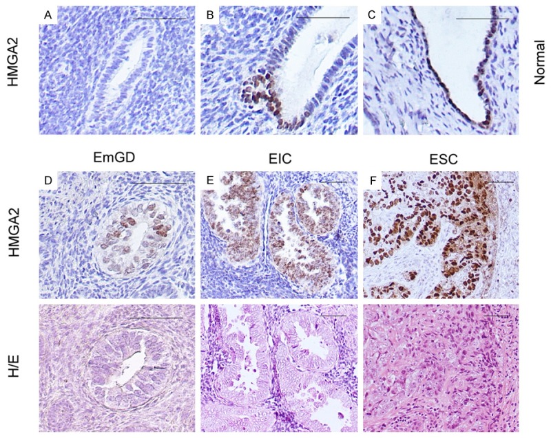
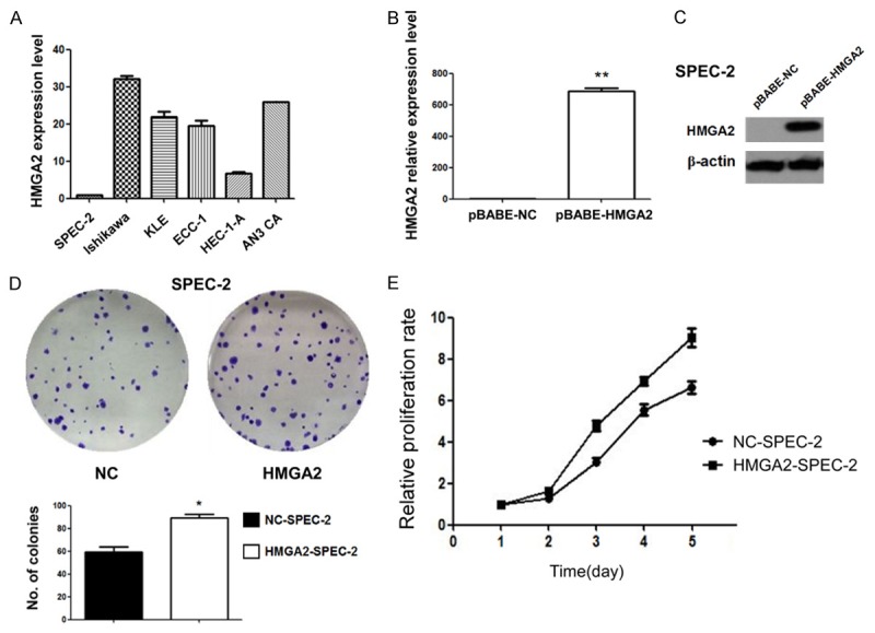
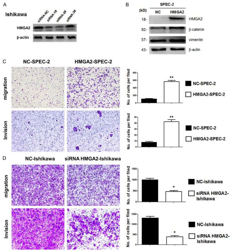
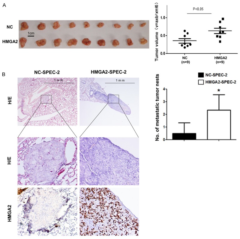
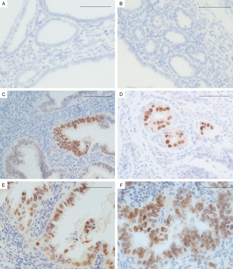
The high-mobility group A protein 2 (HMGA2) is a non-histone chromatin factor highly expressed in fetal tissue and malignant tumors but rarely detected within normal adult tissues. The clinical implications and biological functions of HMGA2 in endometrial carcinoma are largely unknown. Here we report that HMGA2 expression was barely detected in benign endometrium samples (2 of 28 samples). However, HMGA2 expression increased significantly from precancerous lesion endometrial glandular dysplasia (7 of 17, 41.2%), to serous endometrial intraepithelial carcinoma (5 of 8, 62.5%) and to full blown endometrial serous carcinoma (39 of 59, 66.1%). Functional characterization of HMGA2 revealed that the gene has both tumor growth promotion and metastasis. In addition, HMGA2 induced epithelial-mesenchymal transition (EMT) through modulation vimentin and β-catenin. Furthermore, HMGA2 overexpression started from endometrial serous precancers, non-invasive cancers, as well as in full blown carcinomas in a p53 knockout mouse model we recently established in our laboratory. Our findings suggest that HMGA2 may serve as a useful diagnostic marker in the assessment of endometrial serous cancer and its precursor lesions.

Keywords: EMT; HMGA2; endometrial serous carcinoma; metastasis; tumor growth.


Figures

Figure 1
Figure 1
Photomicrographs illustrate some examples of intensity of HMGA2 expression in benign endometrium, endometrial serous carcinomas (ESCs) and their precursor lesions. A-C. HMGA2 expression in cases of benign endometrium. D. Immunohistochemical and hematoxylin and eosin (H/E) staining on serous endometrial glandular dysplasia (EmGD). E. Immunohistochemical and hematoxylin and eosin (H/E) staining on serous endometrial intraepithelial carcinoma (EIC). F. Immunohistochemical and hematoxylin and eosin (H/E) staining on endometrial serous carcinoma (ESC). Scale bars=50 μm.
Figure 2
Figure 2
HMGA2 overexpression promote proliferation of endometrial cancer cells in vitro. A. HMGA2 baseline expression in six endometrial cancer cell lines were measured by real-time PCR. B. The overexpression of HMGA2 was validated by qPCR in SPEC-2 cells. C. The overexpression of HMGA2 was validated by Western blot in SPEC-2 cells. D. Clonogenic assay was performed on SPEC-2 cells with HMGA2 overexpression compared to control cells. E. MTT assay was performed on SPEC-2 cells with HMGA2 overexpression compared to control cells.
Figure 3
Figure 3
HMGA2 promote migration and invasion of endometrial cancer cells in vitro. A. HMGA2 repression by siRNAs in ishikawa cells was measured by Western blot. B. Western blot analysis confirmed MET related markers showed different expression in SPEC-2 cells with and without HMGA2 overexpression. C. Migration and invasion were measured by transwell assay in SPEC-2 cell lines with and without HMGA2 overexpression. D. Migration and invasion were performed in ishikawa cells with and without HMGA2 depletion by siRNA-2#. *P<0.05, **P<0.01.
Figure 4
Figure 4
HMGA2 promote proliferation and invasion of endometrial cancer cells in vivo. A. Photographs illustrate representative tumors in xenografts of SPEC-2 cell lines with and without HMGA2 overexpression. B. Hematoxylin and eosin (HE)-staining and IHC of lungs isolated from mice that received tail vein injection of SPEC-2 cell line with and without HMGA2 overexpression (left). Each group contains 6 mice. Plot graph illustrates the numbers of pulmonary metastatic nodules under microscope were counted and analyzed with Student’s t-test (right). All data are shown as mean ± SD. *P<0.05.
Figure 5
Figure 5
Photomicrographs illustrate some examples of HMGA2 expression in conditional Trp53 knockout mouse model of type II endometrial carcinoma. A. HMGA2 expression in benign endometrium of wild type mice. B and C. HMGA2 expression in benign endometrium of conditional Trp53 knockout mice. D. HMGA2 expression in serous endometrial glandular dysplasia (EmGD). E. serous endometrial intraepithelial carcinoma (EIC) and F, endometrial serous carcinoma (ESC) of conditional Trp53 knockout mice. Scale bars=50 μm.

References

    1. Murali R, Soslow RA, Weigelt B. Classification of endometrial carcinoma: more than two types. Lancet Oncol. 2014;15:e268–278. - PubMed
    1. Zheng W, Xiang L, Fadare O, Kong B. A proposed model for endometrial serous carcinogenesis. Am J Surg Pathol. 2011;35:e1–e14. - PubMed
    1. Fader AN, Santin AD, Gehrig PA. Early stage uterine serous carcinoma: management updates and genomic advances. Gynecol Oncol. 2013;129:244–250. - PubMed
    1. Wu J, Wei JJ. HMGA2 and high-grade serous ovarian carcinoma. J Mol Med (Berl) 2013;91:1155–1165. - PubMed
    1. Hristov AC, Cope L, Reyes MD, Singh M, Iacobuzio-Donahue C, Maitra A, Resar LM. HMGA2 protein expression correlates with lymph node metastasis and increased tumor grade in pancreatic ductal adenocarcinoma. Mod Pathol. 2009;22:43–49. - PMC - PubMed

LinkOut - more resources