. 2016 May 17;315(19):2104-12.
doi: 10.1001/jama.2016.5657.

Association of Acute Gastroesophageal Reflux Disease With Esophageal Histologic Changes

Affiliations

Association of Acute Gastroesophageal Reflux Disease With Esophageal Histologic Changes

Kerry B Dunbar et al. JAMA. .

Abstract

Importance: The histologic changes associated with acute gastroesophageal reflux disease (GERD) have not been studied prospectively in humans. Recent studies in animals have challenged the traditional notion that reflux esophagitis develops when esophageal surface epithelial cells are exposed to lethal chemical injury from refluxed acid.

Objective: To evaluate histologic features of esophageal inflammation in acute GERD to study its pathogenesis.

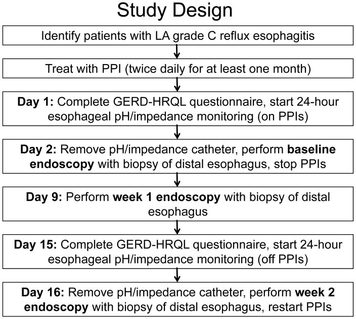
Design, setting, and participants: Patients from the Dallas Veterans Affairs Medical Center who had reflux esophagitis successfully treated with proton pump inhibitors (PPIs) began 24-hour esophageal pH and impedance monitoring and esophagoscopy (including confocal laser endomicroscopy [CLE]) with biopsies from noneroded areas of distal esophagus at baseline (taking PPIs) and at 1 week and 2 weeks after stopping the PPI medication. Enrollment began May 2013 and follow-up ended July 2015.

Interventions: PPIs stopped for 2 weeks.

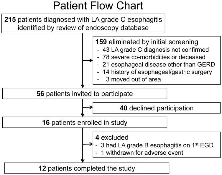
Main outcomes and measures: Twelve patients (men, 11; mean age, 57.6 year [SD, 13.1]) completed the study. Primary outcome was change in esophageal inflammation 2 weeks after stopping the PPI medication, determined by comparing lymphocyte, eosinophil, and neutrophil infiltrates (each scored on a 0-3 scale) in esophageal biopsies. Also evaluated were changes in epithelial basal cell and papillary hyperplasia, surface erosions, intercellular space width, endoscopic grade of esophagitis, esophageal acid exposure, and mucosal impedance (an index of mucosal integrity).

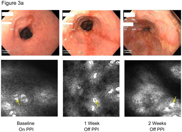
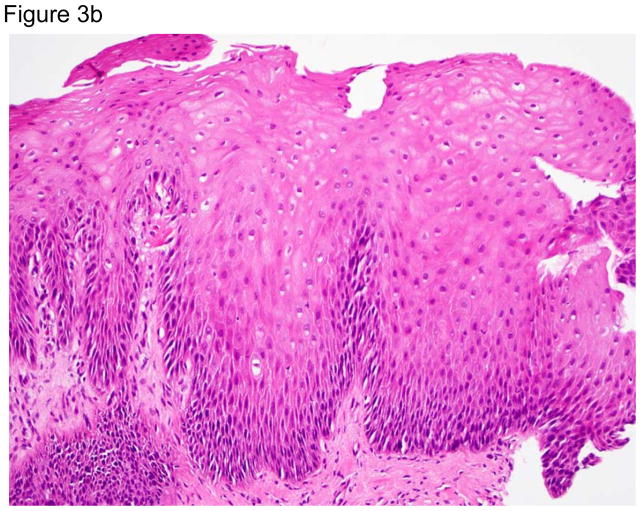
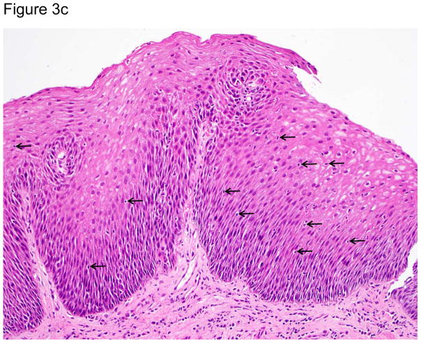
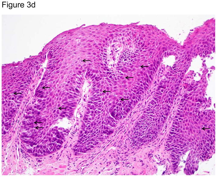
Results: At 1 week and 2 weeks after discontinuation of PPIs, biopsies showed significant increases in intraepithelial lymphocytes, which were predominantly T cells (median [range]: 0 (0-2) at baseline vs 1 (1-2) at both 1 week [P = .005] and 2 weeks [P = .002]); neutrophils and eosinophils were few or absent. Biopsies also showed widening of intercellular spaces (confirmed by CLE), and basal cell and papillary hyperplasia developed without surface erosions. Two weeks after stopping the PPI medication, esophageal acid exposure increased (median: 1.2% at baseline to 17.8% at 2 weeks; Δ, 16.2% [95% CI, 4.4%-26.5%], P = .005), mucosal impedance decreased (mean: 2671.3 Ω at baseline to 1508.4 Ω at 2 weeks; Δ, 1162.9 Ω [95% CI, 629.9-1695.9], P = .001), and all patients had evidence of esophagitis.

Conclusions and relevance: In this preliminary study of 12 patients with severe reflux esophagitis successfully treated with PPI therapy, stopping PPI medication was associated with T lymphocyte-predominant esophageal inflammation and basal cell and papillary hyperplasia without loss of surface cells. If replicated, these findings suggest that the pathogenesis of reflux esophagitis may be cytokine-mediated rather than the result of chemical injury.

Trial registration: clinicaltrials.gov Identifier: NCT01733810.


Figures

Figure 1
Figure 1
Study design.
Figure 2
Figure 2
Patient flow chart. LA=Los Angeles grade of esophagitis, EGD=endoscopy.
Figure 3a
Figure 3a
Representative images of the distal esophagus at baseline, and at 1 and 2 weeks off PPI therapy. All images are from the same patient. Top row: high definition white light endoscopy (HD-WLE), bottom row: confocal laser endomicroscopy (CLE) images. At baseline, HD-WLE reveals an irregular tongue of columnar mucosa (Barrett’s esophagus) in the 12 o’clock position, but no esophagitis. At week 1, HD-WLE shows long linear mucosal breaks (4 and 6 o’clock positions) extending up the esophagus from the gastroesophageal junction (LA-B esophagitis). At week 2, HD-WLE shows long mucosal breaks continuous between the tops of mucosal folds (LA-C esophagitis). CLE reveals fluorescein within bright intraepithelial capillaries (yellow arrows), with fluorescein that leaked from blood vessels into intercellular spaces surrounding individual cells, creating a reticular appearance characteristic of squamous epithelium. CLE measurements revealed widened intercellular spaces with increased intercellular fluorescein at weeks 1 and 2. 3b–d. Photomicrographs of biopsies of the distal esophagus in the same patient: 3b) at baseline, 3c) at one week after stopping PPIs; arrows point to some of the numerous intraepithelial lymphocytes, and 3d) at two weeks after stopping PPIs; note the prominent lymphocytosis, basal cell hyperplasia and papillary elongation (all photomicrographs H&E, original magnification ×20). All images were manipulated in Photoshop to remove patient identification data, and to enhance clarity. Any adjustments in contrast, color balance, brightness or sharpness were applied to the entire image.
Figure 3a
Figure 3a
Representative images of the distal esophagus at baseline, and at 1 and 2 weeks off PPI therapy. All images are from the same patient. Top row: high definition white light endoscopy (HD-WLE), bottom row: confocal laser endomicroscopy (CLE) images. At baseline, HD-WLE reveals an irregular tongue of columnar mucosa (Barrett’s esophagus) in the 12 o’clock position, but no esophagitis. At week 1, HD-WLE shows long linear mucosal breaks (4 and 6 o’clock positions) extending up the esophagus from the gastroesophageal junction (LA-B esophagitis). At week 2, HD-WLE shows long mucosal breaks continuous between the tops of mucosal folds (LA-C esophagitis). CLE reveals fluorescein within bright intraepithelial capillaries (yellow arrows), with fluorescein that leaked from blood vessels into intercellular spaces surrounding individual cells, creating a reticular appearance characteristic of squamous epithelium. CLE measurements revealed widened intercellular spaces with increased intercellular fluorescein at weeks 1 and 2. 3b–d. Photomicrographs of biopsies of the distal esophagus in the same patient: 3b) at baseline, 3c) at one week after stopping PPIs; arrows point to some of the numerous intraepithelial lymphocytes, and 3d) at two weeks after stopping PPIs; note the prominent lymphocytosis, basal cell hyperplasia and papillary elongation (all photomicrographs H&E, original magnification ×20). All images were manipulated in Photoshop to remove patient identification data, and to enhance clarity. Any adjustments in contrast, color balance, brightness or sharpness were applied to the entire image.

Comment in

References

    1. Locke GR, 3rd, Talley NJ, Fett SL, Zinsmeister AR, Melton LJ., 3rd Prevalence and clinical spectrum of gastroesophageal reflux: a population-based study in Olmsted County, Minnesota. Gastroenterology. 1997;112(5):1448–56. - PubMed
    1. Winkelstein A. Peptic esophagitis: a new clinical entity. JAMA. 1935;104(11):906–9.
    1. Ismail-Beigi F, Horton PF, Pope CE., 2nd Histological consequences of gastroesophageal reflux in man. Gastroenterology. 1970;58(2):163–74. - PubMed
    1. Fiocca R, Mastracci L, Riddell R, Takubo K, Vieth M, Yerian L, Sharma P, Fernström P, Ruth M. Development of consensus guidelines for the histologic recognition of microscopic esophagitis in patients with gastroesophageal reflux disease: the Esohisto project. Hum Pathol. 2010;41(2):223–31. - PubMed
    1. Eastwood GL. Histologic changes in gastroesophageal reflux. J Clin Gastroenterol. 1986;8(Suppl 1):45–51. - PubMed

Publication types

MeSH terms

Associated data