

cDNA and Gene Structure of MytiLec-1, A Bacteriostatic R-Type Lectin from the Mediterranean Mussel (Mytilus galloprovincialis)
- PMID: 27187419
- PMCID: PMC4882566
- DOI: 10.3390/md14050092
cDNA and Gene Structure of MytiLec-1, A Bacteriostatic R-Type Lectin from the Mediterranean Mussel (Mytilus galloprovincialis)
Abstract
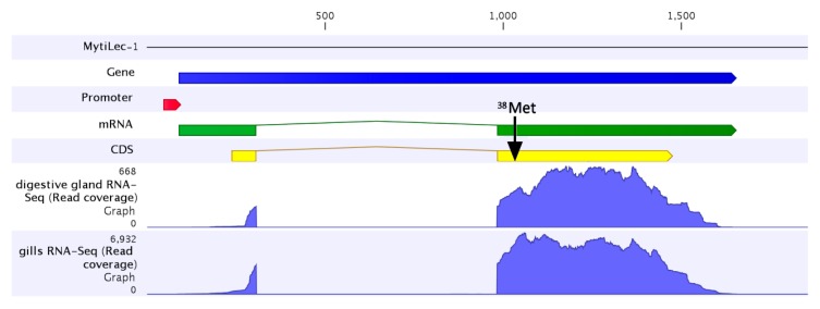
MytiLec is an α-d-galactose-binding lectin with a unique primary structure isolated from the Mediterranean mussel (Mytilus galloprovincialis). The lectin adopts a β-trefoil fold that is also found in the B-sub-unit of ricin and other ricin-type (R-type) lectins. We are introducing MytiLec(-1) and its two variants (MytiLec-2 and -3), which both possess an additional pore-forming aerolysin-like domain, as members of a novel multi-genic "mytilectin family" in bivalve mollusks. Based on the full length mRNA sequence (911 bps), it was possible to elucidate the coding sequence of MytiLec-1, which displays an extended open reading frame (ORF) at the 5' end of the sequence, confirmed both at the mRNA and at the genomic DNA sequence level. While this extension could potentially produce a polypeptide significantly longer than previously reported, this has not been confirmed yet at the protein level. MytiLec-1 was revealed to be encoded by a gene consisting of two exons and a single intron. The first exon comprised the 5'UTR and the initial ATG codon and it was possible to detect a putative promoter region immediately ahead of the transcription start site in the MytiLec-1 genomic locus. The remaining part of the MytiLec-1 coding sequence (including the three sub-domains, the 3'UTR and the poly-A signal) was included in the second exon. The bacteriostatic activity of MytiLec-1 was determined by the agglutination of both Gram-positive and Gram-negative bacteria, which was reversed by the co-presence of α-galactoside. Altogether, these data support the classification of MytiLec-1 as a member of the novel mytilectin family and suggest that this lectin may play an important role as a pattern recognition receptor in the innate immunity of mussels.
Keywords: MytiLec-1; Mytilus galloprovincialis; R-type lectin; bacteriostatic activity; cDNA; gene; innate immunity; mRNA-sequence; mytilectin family.
Figures


Similar articles
-
Purification and Functional Characterization of the Effects on Cell Signaling of Mytilectin: A Novel β-Trefoil Lectin from Marine Mussels.Methods Mol Biol. 2020;2132:201-213. doi: 10.1007/978-1-0716-0430-4_21. Methods Mol Biol. 2020. PMID: 32306329
-
MytiLec, a Mussel R-Type Lectin, Interacts with Surface Glycan Gb3 on Burkitt's Lymphoma Cells to Trigger Apoptosis through Multiple Pathways.Mar Drugs. 2015 Dec 14;13(12):7377-89. doi: 10.3390/md13127071. Mar Drugs. 2015. PMID: 26694420 Free PMC article.
-
A lectin from the mussel Mytilus galloprovincialis has a highly novel primary structure and induces glycan-mediated cytotoxicity of globotriaosylceramide-expressing lymphoma cells.J Biol Chem. 2012 Dec 28;287(53):44772-83. doi: 10.1074/jbc.M112.418012. Epub 2012 Oct 23. J Biol Chem. 2012. PMID: 23093409 Free PMC article.
-
Activity Dependence of a Novel Lectin Family on Structure and Carbohydrate-Binding Properties.Molecules. 2019 Dec 30;25(1):150. doi: 10.3390/molecules25010150. Molecules. 2019. PMID: 31905927 Free PMC article. Review.
-
[Cell Function Research of β-Trefoil Lectins from Mytilidae].Yakugaku Zasshi. 2021;141(4):481-488. doi: 10.1248/yakushi.20-00215. Yakugaku Zasshi. 2021. PMID: 33790114 Review. Japanese.
Cited by
-
Glabralysins, Potential New β-Pore-Forming Toxin Family Members from the Schistosomiasis Vector Snail Biomphalaria glabrata.Genes (Basel). 2020 Jan 7;11(1):65. doi: 10.3390/genes11010065. Genes (Basel). 2020. PMID: 31936048 Free PMC article.
-
A GM1b/asialo-GM1 oligosaccharide-binding R-type lectin from purplish bifurcate mussels Mytilisepta virgata and its effect on MAP kinases.FEBS J. 2020 Jun;287(12):2612-2630. doi: 10.1111/febs.15154. Epub 2019 Dec 24. FEBS J. 2020. PMID: 31769916 Free PMC article.
-
MytiLec-1 Shows Glycan-Dependent Toxicity against Brine Shrimp Artemia and Induces Apoptotic Death of Ehrlich Ascites Carcinoma Cells In Vivo.Mar Drugs. 2019 Aug 28;17(9):502. doi: 10.3390/md17090502. Mar Drugs. 2019. PMID: 31466257 Free PMC article.
-
A Genomic and Transcriptomic Analysis of the C-Type Lectin Gene Family Reveals Highly Expanded and Diversified Repertoires in Bivalves.Mar Drugs. 2023 Apr 20;21(4):254. doi: 10.3390/md21040254. Mar Drugs. 2023. PMID: 37103393 Free PMC article.
-
Taxonomic Distribution and Molecular Evolution of Mytilectins.Mar Drugs. 2023 Nov 27;21(12):614. doi: 10.3390/md21120614. Mar Drugs. 2023. PMID: 38132935 Free PMC article. Review.
References
MeSH terms
Substances
LinkOut - more resources
Full Text Sources
Other Literature Sources
