Review
. 2016 May 13;6(2):27.
doi: 10.3390/biom6020027.

The Diversity of Ribonuclease P: Protein and RNA Catalysts with Analogous Biological Functions

Affiliations
Review

The Diversity of Ribonuclease P: Protein and RNA Catalysts with Analogous Biological Functions

Bradley P Klemm et al. Biomolecules. .

Abstract

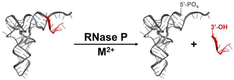
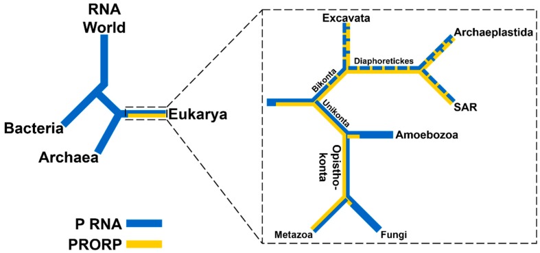
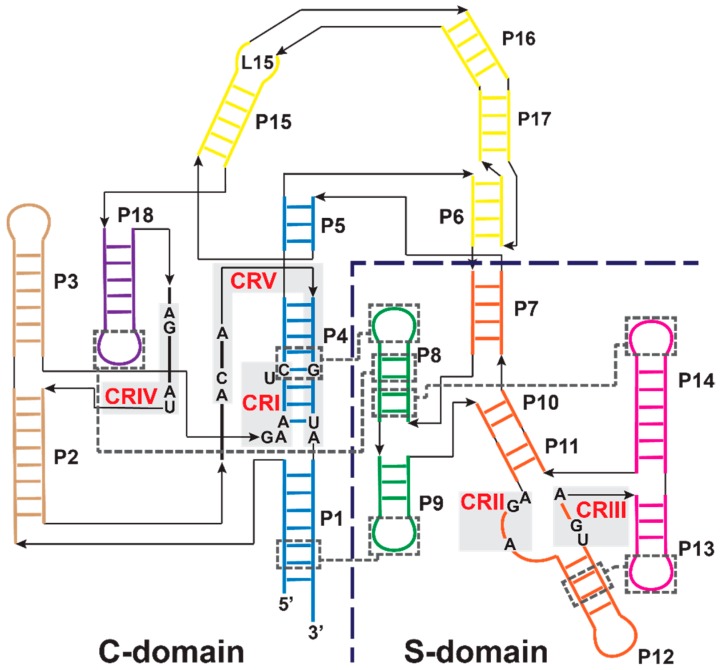
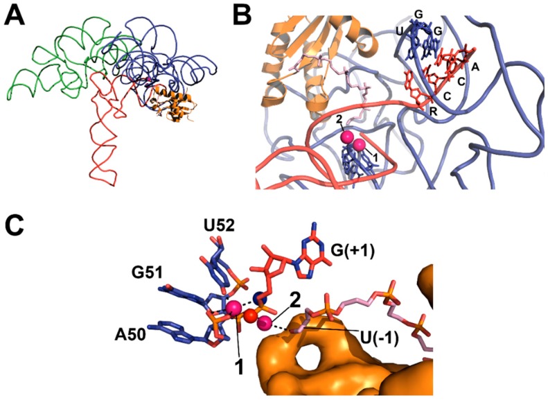
Ribonuclease P (RNase P) is an essential endonuclease responsible for catalyzing 5' end maturation in precursor transfer RNAs. Since its discovery in the 1970s, RNase P enzymes have been identified and studied throughout the three domains of life. Interestingly, RNase P is either RNA-based, with a catalytic RNA subunit, or a protein-only (PRORP) enzyme with differential evolutionary distribution. The available structural data, including the active site data, provides insight into catalysis and substrate recognition. The hydrolytic and kinetic mechanisms of the two forms of RNase P enzymes are similar, yet features unique to the RNA-based and PRORP enzymes are consistent with different evolutionary origins. The various RNase P enzymes, in addition to their primary role in tRNA 5' maturation, catalyze cleavage of a variety of alternative substrates, indicating a diversification of RNase P function in vivo. The review concludes with a discussion of recent advances and interesting research directions in the field.

Keywords: PRORP; RNase P; endonuclease; ribozyme; tRNA maturation; tRNA recognition.


Figures

Figure 1
Figure 1
RNase P enzymes catalyze metal-dependent, endonucleolytic cleavage of pre-tRNA (adapted with permission from [7]).
Figure 2
Figure 2
Distribution of RNase P enzymes across evolutionary lineages. Presence of a P RNA (blue) or PRORP (yellow) is indicated. Left: RNA-based RNase Ps are found in all domains of life and are proposed to have evolved from an RNA predecessor lacking protein components. PRORPs exist only in Eukarya and likely evolved after the divergence with Archaea. Right: Distribution of RNase Ps in Eukarya using the five supergroup model (aspects of the branching order remain controversial) [12,13]. PRORPs are found in four supergroups: Excavata, Archaeplastida, SAR [Stramenopiles, Aveolata, and Rhizaria], and Opisthokonta, but not in the fifth: Amoebozoa [11]. Dashed blue or yellow lines indicate that some clades within the supergroup lack RNase P RNA or PRORP sequences, respectively.
Figure 3
Figure 3
The secondary structure of type A RNase P RNA from T. maritima (adapted with permission from [21]) [22,23]. Individually-folding catalytic (C)- and specificity (S)-domains are divided by the dashed line. Conserved regions (CRI–V, red) are indicated by gray shading and numbered in order of occurrence from the 5’ end [4]. Tertiary interactions in the T. maritima P RNA are indicated by dashed gray boxes and lines. Helices are colored by coaxial-stack and are numbered as P1–P18 in order of occurrence from the 5’ end. The C-domain includes P1/P4/P5 (blue), P2/P3 (brown), P6/P15/P16/P17 (yellow,) and P18 (purple). The S-domain includes P7/P10/P11/P12 (orange), P8/P9 (green), and P13/P14 (pink).
Figure 4
Figure 4
X-ray crystal structure of T. maritima RNase P-tRNA-leader product complex (PDB 3q1r, PyMOL) [22]. (A) The holoenzyme-product complex of RNase P is shown, including C- (blue backbone) and S- (green backbone) domains, tRNA (red backbone), and RnpA (orange cartoon); (B) Topology of substrate contact sites in the catalytic domain (colored as in A), including the 5’ leader (light pink sticks) bound to RnpA and proposed divalent metal ions (pink spheres). Base-pairing between GGU residues in P RNA and the 3’ RCCA of tRNA (U256–R(73), G255–C(74), G254–C(75)) is shown; (C) Topology of the active site (colored as in A), including the active site residues (blue carbon atoms), product G(+1) (red carbon atoms), the 5’ leader (light pink carbon atoms) bound to RnpA (orange surface), proposed metal contacts (black dashed lines), and positions of the pro-RP (red sphere) and pro-SP (blue sphere) oxygens of the product 5’ phosphate.
Figure 5
Figure 5
Crystal structures of AtPRORP1 (PDB 4g24) and AtPRORP2 (PDB 5diz) [64,103]. (A) Three-dimensional alignment of AtPRORP1 and AtPRORP2 structures via their active sites (dashed box). PRORP1 (red) topology includes the NYN metallonuclease domain bound to Mn2+ (purple spheres). PRORP2 (green) is in a more “open” conformation, resulting in a ~35 Å difference in the position of the first PPR helix; (B) AtPRORP2 crystallization dimer (left), with subunits labeled in green and cyan (NYN domains in brighter colors); (C) Expanded views of the dashed boxes in panel A (left) or B (right). The AtPRORP1 active site (left) includes four fully-conserved aspartates and one partially-conserved aspartate (sticks). Close-up of intersubunit interaction (right) between AtPRORP2 molecules, with active site aspartates of one molecule interacting with Lys 42 (sticks) of the second (red dashed lines).
Figure 6
Figure 6
Alignment of two MRPP3 structures (PDB 4xgl, yellow; PDB 4rou, orange) with the AtPRORP1 active site (PDB 4g24, only metal ions shown) [66,69,107]. Left: Alignment of two MRPP3 structures with the PRORP1 active site (purple spheres). MRPP3 NYN residues (4xgl: S361/P362, D409, D479, and D499, or 4rou: residues S361/P362, D409, D478, and D479) were aligned to the equivalent PRORP1 residues. The dashed box is expanded on the right. Middle and Right panels: Dashed box contains the aligned MRPP3 active site residues from 4xgl (middle, yellow) or 4rou (right, orange) with Mn2+ from PRORP1 (transparent purple spheres). Residues with the potential to occlude the metal sites, N412, R445, and R498, are shown if coordinates are available. Hydrogen bonds between residues are shown as dashed red lines.
Scheme 1
Scheme 1
Minimal kinetic mechanism of bacterial RNase P ribozyme catalysis. The initial binding step is bimolecular and dependent on the concentrations of both E and S. Binding, conformational change, and substrate hydrolysis are all dependent on divalent metal ions (M2+), while hydrolysis is also dependent on pH.
Scheme 2
Scheme 2
The concerted hydrolytic mechanism proposed for bacterial RNase P. The acid (A) may be a water or metal-bound water.
Figure 7
Figure 7
Active site coordination of substrate by T. maritima RNase P (top, x-ray crystal structure) and A. thaliana PRORP1 (bottom, complex modeled in PyMOL). The minor groove width was measured as the distance between the non-bridging phosphate oxygens. Top: T. maritima product complex crystal structure (PDB 3q1r) [22]. Pro-RP (blue spheres) and pro-SP (red spheres) oxygen atoms of tRNA product (yellow cartoon) shown for N(+1)–N(+3). Active site metal atoms (purple spheres) and metal-coordinating residues A50, G51, and U52 are visualized (teal sticks). Bottom: The PRORP1 active site (PDB 4g24) was aligned to S. cerevisiae tRNAAsp (PDB 2tra) using the human DNA exonuclease I active site bound to DNA (PDB 3qeb) as a guide [64,153,154]. The tRNA, backbone oxygen atoms, active site metal atoms, and active site residues D399, D474/475, D493, and D497 are colored as in the top panel.
Figure 8
Figure 8
Proposed transition state structure of the catalytic mechanisms of (A) bacterial RNase P (as proposed in [22]) and (B) AtPRORP1 (adapted from [110]).
Figure 9
Figure 9
Secondary and tertiary structures of canonical tRNA (adapted with permission from [7]). (A) and (B) Secondary structure of B. subtilis tRNAAsp with tertiary interactions denoted by dashed lines. (C) A crystal structure of an unmodified E. coli tRNAPhe (PDB 3l0u) [161].
Figure 10
Figure 10
Sequence specific interactions between 5’ and 3’ sequences of pre-tRNA and bacterial RNase P (adapted with permission from [7]). The 3’ RCCA and U(-1) base pair with the GGU motif in the L15 loop and A213, respectively, while A(-2) makes a non-Watson-Crick interaction with U294 [23,120,175].

Similar articles

Cited by

References

    1. The Nobel Prize in Chemistry 1989. [(accessed on 15 January 2016)]. Available online: http://www.nobelprize.org/nobel_prizes/chemistry/laureates/1989/
    1. Robertson H.D., Altman S., Smith J.D. Purification and properties of a specific Escherichia coli ribonuclease which cleaves a tyrosine transfer ribonucleic acid precursor. J. Biol. Chem. 1972;247:5243–5251. - PubMed
    1. Guerrier-Takada C., Gardiner K., Marsh T., Pace N.R., Altman S. The RNA moiety of Ribonuclease P is the catalytic subunit of the enzyme. Cell. 1983;35:849–857. doi: 10.1016/0092-8674(83)90117-4. - DOI - PubMed
    1. Frank D.N., Pace N.R. Ribonuclease P: Unity and diversity in a trna processing ribozyme. Annu Rev Biochem. 1998;67:153–180. doi: 10.1146/annurev.biochem.67.1.153. - DOI - PubMed
    1. Walker S.C., Engelke D.R. Ribonuclease P: The evolution of an ancient RNA enzyme. Crit. Rev. Biochem. Mol. Biol. 2006;41:77–102. doi: 10.1080/10409230600602634. - DOI - PMC - PubMed

Publication types

LinkOut - more resources