

Evaluation of diffusivity in pituitary adenoma: 3D turbo field echo with diffusion-sensitized driven-equilibrium preparation
- PMID: 27187598
- PMCID: PMC5257300
- DOI: 10.1259/bjr.20150755
Evaluation of diffusivity in pituitary adenoma: 3D turbo field echo with diffusion-sensitized driven-equilibrium preparation
Abstract
Objective: Diffusivity of pituitary adenoma has not been investigated fully. The purpose of this study was to evaluate the feasibility of turbo field echo with diffusion-sensitized driven-equilibrium (DSDE-TFE) preparation for pituitary adenoma in the sella turcica and unaffected anterior lobe of the pituitary gland.
Methods: This retrospective study included 23 adult patients with pituitary adenomas. Among them, 6 each were prolactin-producing adenomas and growth hormone-producing adenomas (GH) and the remaining 11 were non-functioning adenomas (NON). The apparent diffusion coefficients (ADCs) were measured in the pituitary adenoma and in the unaffected pituitary gland using coronal reformatted plane.
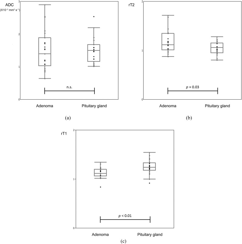
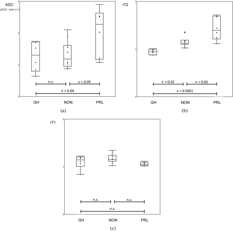
Results: All pituitary adenomas were clearly visualized on DSDE-TFE and ADC maps without obvious geometrical distortion. There were no statistically significant differences in ADC of the all pituitary adenoma (1.50 ± 0.61 × 10(-3) mm(2) s(-1)) and the unaffected anterior lobe of the pituitary gland (1.49 ± 0.37 × 10(-3) mm(2) s(-1), p = 0.99). The ADC in prolactin-producing adenomas (2.04 ± 0.76 × 10(-3) mm(2) s(-1)) was significantly higher than that in GH (1.26 ± 0.47 × 10(-3) mm(2) s(-1); p < 0.05) and NON (1.33 ± 0.42 × 10(-3) mm(2) s(-1); p = 0.04). There was no statistically significant difference between GH and NON (p = 0.97). The intraclass correlation coefficient for ADC was 0.985 in adenomas and 0.635 in unaffected glands.
Conclusion: With its insensitivity to field inhomogeneity and high spatial resolution, DSDE-TFE proved a feasible method for evaluating the diffusivity in the pituitary gland and adenoma.
Advances in knowledge: DSDE-TFE could enable us to assess ADC of pituitary adenoma in the sella turcica with high resolution and few susceptibility artefacts.
Figures
Similar articles
-
3D turbo field echo with diffusion-sensitized driven-equilibrium preparation technique (DSDE-TFE) versus echo planar imaging in evaluation of diffusivity of retinoblastoma.Br J Radiol. 2016 Nov;89(1067):20160074. doi: 10.1259/bjr.20160074. Epub 2016 Sep 16. Br J Radiol. 2016. PMID: 27636188 Free PMC article.
-
Evaluation of diffusivity in the anterior lobe of the pituitary gland: 3D turbo field echo with diffusion-sensitized driven-equilibrium preparation.AJNR Am J Neuroradiol. 2014 Jan;35(1):95-8. doi: 10.3174/ajnr.A3620. Epub 2013 Jul 18. AJNR Am J Neuroradiol. 2014. PMID: 23868152 Free PMC article.
-
High-resolution three-dimensional diffusion-weighted imaging of middle ear cholesteatoma at 3.0 T MRI: usefulness of 3D turbo field-echo with diffusion-sensitized driven-equilibrium preparation (TFE-DSDE) compared to single-shot echo-planar imaging.Eur J Radiol. 2013 Sep;82(9):e471-5. doi: 10.1016/j.ejrad.2013.04.018. Epub 2013 May 20. Eur J Radiol. 2013. PMID: 23701953
-
The radiology of pituitary adenomas.Semin Roentgenol. 1984 Jan;19(1):53-69. doi: 10.1016/0037-198x(84)90043-9. Semin Roentgenol. 1984. PMID: 6322348 Review. No abstract available.
-
The current state of MRI-based radiomics in pituitary adenoma: promising but challenging.Front Endocrinol (Lausanne). 2024 Sep 20;15:1426781. doi: 10.3389/fendo.2024.1426781. eCollection 2024. Front Endocrinol (Lausanne). 2024. PMID: 39371931 Free PMC article. Review.
Cited by
-
Application of Reduced-FOV Diffusion-Weighted Imaging in Evaluation of Normal Pituitary Glands and Pituitary Macroadenomas.AJNR Am J Neuroradiol. 2018 Aug;39(8):1499-1504. doi: 10.3174/ajnr.A5735. Epub 2018 Jul 19. AJNR Am J Neuroradiol. 2018. PMID: 30026383 Free PMC article.
-
Whole-Brain Functional and Diffusion Tensor MRI in Human Participants with Metallic Orthodontic Braces.Radiology. 2020 Jan;294(1):149-157. doi: 10.1148/radiol.2019190070. Epub 2019 Nov 12. Radiology. 2020. PMID: 31714192 Free PMC article.
-
Assessment of microvessel perfusion of pituitary adenomas: a feasibility study using turbo spin-echo-based intravoxel incoherent motion imaging.Eur Radiol. 2020 Apr;30(4):1908-1917. doi: 10.1007/s00330-019-06443-x. Epub 2019 Dec 10. Eur Radiol. 2020. PMID: 31822973
-
Time-dependent MR diffusion analysis of functioning and nonfunctioning pituitary adenomas/pituitary neuroendocrine tumors.J Neuroimaging. 2025 Jan-Feb;35(1):e13254. doi: 10.1111/jon.13254. J Neuroimaging. 2025. PMID: 39636086 Free PMC article.
-
Diffusivity of intraorbital lymphoma vs. inflammation: comparison of single shot turbo spin echo and multishot echo planar imaging techniques.Eur Radiol. 2018 Jan;28(1):325-330. doi: 10.1007/s00330-017-4995-5. Epub 2017 Aug 4. Eur Radiol. 2018. PMID: 28779398
References
MeSH terms
LinkOut - more resources
Full Text Sources
Other Literature Sources
Medical
