. 2016 May 17;12(5):e1006037.
doi: 10.1371/journal.pgen.1006037. eCollection 2016 May.

Molecular Characterization of Three Canine Models of Human Rare Bone Diseases: Caffey, van den Ende-Gupta, and Raine Syndromes

Affiliations

Molecular Characterization of Three Canine Models of Human Rare Bone Diseases: Caffey, van den Ende-Gupta, and Raine Syndromes

Marjo K Hytönen et al. PLoS Genet. .

Abstract

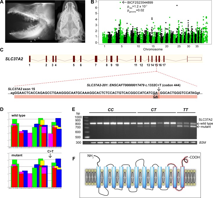
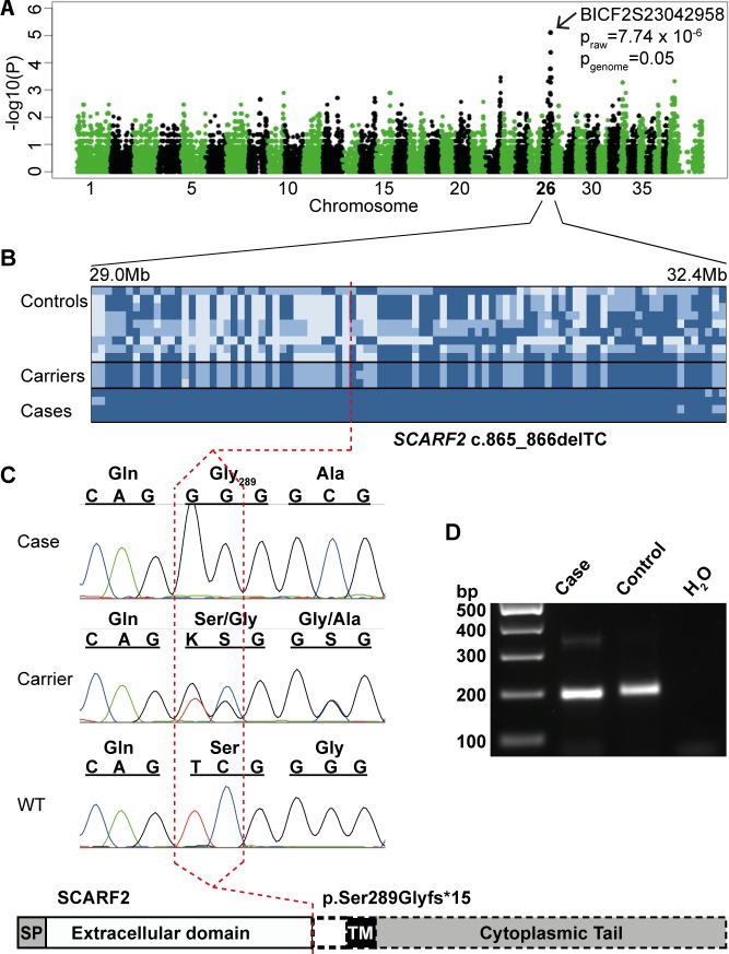
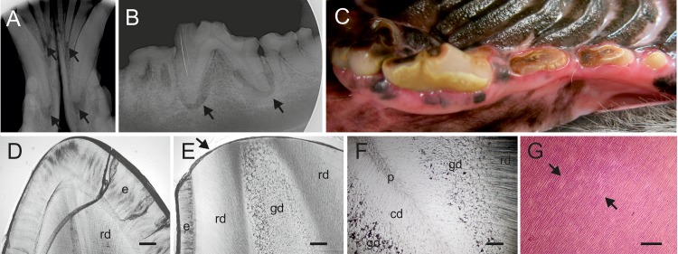
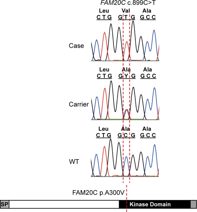
One to two percent of all children are born with a developmental disorder requiring pediatric hospital admissions. For many such syndromes, the molecular pathogenesis remains poorly characterized. Parallel developmental disorders in other species could provide complementary models for human rare diseases by uncovering new candidate genes, improving the understanding of the molecular mechanisms and opening possibilities for therapeutic trials. We performed various experiments, e.g. combined genome-wide association and next generation sequencing, to investigate the clinico-pathological features and genetic causes of three developmental syndromes in dogs, including craniomandibular osteopathy (CMO), a previously undescribed skeletal syndrome, and dental hypomineralization, for which we identified pathogenic variants in the canine SLC37A2 (truncating splicing enhancer variant), SCARF2 (truncating 2-bp deletion) and FAM20C (missense variant) genes, respectively. CMO is a clinical equivalent to an infantile cortical hyperostosis (Caffey disease), for which SLC37A2 is a new candidate gene. SLC37A2 is a poorly characterized member of a glucose-phosphate transporter family without previous disease associations. It is expressed in many tissues, including cells of the macrophage lineage, e.g. osteoclasts, and suggests a disease mechanism, in which an impaired glucose homeostasis in osteoclasts compromises their function in the developing bone, leading to hyperostosis. Mutations in SCARF2 and FAM20C have been associated with the human van den Ende-Gupta and Raine syndromes that include numerous features similar to the affected dogs. Given the growing interest in the molecular characterization and treatment of human rare diseases, our study presents three novel physiologically relevant models for further research and therapy approaches, while providing the molecular identity for the canine conditions.


Conflict of interest statement

I have read the journal's policy and the authors of this manuscript have the following competing interests: Genetic tests will be available from Genoscoper Ltd, which is partly owned by HL. CD is affiliated at the Institute of Genetics, University of Bern, Switzerland, which currently offers a diagnostic genetic test for CMO.

Figures

Fig 1
Fig 1. Genetic analyses in canine craniomandibular osteopathy identify a leaky splicing variant in exon 15 of SLC37A2, resulting in a frameshift that truncates the protein.
A. Laterolateral (left) and ventrodorsal (right) radiograph of the skull of two different WHWT affected with CMO. The typical palisade-like periosteal new bone formation is commonly located along the corpus mandibulae (left, white arrowheads) or around the Bulla tympanica (right, black arrowheads). B. Manhattan plot from GWAS indicates the associated region on chromosome 5 (pgenome = 0.02). C. A synonymous variant, c. 1332C>T, in exon 15 of SLC37A2. D. ESEfinder suggests that the synonymous variant affects a splicing enhancer site. The binding scores for the different splicing factors (colored blocks) for 79 nucleotides of exon 15 are shown for the wild-type and mutant allele. The c.1332C>T mutation eliminates a potential binding site for the SF2/ASF splicing factor (shown in purple). Binding scores for different splicing proteins (shown on the y-axis) are based on different weight matrices and cannot be compared directly to each other. E. A semi-quantitative RT-PCR in the wild-type (CC), carrier (CT) and affected (TT) WHWTs confirms the splicing defect (79 bp deletion) caused by the variant, and reveals a splicing leakage in the carrier and affected dogs. B2M was used as a loading control. F. A schematic 12-transmembrane secondary structure of SLC37A2 predicted by TMHMM. The truncated region in the mutated protein is indicated by a dashed line.
Fig 2
Fig 2. Radiographic and CT findings in a novel canine developmental syndrome.
A. Lateral radiograph of a 7-week-old affected Wire Fox Terrier puppy, where prominent underbite (long arrow) and dorsally convex maxilla (short arrow) are clearly visible. B. Healthy littermate with normal scissor bite and flat dorsal border of the maxilla. C. Transverse CT image of the nasal cavities of an affected puppy. Marked lateral deviation of the nasal septum is evident (arrow). D. Lateral radiograph of stifle joints of an affected puppy. The ossification centers are poorly mineralized (long arrows) and the proximal fibular epiphyses are non-mineralized (short arrows). E. Lateral image of an unaffected puppy indicates that the ossification centers are normal for the age (long arrows) and proximal epiphysis of the fibula is clearly seen (short arrow). F. Ventrodorsal radiograph of an affected puppy. Lateral luxation of the left radial head is evident.
Fig 3
Fig 3. Genetic analyses in a developmental syndrome in Wire Fox Terriers reveal a deletion in exon 6 of SCARF2, resulting in an early truncation of the predicted protein.
A. A Manhattan plot from a GWAS indicates a locus on chromosome 26 (pgenome = 0.05). B. A 3.3-Mb disease-associated haplotype in the affected dogs. C. Targeted resequencing revealed a 2-bp deletion in exon 6 (c.865-866delTC), resulting in a severe truncation at Ser289 that removes the transmembrane and cytoplasmic domains of the predicted protein as indicated by a dashed line in the schematic structure. D. RT-PCR in the affected and control Wire Fox Terrier indicate expression of SCARF2 mRNA and suggest that the mutated SCARF2 transcript is not directed to nonsense-mediated RNA decay.
Fig 4
Fig 4. The dental phenotype in the Border Collies with a homozygous FAM20C variant.
A. X-ray of the anterior mandibular teeth of a 9-year-old female Border Collie. Arrows indicate apical inflammations. B. X-ray of the anterior mandibular molars and premolars of a 10 year old male Border Collie indicating dental wear and apical inflammations (arrows). C. Anterior mandibular molars and premolars of the same dog indicating dental wear causing pulpal exposure. D. Ground section of a deciduous incisor from an unaffected Border Collie showing intact enamel and normal dentin, the crack is artefactual. E. Ground section of a permanent incisor of an affected 9-year-old female Border Collie. The enamel layer (e) preserved cervically is thin as compared to the control tooth (D). Wear at the edge (arrow) of tooth extends to the peripheral regular dentin (rd) and deeper to the abnormal globular dentin (gd), not present in the control (D). F. Ground section from deeper position of the same tooth as in (E) showing the layerwise alteration in the dentin structure. Peripheral dentin (rd) has a normal tubular pattern with no globules, the middlemost zone is pronouncedly globular (gd), while the central zone (cd) next to the diminished or almost completely obliterated pulp chamber (p) shows a slightly irregular tubular pattern but no globules. G. Haematoxylin-eosin stained paraffin section of a premolar from a 9-year old female. Contiguous interglobular spaces (arrows) in the middlemost dentin zone stain lightly with eosin. Tubules run uninterruptedly in stained areas. Bars, 50 um (D,E,F) and 200 um (G).
Fig 5
Fig 5. Whole genome sequencing of three Border Collies affected with tooth attrition reveals a missense variant, FAM20C c.899C>T, in the conserved kinase domain of the protein (p.A300V).

References

    1. Scriver CR. (1995) Disease, war, and biology: Languages for medicine—and pediatrics. Pediatr Res 38(6): 819–829. - PubMed
    1. Rabbani B, Mahdieh N, Hosomichi K, Nakaoka H, Inoue I. (2012) Next-generation sequencing: Impact of exome sequencing in characterizing mendelian disorders. J Hum Genet 57(10): 621–632. 10.1038/jhg.2012.91 - DOI - PubMed
    1. Tønnessen R, Borge KS, Nødtvedt A, Indrebø A. (2012) Canine perinatal mortality: A cohort study of 224 breeds. Theriogenology 77(9): 1788–1801. 10.1016/j.theriogenology.2011.12.023 - DOI - PubMed
    1. Kyöstilä K, Syrjä P, Jagannathan V, Chandrasekar G, Jokinen TS, et al. (2015) A missense change in the ATG4D gene links aberrant autophagy to a neurodegenerative vacuolar storage disease. PLoS Genet 11(4):e1005169 10.1371/journal.pgen.1005169 - DOI - PMC - PubMed
    1. Drögemüller M, Jagannathan V, Becker D, Drögemüller C, Schelling C et al. (2014) A mutation in the FAM83G gene in dogs with hereditary footpad hyperkeratosis (HFH). PLoS Genet. 10(5):e1004370 10.1371/journal.pgen.1004370 - DOI - PMC - PubMed

Publication types

MeSH terms

Supplementary concepts