. 2016 May 17;11(5):e0155650.
doi: 10.1371/journal.pone.0155650. eCollection 2016.

Powerful Complex Immunoadjuvant Based on Synergistic Effect of Combined TLR4 and NOD2 Activation Significantly Enhances Magnitude of Humoral and Cellular Adaptive Immune Responses

Affiliations

Powerful Complex Immunoadjuvant Based on Synergistic Effect of Combined TLR4 and NOD2 Activation Significantly Enhances Magnitude of Humoral and Cellular Adaptive Immune Responses

Amir I Tukhvatulin et al. PLoS One. .

Abstract

Binding of pattern recognition receptors (PRRs) by pathogen-associated molecular patterns (PAMPs) activates innate immune responses and contributes to development of adaptive immunity. Simultaneous stimulation of different types of PRRs can have synergistic immunostimulatory effects resulting in enhanced production of molecules that mediate innate immunity such as inflammatory cytokines, antimicrobial peptides, etc. Here, we evaluated the impact of combined stimulation of PRRs from different families on adaptive immunity by generating alum-based vaccine formulations with ovalbumin as a model antigen and the Toll-like receptor 4 (TLR4) agonist MPLA and the Nucleotide-binding oligomerization domain-containing protein 2 (NOD2) agonist MDP adsorbed individually or together on the alum-ovalbumin particles. Multiple in vitro and in vivo readouts of immune system activation all showed that while individual PRR agonists increased the immunogenicity of vaccines compared to alum alone, the combination of both PRR agonists was significantly more effective. Combined stimulation of TLR4 and NOD2 results in a stronger and broader transcriptional response in THP-1 cells compared to individual PRR stimulation. Immunostimulatory composition containing both PRR agonists (MPLA and MDP) in the context of the alum-based ovalbumin vaccine also enhanced uptake of vaccine particles by bone marrow derived dendritic cells (BMDCs) and promoted maturation (up-regulation of expression of CD80, CD86, MHCII) and activation (production of cytokines) of BMDCs. Finally, immunization of mice with vaccine particles containing both PRR agonists resulted in enhanced cellular immunity as indicated by increased proliferation and activation (IFN-γ production) of splenic CD4+ and CD8+ T cells following in vitro restimulation with ovalbumin and enhanced humoral immunity as indicated by higher titers of ovalbumin-specific IgG antibodies. These results indicate that combined stimulation of TLR4 and NOD2 receptors dramatically enhances activation of both the humoral and cellular branches of adaptive immunity and suggests that inclusion of agonists of these receptors in standard alum-based adjuvants could be used to improve the effectiveness of vaccination.


Conflict of interest statement

Competing Interests: Commercial affiliation of A.V.G. and P.S-B. to Cleveland BioLabs, Inc, does not alter the authors' adherence to PLOS ONE policies on sharing data and materials.

Figures

Fig 1
Fig 1. Combination of MPLA and MDP strongly enhances the transcriptional response in THP-1 cells in comparison to individual PRR agonists.
(A) Venn diagram demonstrating overlap in the sets of genes found to be MDP-, MPLA- and/or MDP+MPLA-induced in THP-1 cells. Cells were treated with MDP (20μg/ml) and MPLA (1μg/ml) individually or in combination or left unstimulated. RNA was harvested after 3 h and analyzed by hybridization to Affymetrix Human Gene 1.0 ST genechip microarrays. Genes induced by MDP (white circle), MPLA (gray circle) or their combination (black circle) by at least 3-fold over untreated cells are shown. (B) List of MDP+MPLA-induced genes with potentiated expression in comparison to gene expression levels in untreated cells and cells treated with either MPLA or MDP alone. Cells were left untreated or treated with MDP (20μg/ml) and MPLA (1μg/ml) individually or in combination. RNA was harvested after 3 h and analyzed by hybridization to Affymetrix Human Gene 1.0 ST Genechip microarrays. The figure lists 72 genes that showed potentiated expression with MDP+MPLA treatment compared to MPLA or MDP alone (potentiated = fold induction of gene expression level with combined treatment over the untreated control was greater than the sum of the fold induction seen with MPLA alone and MDP alone). Statistical significance was determined by a random-variance t-test with P<0.01 as a cut-off for signficance. The magnitude of the fold-change in gene expression relative to untreated cells is indicated by color. (C) Multiplex analysis of molecular pathways activated via phosphorylation in THP-1 cells in response to individual or combined TLR4 and NOD2 stimulation. Cells were left untreated or treated for 20 min with MDP (20 μg/ml) and MPLA (1 μg/ml) alone or in combination. Phosphoprotein levels of p65 subunit NF-κB (Ser536), IκB (Ser32), IKKα/β (Ser177/Ser181), p38 (Thr180/Tyr182), ERK/MAP kinase 1/2 (Thr185/Tyr187), JNK (Thr183/Tyr185), Akt (Ser473), CREB (Ser133), STAT3 (Ser727), p70 S6 kinase (Thr412), and STAT5A/B (Tyr694/699), as well as total protein levels of TNFR1 and c-Myc were evaluated using MILLIPLEX Magnetic Bead Signaling kits and a Bio-Plex MAGPIX multiplex reader (Bio-Rad). Data was collected from triplicate samples in two independent experiments and is presented as mean fluorescent intensity (MFI) ± SD. * indicates significant difference (P≤0.05) between formulations containing MDP or MPLA individually and the formulation without PRR agonists. # indicates significant difference (P≤0.05) between group treated with Alum+OVA+MDP+MPLA and Alum+OVA+MPLA or Alum+OVA+MDP (Student’s t-test).
Fig 2
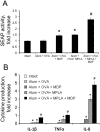
Fig 2. Vaccine formulations containing a combination of TLR4 and NOD2 agonists enhance NF-κB/AP-1 activation in THP-1 cells compared to formulations containing individual agonists.
(A) Addition of the vaccine formulation containing both TLR4 and NOD2 agonists to THP1-XBlue-CD14 cells leads to enhanced NF-κB/AP-1-dependent SEAP activity compared to formulations with individual PRR agonists. SEAP activity was measured in cell-free culture supernatants 18 h after vaccine formulation addition. Results are expressed as the fold-increase in SEAP activity relative to untreated (intact) cells; mean values ± SD from three independent experiments, each performed in duplicate. * indicates significant difference (P≤0.05) between formulations containing MDP or MPLA individually and the formulation without PRR agonists. # indicates significant difference (P≤0.05) between Alum+OVA+MDP+MPLA treatment and Alum+OVA+MPLA or Alum+OVA+MDP (Student’s t-test). (B) Addition of the vaccine formulation containing both TLR4 and NOD2 agonists to THP1 cells leads to enhanced cytokine production in comparison to vaccine formulations with individual PRR agonists. Cells were left untreated or treated with Alum+OVA, Alum+OVA+MDP, Alum+OVA+MPLA, or Alum+OVA+MDP+MPLA formulations for 18 hrs. Cell-free supernatants were prepared and analyzed by multiplex-bead ELISA Bio-Plex Pro kit (BioRad, USA) for production of IL-1β, TNF-α, and IL-8. Results are representative of two separate experiments, each performed in triplicate. Mean ± SD is shown for triplicate samples. * and # indicate significant differences as described for (A).
Fig 3
Fig 3. Vaccine formulations containing a combination of TLR4 and NOD2 agonists enhance the phagocytic activity of BMDCs compared to formulations containing individual agonists.
Immature BMDCs isolated from naïve C57BL/6 mice were treated with Alum+OVA, Alum+OVA+MDP, Alum+OVA+MPLA and Alum+OVA+MDP+MPLA formulations produced with ovalbumin labeled with FITC and pHRodo dye for 40 min. Cells were then washed with PBS twice and evaluated by fluorescent microscopy and flow cytometry. (A) Representative fluorescence microscopy images of BMDCs treated with vaccine formulations. Each column shows images obtained with different filters to visualize FITC (green), pHRodo (red) and DAPI (blue) fluorescence. Phagocytosed particles are specifically visualized by pHRodo (red) fluorescence, whereas total (Extra- and intracellular) quantity of vaccine particles show FITC fluorescence. Images with all three fluorescence signals merged and bright field images also are shown. Scale bar represents 20μm. (B) % of cells that were pHRodo-positive; (C) fold-increase in mean fluorescence intensity (MFI) relative to untreated BMDCs; and (D) phagocytic index calculated by multiplying the percentage of pHRodo-positive cells by the MFI. Each bar represents the mean ± SD from at least three independent experiments, each performed with triplicates. Statistically significant differences were determined by Student’s t-test with P≤0.05 cut-off. * indicates significant difference (P≤0.05) between formulations containing MDP or MPLA individually and the formulation without PRR agonists. # indicates significant difference (P≤0.05) between Alum+OVA+MDP+MPLA treatment and Alum+OVA+MPLA or Alum+OVA+MDP (Student’s t-test).
Fig 4
Fig 4. Vaccine formulations containing a combination of TLR4 and NOD2 agonists significantly enhance maturation of BMDC compared to formulations containing individual PRR agonists.
BMDCs were harvested on day 8 of culture and incubated in 24-well plates for 24 hours with vaccine formulations. Expression of the maturation markers, CD80 (A), CD86 (B), and major histocompatibility complex (MHC) class II (C) was assessed by flow cytometric analysis of 5x10⁴ CD11c+ cells. Mean MFI values (indicating expression level) ± SEM from two independent experiments with 5 replicates each are shown. * indicates significant difference (P≤0.05) between formulations containing MDP or MPLA individually and the formulation without PRR agonists. # indicates significant difference (P≤0.05) between Alum+OVA+MDP+MPLA treatment and Alum+OVA+MPLA or Alum+OVA+MDP (Student’s t-test). (D) Cytokine levels were measured in cell-free culture supernatants collected 24 hours after addition of vaccine formulations to BMDCs (4x10⁴ cells/well) using bead-based immunoassay. Data represent mean ± SD. * indicates significant difference (P≤0.05) between formulations containing MDP or MPLA individually and the formulation without PRR agonists. # indicates significant difference (P≤0.05) between Alum+OVA+MDP+MPLA treatment and Alum+OVA+MPLA or Alum+OVA+MDP (Student’s t-test).
Fig 5
Fig 5. Vaccine formulations containing a combination of TLR4 and NOD2 agonists induce stronger antigen-specific CD4 and CD8 T-cell responses in mice than formulations containing individual PRR agonists.
Mice (n = 5/group) were immunized s.c. twice with alum-based vaccine formulations: Alum+OVA, Alum+OVA+MDP, Alum+OVA+MPLA, and Alum+OVA+MDP+MPLA. Controls included naıve (untreated) mice and mice immunized with soluble OVA (no Alum or PRR agonist). Splenocytes were harvested from mice 14 days after the last immunization. (A-B) T-cell proliferation in response to ovalbumin restimulation. Splenocytes were CFSE-labeled, restimulated with whole OVA protein (1μg/ml) for 72h, stained with fluorescently-tagged antibodies against CD3, CD4 and CD8 and analyzed by flow cytometry. A. Gating strategy used for determination of antigen-specific CD4 and CD8 T-cell responses. B. Representative dot plots for the naïve (intact) group and each immunized group showing CFSE fluorescence (x-axis) of CD4 and CD8 T cells (distinguished by CD8 expression shown on the y-axis). The percentage of proliferating CD4+ and CD8+ T cells in response to antigen represent mean from two independent experiments with 5 mice/group each. (C-D) T-cell activation indicated by IFN-γ production in response to ovalbumin restimulation. Splenocytes were restimulated for 18 hours with whole OVA antigen at 1μg/ml in the presence of BD GolgiPlug solution (BD biosciences), stained with fluorescently-tagged antibodies against CD3, CD4 and CD8, permeabilized, stained with a fluorescently-tagged antibody against IFN-γ and then analyzed by flow cytometry. The percentage of CD4 (C) or CD8 (D) T cells expressing IFN-γ is shown as the mean ± SD for 5 mice per group. Two additional experiments yielded similar results * indicates significant difference (P≤0.05) between formulations containing MDP or MPLA individually and the formulation without PRR agonists (Alum+OVA). # indicates significant difference (P≤0.05) between Alum+OVA+MDP+MPLA treatment and Alum+OVA+MPLA or Alum+OVA+MDP (Student’s t-test).
Fig 6
Fig 6. Vaccine formulations containing a combination of TLR4 and NOD2 agonists lead to a stronger ovalbumin-specific antibody response in mice than formulations containing individual PRR agonists.
Mice (n = 5/group) were immunized s.c. twice with alum-based vaccine formulations: Alum+OVA, Alum+OVA+MDP, Alum+OVA+MPLA, and Alum+OVA+MDP+MPLA. Controls included naıve (untreated) mice and mice immunized with soluble OVA (no Alum or PRR agonist). Blood was collected from mice 14 days after the last immunization and serum levels of ovalbumin-specific total IgG (A), IgG1 (B), IgG2a (C), IgG2b (D) and IgG2c (E) antibodies were detected by ELISA. The mean for 5 mice/group ± SEM is shown. Experiment was repeated three times with analogous results.

Similar articles

Cited by

References

    1. Coffman RL, Sher A, Seder RA. Vaccine adjuvants: putting innate immunity to work. Immunity. 2010. October 29;33(4):492–503. 10.1016/j.immuni.2010.10.002 - DOI - PMC - PubMed
    1. Brewer JM, Conacher M, Hunter CA, Mohrs M, Brombacher F, Alexander J. Aluminium hydroxide adjuvant initiates strong antigen-specific Th2 responses in the absence of IL-4- or IL-13-mediated signaling. J Immunol. 1999. December 15;163(12):6448–54. - PubMed
    1. Seder RA, Paul WE. Acquisition of lymphokine-producing phenotype by CD4+ T cells. Annu Rev Immunol. 1994;12:635–73. - PubMed
    1. Steinhagen F, Kinjo T, Bode C, Klinman DM. TLR-based immune adjuvants. Vaccine. 2011. April 12;29(17):3341–55. 10.1016/j.vaccine.2010.08.002 - DOI - PMC - PubMed
    1. Wang L, Smith D, Bot S, Dellamary L, Bloom A, Bot A. Noncoding RNA danger motifs bridge innate and adaptive immunity and are potent adjuvants for vaccination. J Clin Invest. 2002. October;110(8):1175–84. - PMC - PubMed

Publication types

MeSH terms