Review
. 2016 Jun;7(3):449-59.
doi: 10.1007/s13244-016-0487-4. Epub 2016 May 17.

Spectrum of early lung cancer presentation in low-dose screening CT: a pictorial review

Affiliations
Review

Spectrum of early lung cancer presentation in low-dose screening CT: a pictorial review

Cristiano Rampinelli et al. Insights Imaging. 2016 Jun.

Abstract

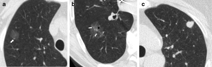
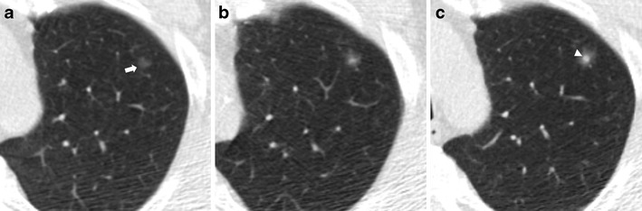
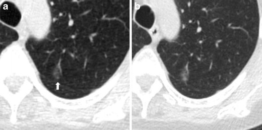
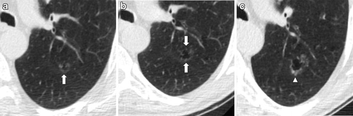
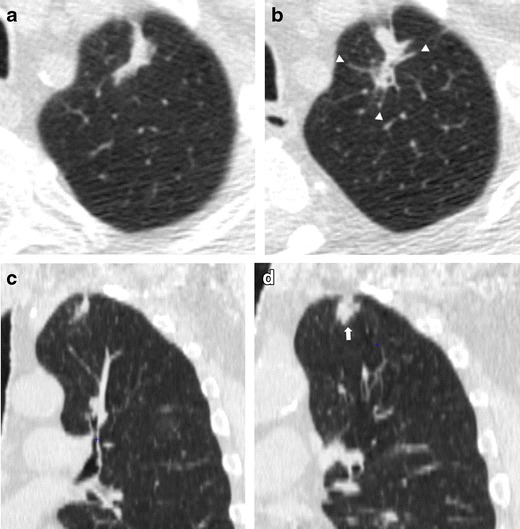
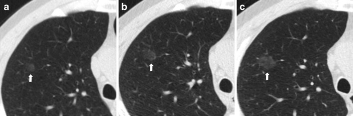
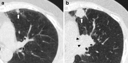
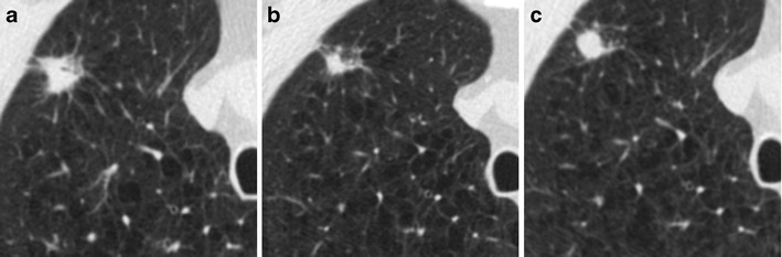
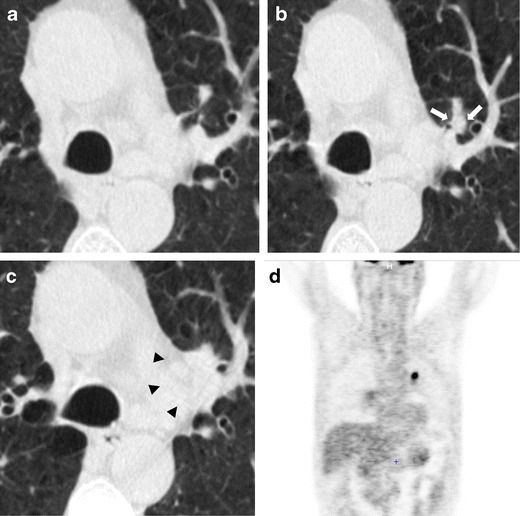
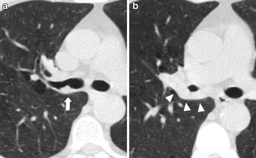
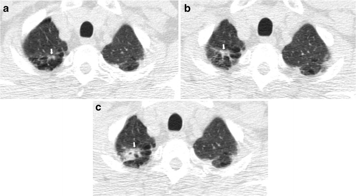
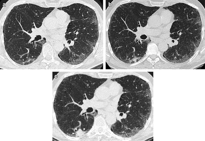
The typical presentation of early stage lung cancers on low-dose CT screening are non-calcified pulmonary nodules. However, there is a wide spectrum of unusual focal abnormalities that can be early presentations of lung cancer. These abnormalities include, for example, cancers associated with 'cystic airspaces' or scar-like cancers. The detection of lung cancer with low-dose CT can be affected by the absence of intravenous contrast medium. As a consequence, endobronchial and central lesions can be difficult to recognize, raising the potential for missed cancers. Focal lesions arising within pre-existing lung disease, such as lung fibrosis or apical scars, can also be early lung cancer manifestations and deserve particular consideration as recognition of these lesions may be hindered by the underlying disease. Furthermore, the unpredictable growth rate of lung cancer, which ranges from indolent to aggressive cancers, necessitates attention to the wide spectrum of progression in lung cancer appearance on serial low-dose CT scans. In this pictorial review we discuss the spectrum of early lung cancer presentation in low-dose CT screening, highlighting typical as well as unusual radiological features and the varied growth rates of early lung cancer. Teaching Points • There is a wide spectrum of early presentations of lung cancer on LDCT. • Low radiation dose and the absence of contrast medium injection can affect lung cancer detection. • Lung cancer growth shows various behaviours, ranging from indolent to aggressive cancers. • Familiarity with LDCT technique can improve CT screening effectiveness and avoid missed diagnosis.

Keywords: Low-dose CT; Lung cancer; Pulmonary nodule; Screening.


Figures

Fig. 1
Fig. 1
Primary lung adenocarcinomas. Radiological features of pulmonary nodules according to their attenuation. (a) A low-dose CT thin-section shows a 15-mm non-solid nodule (pure ground-glass attenuation) of the right upper lobe: the nodule density is slightly higher than that of normal lung parenchyma, and vessels are visualized within the nodule (arrows). (b) Low-dose CT scan shows a 25-mm part-solid nodule of the right lower lobe. This nodule is characterized by a 3-mm central solid component (arrowhead) surrounded by a pure ground-glass opacity. The vessels and a small peripheral airway (arrow) can be clearly depicted within the ground-glass opacity. (c) Axial low-dose CT scan shows a 12-mm subpleural solid nodule of the left upper lobe. The spectrum of early stage lung cancer presentation at low-dose CT includes solid nodules as well as part-solid nodules and ground-glass nodules
Fig. 2
Fig. 2
Increased attenuation of a small lung adenocarcinoma over 3 years in a 62-year-old man. (a) Low-dose CT image show a 5-mm non-solid nodule of the upper left lobe (arrow). (b) Follow-up CT obtained 1 year later show a minimum increment of axial diameter and a slight increase of nodule attenuation. (c) Two years later the progression to a part-solid nodule is obvious. The development of a central solid component (arrowhead) within the nodule can be observed. An increase in pulmonary nodule attenuation at LCDT follow-up, the development or increment of a solid component within the pulmonary nodule should raise suspicions of malignancy, even if the nodule size is stable. Typically, this appearance is suggestive of a peripheral lung adenocarcinoma
Fig. 3
Fig. 3
Progression at the 1-year interval of a non-solid nodule in a 66-year-old woman. (a) Low-dose CT section shows a 12-mm nodule with ground-glass attenuation of the upper left lobe (arrow). (b) CT scan obtained at annual repeat LDCT demonstrates a substantial stability in size but with increased density, that indicates an increased risk for malignancy. The lesion was surgically removed with a diagnosis of lung adenocarcinoma. Sub-solid nodules that show an increment in attenuation should be considered suspicious for malignancy
Fig. 4
Fig. 4
Progressive changes of a lung adenocarcinoma associated with a cystic airspace in a 60-year-old woman. (a) Axial low-dose CT shows an 8-mm cystic airspace with thin irregular non-solid wall in the upper left lobe (arrow). (b) One year later the lesion has increased in size to 12 mm and 2 years later, (c) a consistent irregular thickening with the development of solid components along the wall was evident (arrowhead). The histologic diagnosis was lung adenocarcinoma. This kind of presentation is usually slow growing on serial low-dose CT scans and the diagnosis of lung cancer is suggested by the circumferential wall thickening of the cystic airspaces; with tumor growth, airspaces size could increase, decrease or remain unchanged
Fig. 5
Fig. 5
Low-dose CT images show both an increase in size and density of a scar-like lesion of the left lung apex, consisting of a lung adenocarcinoma. (a, c) Axial and coronal LDCT sections of the left apex show an irregular subpleural opacity that was interpreted as apical fibrotic changes. (b, d) LDCT repeated at a 1-year interval shows spiculations (arrowhead) and a significant thickening of the lesion (arrow). A PET/CT scan was positive and the patient went to lobectomy with histologic diagnosis of adenocarcinoma. Lung cancers presenting as fibrotic changes or as scar-like lesions are relatively uncommon but could be encountered in LDCT screening
Fig. 6
Fig. 6
Slow-growing lung adenocarcinoma in a 61-year-old man. (a) Low-dose CT axial section in the right upper lobe shows a 6-mm ground-glass nodule (arrow). (b) Low-dose CT scan after 2 years shows an increase in size of the nodule to 10 mm (arrow). (c) After 4 years from the first low-dose CT scan, the nodule showed the same ground-glass attenuation but a progressive increase in size from 10 to 14 mm (arrow). Typically, early lung cancer presenting as non-solid nodules are slow-growing forms
Fig. 7
Fig. 7
Fast-growing lung adenocarcinoma in a 79-year-old man. (a) Axial LDCT image of the chest shows a 7-mm sub-pleural solid nodule in the left upper lobe (arrow). (b) LDCT scan obtained at 1-year interval shows a rapid growth of the lesion into a 23-mm solid nodule (arrow) with an aggressive behavior documented by the concomitant enlargement of hylar lymph nodes (arrowheads)
Fig. 8
Fig. 8
Transient decrease in size of a squamous cell lung cancer in a 74-year-old man. (a) Unenhanced axial LDCT image of the chest shows a solid nodule in the upper right lobe with an irregular shape and a maximum diameter of 19 mm. (b) Follow-up LDCT scan obtained 1 month after antibiotic therapy demonstrated a significant regression of the lesion. (c) At LDCT obtained 3 months later, the nodule grew in size again. Biopsy was performed, and results of histology revealed lung adenocarcinoma. It is reported that adenocarcinomas can show a transient decrease in size, maybe related to fibrosis or atelectasis. In this cases, a further LDCT scan is suggested to assess the complete resolution or stability of the findings and to avoid missed diagnosis
Fig. 9
Fig. 9
Lung cancer of the left hylum in a 67-year-old man with history of sarcoidosis. (a) Axial low-dose CT image through lung hyla does not show any significant findings at baseline CT screening. (b) Low-dose CT image obtained at the same level 1 year later shows the appearance of a 6-mm pulmonary nodule (arrow) surrounded by vascular and airways structures of the left hylum. This finding without contrast material injection can be misinterpreted as a pulmonary vessel. (c) The following year, the low-dose CT scan reveals the presence of a large lesion of the left hylum (arrows) that was characterize by increased metabolic activity via PET scan (d). The histological diagnosis was a T2N1 squamous cell carcinoma
Fig. 10
Fig. 10
Endobronchial lung cancer. (a) Axial low-dose CT shows a small lesion abutting the posterior wall of the right main bronchus, interpreted as non-suspicious bronchial mucus (arrow). (b) Low-dose CT performed 1 year later shows a significant reduction of bronchial caliber due to the presence of neoplastic tissue (arrowheads)
Fig. 11
Fig. 11
Right apical lung cancer in a 71-year-old man. (a) Low-dose axial CT scan shows small irregular fibrotic changes of the right apex (arrow). (b) One year later, LDCT shows a slight increment of the focal abnormalities with the typical aspect of apical fibrotic changes, with a pleural tag and concomitant emphysema (arrow). (c) Two years later, a remarkable increment in size was clearly depicted (arrow) and the lesion has been surgically removed with a diagnosis of lung adenocarcinoma
Fig. 12
Fig. 12
Lung cancer arising within a pre-existing fibrotic lung disease. (a) Low-dose CT demonstrates a bilateral subpleural reticular pattern and a ground-glass opacity, with a subpleural curvilinear opacity in the right lower lobe (arrowheads). (b) Low-dose CT scan obtained 1 year later confirmed the fibrotic disease with an increased density of the subpleural opacity in the right lower lobe (arrowheads), interpreted as a progression of the fibrosing pattern. (c) At follow-up CT performed 1 year later, there is a remarkable increase of subpleural opacity in the right lobe with the development of a 32-mm mass, resulting in lung adenocarcinoma (arrow)

References

    1. Jemal A, Bray F, Center MM, et al. Global cancer statistics. CA Cancer J Clin. 2011;61:69–90. doi: 10.3322/caac.20107. - DOI - PubMed
    1. Aberle DR, Adams AM, Berg CD, Black WC, Clapp JD, Fagerstrom RM, Gareen IF, Gatsonis C, Marcus PM, Sicks JD, National Lung Screening Trial Research Team Reduced lung-cancer mortality with low-dose computed tomographic screening. N Engl J Med. 2011;365(5):395–409. doi: 10.1056/NEJMoa1102873. - DOI - PMC - PubMed
    1. Detterbeck FC, Mazzone PJ, Naidich DP, Bach PB. Screening for lung cancer: Diagnosis and management of lung cancer, 3rd ed: American College of Chest Physicians evidence-based clinical practice guidelines. Chest. 2013;143(5 Suppl):78S–92S. doi: 10.1378/chest.12-2350. - DOI - PMC - PubMed
    1. Jaklitsch MT, Jacobson FL, Austin JH, Field JK, Jett JR, Keshavjee S, MacMahon H, Mulshine JL, Munden RF, Salgia R, Strauss GM, Swanson SJ, Travis WD, Sugarbaker DJ. The American Association for Thoracic Surgery guidelines for lung cancer screening using low-dose computed tomography scans for lung cancer survivors and other high-risk groups. J Thorac Cardiovasc Surg. 2012;144(1):33–38. doi: 10.1016/j.jtcvs.2012.05.060. - DOI - PubMed
    1. Wender R, Fontham ET, Barrera E, Jr, Colditz GA, Church TR, Ettinger DS, Etzioni R, Flowers CR, Gazelle GS, Kelsey DK, LaMonte SJ, Michaelson JS, Oeffinger KC, Shih YC, Sullivan DC, Travis W, Walter L, Wolf AM, Brawley OW, Smith RA. American Cancer Society lung cancer screening guidelines. CA Cancer J Clin. 2013;63(2):107–117. doi: 10.3322/caac.21172. - DOI - PMC - PubMed