. 2016 May 17;16(1):17.
doi: 10.1186/s12861-016-0114-0.

Mediator subunit Med12 contributes to the maintenance of neural stem cell identity

Affiliations

Mediator subunit Med12 contributes to the maintenance of neural stem cell identity

Nam Hee Kim et al. BMC Dev Biol. .

Abstract

Background: The RNA polymerase II transcriptional Mediator subunit Med12 is broadly implicated in vertebrate brain development, and genetic variation in human MED12 is associated with X-linked intellectual disability and neuropsychiatric disorders. Although prior studies have begun to elaborate the functional contribution of Med12 within key neurodevelopmental pathways, a more complete description of Med12 function in the developing nervous system, including the specific biological networks and cellular processes under its regulatory influence, remains to be established. Herein, we sought to clarify the global contribution of Med12 to neural stem cell (NSC) biology through unbiased transcriptome profiling of mouse embryonic stem (ES) cell-derived NSCs following RNAi-mediated Med12 depletion.

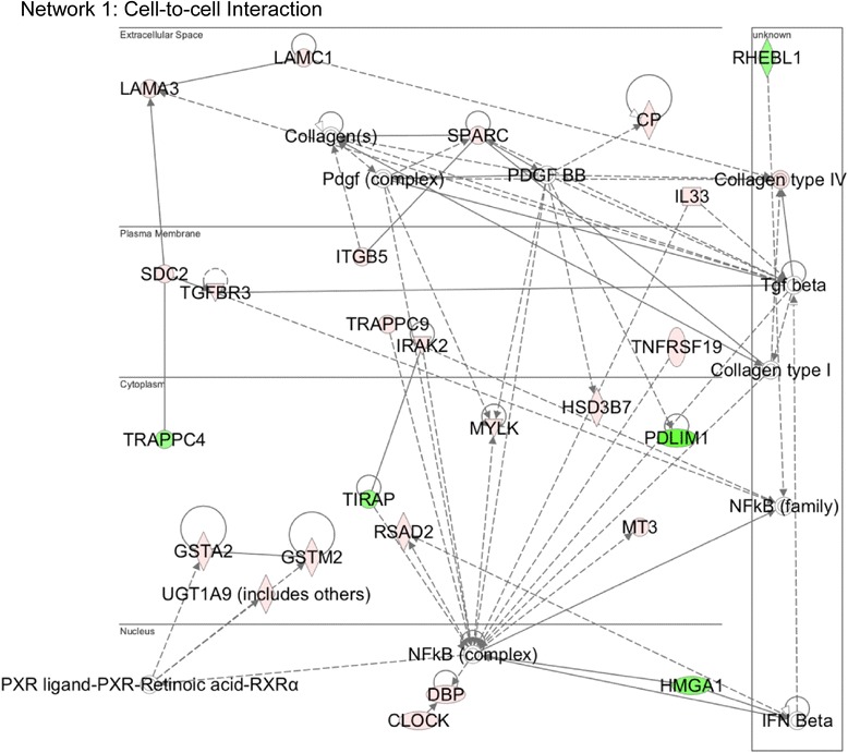
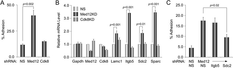
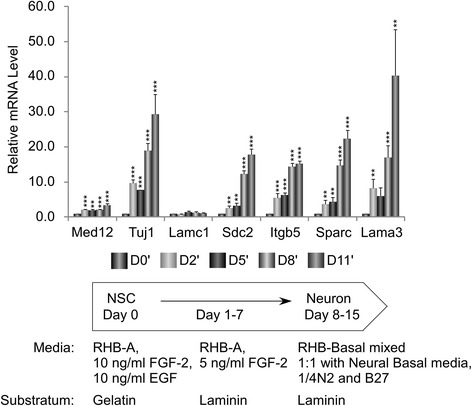
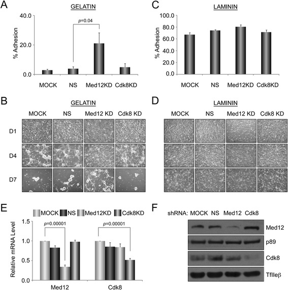
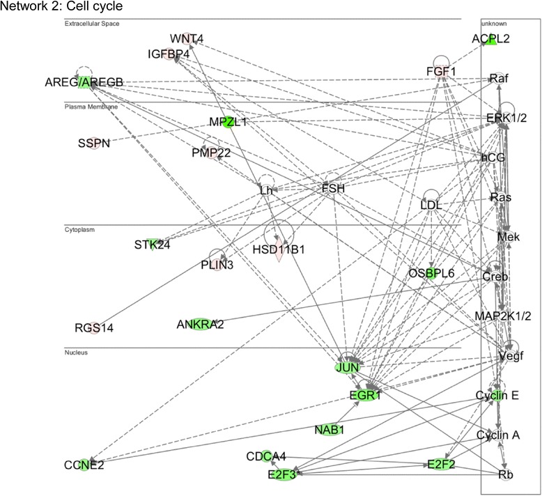
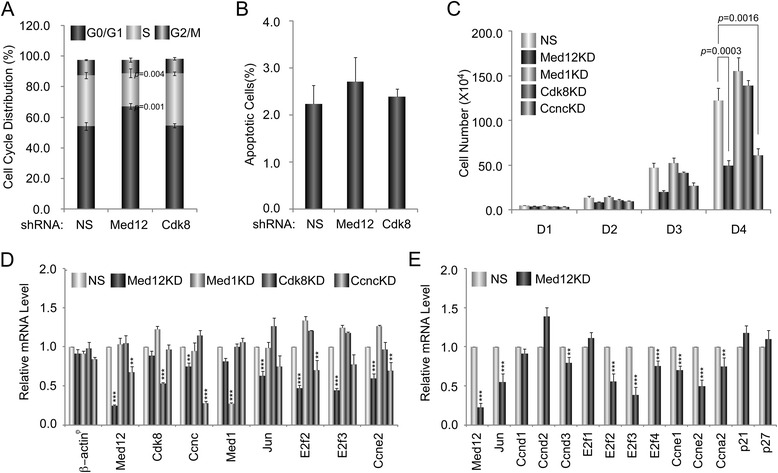
Results: A total of 240 genes (177 up, 73 down) were differentially expressed in Med12-knockdown versus control mouse NS-5 (mNS-5) NSCs. Gene set enrichment analysis revealed Med12 to be prominently linked with "cell-to-cell interaction" and "cell cycle" networks, and subsequent functional studies confirmed these associations. Targeted depletion of Med12 led to enhanced NSC adhesion and upregulation of cell adhesion genes, including Syndecan 2 (Sdc2). Concomitant depletion of both Sdc2 and Med12 reversed enhanced cell adhesion triggered by Med12 knockdown alone, confirming that Med12 negatively regulates NSC cell adhesion by suppressing the expression of cell adhesion molecules. Med12-mediated suppression of NSC adhesion is a dynamically regulated process in vitro, enforced in self-renewing NSCs and alleviated during the course of neuronal differentiation. Accordingly, Med12 depletion enhanced adhesion and prolonged survival of mNS-5 NSCs induced to differentiate on gelatin, effects that were bypassed completely by growth on laminin. On the other hand, Med12 depletion in mNS-5 NSCs led to reduced expression of G1/S phase cell cycle regulators and a concordant G1/S phase cell cycle block without evidence of apoptosis, resulting in a severe proliferation defect.

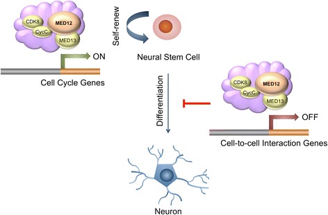
Conclusions: Med12 contributes to the maintenance of NSC identity through a functionally bipartite role in suppression and activation of gene expression programs dedicated to cell adhesion and G1/S phase cell cycle progression, respectively. Med12 may thus contribute to the regulatory apparatus that controls the balance between NSC self-renewal and differentiation, with important implications for MED12-linked neurodevelopmental disorders.

Keywords: Cell adhesion; Cell cycle; Gene expression; Med12; Mediator; Microarray; Neural stem cell.


Figures

Fig. 1
Fig. 1
Med12 mediates suppression of genes involved in “Cell Adhesion”. Functional gene interaction networks were identified by IPA®. Genes are represented as nodes, and the biological relationship between two nodes is represented as a line. The color of the node indicates up- or down- regulation (red, up; green, down); uncolored nodes represent genes that were not identified as differentially expressed in our microarray, but have nonetheless been integrated into the computationally generated networks on the basis of the evidence stored in the IPA knowledge memory indicating a relevance to this network
Fig. 2
Fig. 2
Med12 depletion enhances NSC adhesion in a manner reversible by concurrent depletion of Sdc2. a mNS-5 NSCs infected with lentiviruses expressing non-specific (NS) control, Med12-, or Cdk8-specific shRNAs were harvested and incubated with Calcein-AM prior to seeding onto 96-well plates. After washing, adherent cells were detected by fluorometry. The percentage of adhesion was calculated by dividing the background-subtracted fluorescence of adherent cells by the total corrected fluorescence of cells added to each well and multiplying by 100. b RNA from mNS-5 NSCs infected with lentiviruses expressing NS control, Med12-, or Cdk8-specific shRNAs as indicated were used for RT-qPCR, mRNA levels for each gene were normalized to β-actin mRNA and expressed relative to their corresponding mRNA levels in NS control shRNA-expressing cells. c mNS-5 NSCs co-infected with lentiviruses expressing shRNAs specific for Med12 or the indicated cell adhesion genes were assayed for cell adhesion as described in (a). 7All data represent the mean +/− SEM of at least three independent experiments performed in triplicate. p values were calculated by Student’s t test
Fig. 3
Fig. 3
Expression of Med12-regulated cell adhesion genes increases during neuronal differentiation of mNS-5 NSCs. mNS-5 NSCs were seeded onto laminin-coated plates prior to initiation of neuronal differentiation by sequential withdrawal of growth factors as indicated in the schematic and described in Methods. RNA isolated from cells on 0, 2, 5, 8, and 11 days after initiation of neuronal differentiation was subjected to RT-qPCR. mRNA levels for each gene were normalized to β-actin mRNA and expressed relative to their corresponding mRNA levels on day 0 (D0′) of the differentiation protocol. Data represent the mean +/− SEM of three independent experiments performed in triplicate. Asterisks denote statistically significant differences in the relative mRNA levels for each gene compared to their corresponding levels on D0′ (Student’s t test, **p < 0.05, ***p < 0.01)
Fig. 4
Fig. 4
Med12 knockdown increases cell adhesion and preserves cell viability on limiting (gelatin) substratum during neuronal differentiation. a-d mNS-5 NSCs infected with lentiviruses expressing NS control, Med12-, or Cdk8-specific shRNAs were seeded into T-75 tissue culture flasks coated with gelatin (a, b) or laminin (c, d) prior to initiation of neuronal differentiation as described in Fig. 3. Fluorescence based adhesion assay (a, c) was performed 40 h after initiation of the neuronal differentiation. Data represent the mean +/− SEM of at least three independent experiments performed in triplicate. p values were calculated by Student’s t test. Brightfield images (b, d) were obtained by optical microscopy at 1, 4, and 7 days after initiation of neuronal differentiation. e and f Validation of Med12 and Cdk8 depletion in knockdown cells by RT-qPCR (e) and immunoblot (f) analyses. mRNA levels for each gene in (e) were normalized to β-actin mRNA and expressed relative to their corresponding mRNA levels in untreated (MOCK) cells. Data represent the mean +/− SEM of at least three independent experiments performed in triplicate. p values were calculated by Student’s t test
Fig. 5
Fig. 5
Med12 mediates activation of genes involved in “Cell Cycle Control”. Genes involved in cell cycle control are largely downregulated (green) upon Med12 depletion in mNS-5 NSCs. Functional gene interaction networks were identified by IPA® as described in Fig. 1
Fig. 6
Fig. 6
Med12 depletion reduces mNS-5 NSC proliferation likely through a G1/S phase cell cycle block. a-e mNS-5 NSCs were infected with lentiviruses expressing NS control, Med12-, Med1-, Cdk8-, and/or CycC (Ccnc)-specific shRNAs as indicated. a Five days post-infection, cells were harvested, fixed, and monitored for cell cycle distribution using a FACSCalibur flow cytometer and CellQuest Pro software prior to data analysis using FlowJo software. At least 10,000 single cell events were collected for the analysis. b Five days post-infection, cells were stained with annexin V conjugated to APC and propidium iodide. Stained cells were analyzed by FACSCalibur and data was analyzed using FlowJo software. c Lentivirus-infected cells were seeded at a concentration of 5 × 104 cells/well in 12-well plates. Cell proliferation was monitored by counting viable cells using 0.4 % trypan blue at 24, 48, 72, and 96 h post-seeding. d, e mRNA expression levels for the indicated genes were determined by RT-qPCR analyses as described in Fig. 2. mRNA levels for each gene were normalized to Gapdh mRNA and expressed relative to their corresponding mRNA levels in control NS shRNA-expressing cells. All data represent the mean +/− SEM of at least three independent experiments performed in triplicate. p values were calculated by Student’s t test. Asterisks denote statistically significant differences relative to NS control shRNA (*p < 0.1, **p < 0.05, ***p < 0.01)
Fig. 7
Fig. 7
Schematic model for the role of Med12 in mNSCs. Med12 contributes to the maintenance of NSC identity through a functionally bipartite role in suppression and activation of gene expression programs dedicated to cell adhesion and G1/S phase cell cycle progression, respectively

References

    1. Braun SM, Jessberger S. Adult neurogenesis: mechanisms and functional significance. Development. 2014;141(10):1983–6. doi: 10.1242/dev.104596. - DOI - PubMed
    1. Gage FH, Temple S. Neural stem cells: generating and regenerating the brain. Neuron. 2013;80(3):588–601. doi: 10.1016/j.neuron.2013.10.037. - DOI - PubMed
    1. Kintner C. Neurogenesis in embryos and in adult neural stem cells. J Neurosci. 2002;22(3):639–43. - PMC - PubMed
    1. Baron W, Hoekstra D. On the biogenesis of myelin membranes: sorting, trafficking and cell polarity. FEBS Lett. 2010;584(9):1760–70. doi: 10.1016/j.febslet.2009.10.085. - DOI - PubMed
    1. Freeman MR, Rowitch DH. Evolving concepts of gliogenesis: a look way back and ahead to the next 25 years. Neuron. 2013;80(3):613–23. doi: 10.1016/j.neuron.2013.10.034. - DOI - PMC - PubMed

Publication types