Review
. 2015 Jul 2:1:15016.
doi: 10.1038/nrdp.2015.16.

Varicella zoster virus infection

Affiliations
Review

Varicella zoster virus infection

Anne A Gershon et al. Nat Rev Dis Primers. .

Abstract

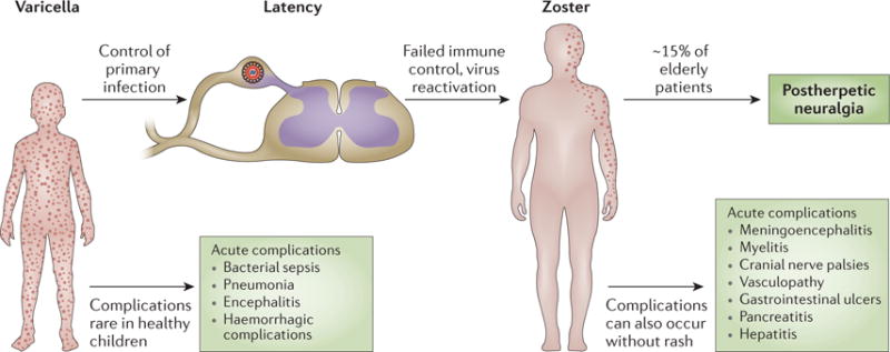
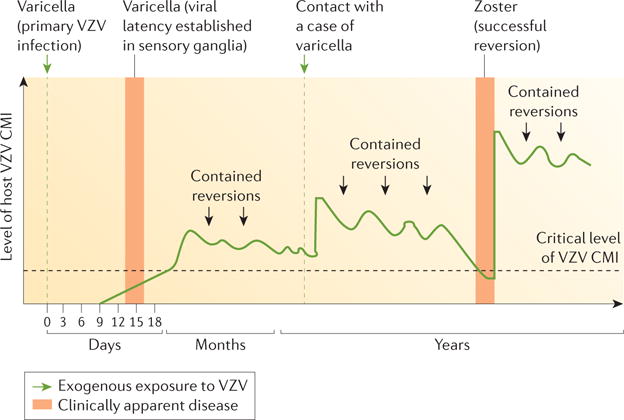
Infection with varicella zoster virus (VZV) causes varicella (chickenpox), which can be severe in immunocompromised individuals, infants and adults. Primary infection is followed by latency in ganglionic neurons. During this period, no virus particles are produced and no obvious neuronal damage occurs. Reactivation of the virus leads to virus replication, which causes zoster (shingles) in tissues innervated by the involved neurons, inflammation and cell death - a process that can lead to persistent radicular pain (postherpetic neuralgia). The pathogenesis of postherpetic neuralgia is unknown and it is difficult to treat. Furthermore, other zoster complications can develop, including myelitis, cranial nerve palsies, meningitis, stroke (vasculopathy), retinitis, and gastroenterological infections such as ulcers, pancreatitis and hepatitis. VZV is the only human herpesvirus for which highly effective vaccines are available. After varicella or vaccination, both wild-type and vaccine-type VZV establish latency, and long-term immunity to varicella develops. However, immunity does not protect against reactivation. Thus, two vaccines are used: one to prevent varicella and one to prevent zoster. In this Primer we discuss the pathogenesis, diagnosis, treatment, and prevention of VZV infections, with an emphasis on the molecular events that regulate these diseases. For an illustrated summary of this Primer, visit: http://go.nature.com/14xVI1.


Conflict of interest statement

Competing interests

J.B., J.I.C., R.J.C., M.D.G., D.G., C.G., S.H., M.N.O. and J.F.S. declare no competing interests. A.A.G. declares service contracts from Merck to investigate the safety of VZV vaccines (identifying VZV in samples from patients with possible adverse reactions), chairs an independent data monitoring committee for GlaxoSmithKline’s Phase III subunit glycoprotein E zoster vaccine trial, consults with Pfizer when invited, and has participated in an educational programme (supported by an unrestricted educational grant) on zoster for GlaxoSmithKline. P.G.E.K. has served on a scientific advisory board on zoster vaccination for Sanofi Pasteur MSD. Y.K. is Director General of the BIKEN foundation (The Research Foundation for Microbial Diseases of Osaka University), which produces varicella vaccines.

Figures

Figure 1
Figure 1. Different phases of varicella zoster virus infection
Primary infection with varicella zoster virus (VZV) in susceptible individuals causes varicella, which usually is harmless in healthy children whose immune system controls the infection. VZV establishes latency in ganglionic neurons, and reactivation of viral replication and spread of the virus to the skin innervated by these neurons causes zoster. Increasing age and compromised immune function are risk factors for complications of VZV infections. However, some of these complications, such as postherpetic neuralgia, can also occur without these predisposing factors.
Figure 2
Figure 2. Epidemiology
The introduction of the varicella vaccine in 1 995 reduced the number of varicella cases substantially (data shown for Illinois, Michigan, Texas and West Virginia, USA). Reprinted with permission from REF , Centers for Disease Control and Prevention (CDC).
Figure 3
Figure 3. Clinical presentation of varicella withsevere rash
Severe varicella in an 11-month-old infant who, during the incubation period, had received dexamethasone as part of therapy for pneumococcal meningitis. Pictured on day 5 of the rash, when he was still systemically unwell and acquiring new lesions. The infant made a full recovery with intravenous acyclovir therapy.
Figure 4
Figure 4. Latent and lytic infection
a | Lytic infection with varicella zoster virus (VZV) starts with attachment, fusion and uncoating of the virion. The virus capsid is then transported to the cell nucleus, where the viral DNA becomes circular. The full set of viral proteins, including immediate early (IE), early (E) and late (L) proteins, are expressed and enter the nucleus. New virions then bud in a two-step process. This full cycle of viral replication leads to substantial cell damage and eventually lysis; the acidic environmental in the endosome damages the virus particles and reduces their infectiousness. The micrograph shows VZV infection of guinea pig enteric neurons showing lytic infection. Isolated neurons were cultured in vitro and infected with cell-free VZV to induce infection. The cultures were fixed and immunostained with antibodies against VZV ORF29p (red) and glycoprotein E (green). The neurons were analysed 48 h after infection with cell-associated virus; after lytic infection, neurons die with 48–72 h. The neuron is filled with cytoplasmic glycoprotein E immunoreactivity and the immunoreactivity of ORF29p has almost entirely translocated to the nucleus (arrow). b | The exact mechanisms of latent infection are unclear but viral replication is thought to stop at the circular DNA stage and no or only limited protein expression occurs. Furthermore, no viral proteins are found in the nucleus. Latent infection causes no easily observable changes of cell morphology (see micrograph). The micrograph shows VZV infection of guinea pig enteric neurons showing latent infection. Isolated neurons were cultured in vitro and infected with cell-free VZV to induce infection. The cultures were fixed and immunostained with antibodies against VZV ORF29p (red) and glycoprotein E (green). The neurons were analysed 2 weeks after infection with cell-free VZV; after latent infection, neurons survive in vitro for as long as cultures can be maintained. Note that ORF29p immunoreactivity is confined to the cytoplasm; there is no nuclear immunoreactivity (arrow). TGN, trans-Golgi network. Adapted from REF. , Nature Publishing Group.
Figure 5
Figure 5. Natural history and pathogenesis of zoster
The classic mod el of zoster pathogenesis first described by Hope-Simpson links viral replication and clinical disease with the levels of cell-mediated immunity (CMI). In primary infection, no immunity to varicella zoster virus (VZV) exists and infection becomes apparent as varicella. CMI then controls replication, and reactivation of the virus remains subclinical and asymptomatic (‘contained reversions’). Exogenous contact with VZV can boost CMI; nevertheless, CMI levels decrease over time and when they fall below a critical level, zoster can develop. Adapted with permission from REF , The Royal Society of Medicine.
Figure 6
Figure 6. Antiviral treatment in VZV disease
a | Antiviral treatment of varicella is indicated in immunocompromised individuals, neonates, patients with chronic skin or lung diseases and in individuals aged >13 years. Patients receive oral acyclovir (ACV), valaciclovir (VACV) or famciclovir (FCV; not approved by the FDA for use in children) unless they are clinically ill or at high risk (most immunocompromised patients are considered to be at high risk, except those who receive long-term, effective immunoglobulin replacement therapy or those who received only mildly immunosuppressive drugs a long time ago). Ill and high risk patients receive intravenous (IV) ACV or foscarnet if the infection is caused by ACV-resistant VZV. Intravenous treatment always needs careful consideration of kidney function. b | The treatment of zoster follows a similar algorithm; here compromised immunity, illness, severe rash, involvement of eyes or face, and other complications are indications for antiviral treatment. In addition to ACV, VACV and FCV, brivudin (BVDU; not approved by the FDA) might be used. Patients who develop varicella or zoster in hospital will generally receive antiviral therapy as part of an infection control strategy. PO, per os (oral administration).
Figure 7
Figure 7. Mode of action of acyclovir
In cells infected with varicella zoster virus (VZV), acyclovir (ACV) is converted by the viral thymidine kinase (TK) to ACV-monophosphate (ACV-MP). The cellular enzymes guanylate (GMP) kinase and nucleoside-diphosphate (NDP) kinase further catalyse the production of ACV-diphosphate (ACV-DP) and ACV-triphosphate (ACV-TP). When the viral DNA polymerase uses ACV-TP, elongation of the DNA chain is terminated. Adapted from REF , Nature Publishing Group.
Figure 8
Figure 8. Autophagosomes in VZV-infected cells
Autophagosomes are present in the skin vesicles during both varicella and zoster. Autophagy is required for the replication of varicella zoster virus (VZV), and replication is enhanced when autophagy is induced. Autophagy can be quantitated by enumeration of autophagosomes. The figure illustrates a monolayer of VZV-infected cells labelled with antibodies against a VZV protein (IE62 protein; red) and autophagosomes (LC3–II protein; green); nuclei were stained blue with Hoechst 33342. The monolayer was imaged by confocal microscopy, after which the confocal images were converted into a 3D animation by Imaris software. A single frame from the animation is shown in the figure. Autophagosomes (~100) appear as green dots against the background of blue nuclei (~70). Many of the nuclei are clustered within a syncytium of VZV-infected cells (red cytoplasm). By contrast, only the nuclei of uninfected cells are visible in this image, as the cytoplasm of uninfected cells is not stained.

References

    1. Gilden D, Cohrs RJ, Mahalingam R, Nagel MA. Neurological disease produced by varicella zoster virus reactivation without rash. Curr Top Microbiol Immunol. 2010;342:243–253. - PMC - PubMed
    1. Gershon AA, Gershon MD. Pathogenesis and current approaches to control of varicella-zoster virus infections. Clin Microbiol Rev. 2013;26:728–743. - PMC - PubMed
    1. Gershon AA, Takahashi M, Seward JF. In: Vaccines. Plotkin S, Orenstein W, Offit P, editors. Saunders Elsevier; 2011. pp. 915–958.
    1. Tsolia M, Gershon AA, Steinberg SP, Gelb L. Live attenuated varicella vaccine: evidence that the virus is attenuated and the importance of skin lesions in transmission of varicella-zoster virus. National Institute of Allergy and Infectious Diseases Varicella Vaccine Collaborative Study Group. J Pediatr. 1990;116:184–189. A study demonstrating that VZV spreads from skin lesions. - PubMed
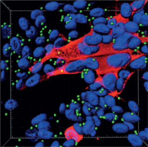
    1. Chen JJ, Zhu Z, Gershon AA, Gershon MD. Mannose 6-phosphate receptor dependence of varicella zoster virus infection in vitro and in the epidermis during varicella and zoster. Cell. 2004;119:915–926. This study demonstrates the importance of the skin in VZV infection and identifies the cellular receptor that the virus uses for infection. - PubMed

Publication types

MeSH terms

Substances