

In vivo dosimetry using Gafchromic films during pelvic intraoperative electron radiation therapy (IOERT)
- PMID: 27188847
- PMCID: PMC5257329
- DOI: 10.1259/bjr.20160193
In vivo dosimetry using Gafchromic films during pelvic intraoperative electron radiation therapy (IOERT)
Abstract
Objective: To characterize in vivo dose distributions during pelvic intraoperative electron radiation therapy (IOERT) for rectal cancer and to assess the alterations introduced by irregular irradiation surfaces in the presence of bevelled applicators.
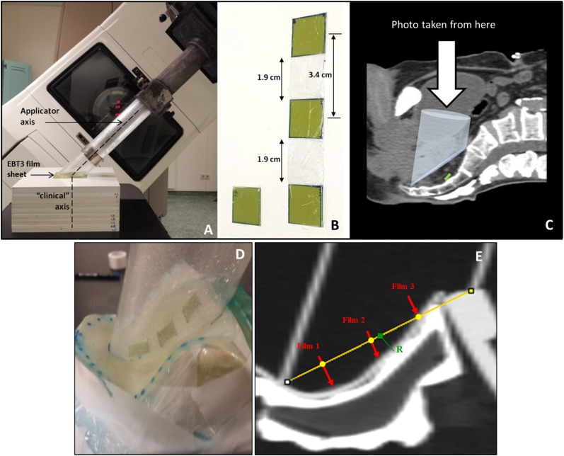
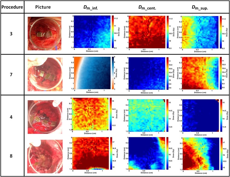
Methods: In vivo measurements were performed with Gafchromic films during 32 IOERT procedures. 1 film per procedure was used for the first 20 procedures. The methodology was then optimized for the remaining 12 procedures by using a set of 3 films. Both the average dose and two-dimensional dose distributions for each film were determined. Phantom measurements were performed for comparison.
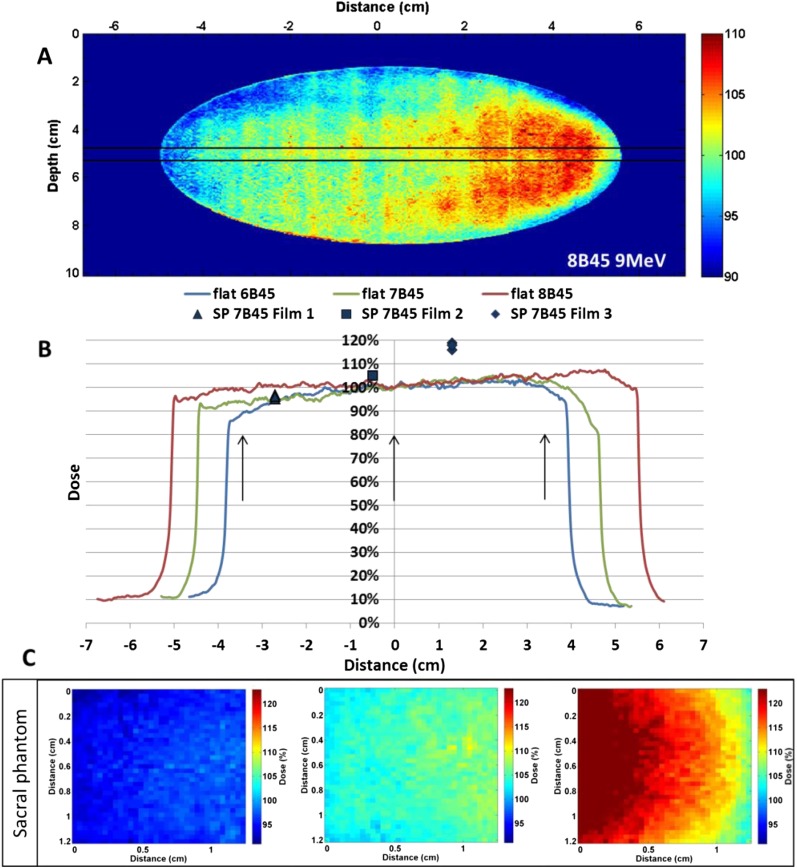
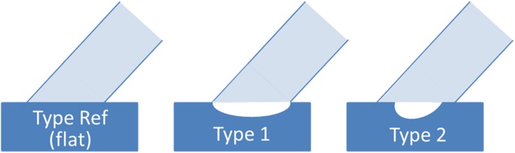
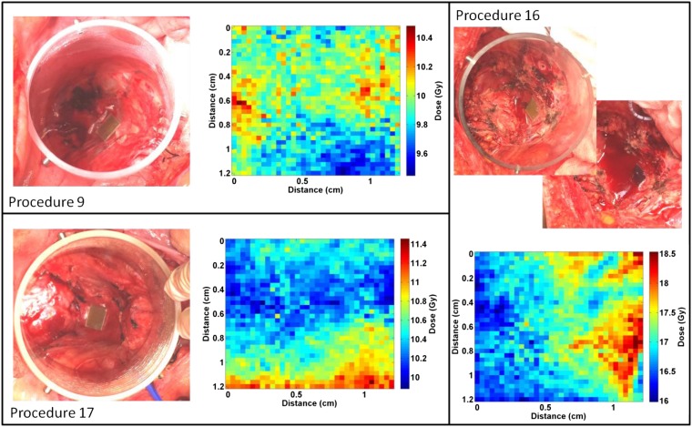
Results: For flat and concave surfaces, the doses measured in vivo agree with expected values. For concave surfaces with step-like irregularities, measured doses tend to be higher than expected doses. Results obtained with three films per procedure show a large variability along the irradiated surface, with important differences from expected profiles. These results are consistent with the presence of surface hotspots, such as those observed in phantoms in the presence of step-like irregularities, as well as fluid build-up.
Conclusion: Clinical dose distributions in the IOERT of rectal cancer are often different from the references used for prescription. Further studies are necessary to assess the impact of these differences on treatment outcomes. In vivo measurements are important, but need to be accompanied by accurate imaging of positioning and irradiated surfaces.
Advances in knowledge: These results confirm that surface irregularities occur frequently in rectal cancer IOERT and have a measurable effect on the dose distribution.
Figures
Similar articles
-
Assessment of clinically relevant dose distributions in pelvic IOERT using Gafchromic EBT3 films.Phys Med. 2015 Nov;31(7):692-701. doi: 10.1016/j.ejmp.2015.05.013. Epub 2015 Jun 12. Phys Med. 2015. PMID: 26078013
-
Optimized method for in vivo dosimetry with small films in pelvic IOERT for rectal cancer.Phys Med. 2021 Jan;81:20-30. doi: 10.1016/j.ejmp.2020.11.019. Epub 2020 Dec 15. Phys Med. 2021. PMID: 33338728
-
Implementation of an intraoperative electron radiotherapy in vivo dosimetry program.Radiat Oncol. 2016 Mar 15;11:41. doi: 10.1186/s13014-016-0621-y. Radiat Oncol. 2016. PMID: 26980076 Free PMC article.
-
In vivo dosimetry and shielding disk alignment verification by EBT3 GAFCHROMIC film in breast IOERT treatment.J Appl Clin Med Phys. 2014 Jan 8;16(1):5065. doi: 10.1120/jacmp.v16i1.5065. J Appl Clin Med Phys. 2014. PMID: 25679150 Free PMC article.
-
In vivo dosimetry in pelvic brachytherapy.Br J Radiol. 2022 Sep 1;95(1137):20220046. doi: 10.1259/bjr.20220046. Epub 2022 Aug 10. Br J Radiol. 2022. PMID: 35635803 Free PMC article. Review.
Cited by
-
Surface scanning for 3D dose calculation in intraoperative electron radiation therapy.Radiat Oncol. 2018 Dec 7;13(1):243. doi: 10.1186/s13014-018-1181-0. Radiat Oncol. 2018. PMID: 30526626 Free PMC article.
-
Dosimetric properties of a newly developed thermoluminescent sheet-type dosimeter for clinical proton beams.J Appl Clin Med Phys. 2021 Apr;22(4):158-165. doi: 10.1002/acm2.13222. Epub 2021 Mar 15. J Appl Clin Med Phys. 2021. PMID: 33720527 Free PMC article.
-
Intraoperative computed tomography imaging for dose calculation in intraoperative electron radiation therapy: Initial clinical observations.PLoS One. 2020 Jan 10;15(1):e0227155. doi: 10.1371/journal.pone.0227155. eCollection 2020. PLoS One. 2020. PMID: 31923183 Free PMC article.
References
-
- Pascau J, Ph D, Santos A, Morillo V, Calvo FA, Bouche A, et al. . An innovative tool for intraoperative electron beam radiotherapy simulation and planning: description and initial evaluation by radiation oncologists. Int J Radiat Oncol Biol Phys 2012; 83: 287–95. doi: 10.1016/j.ijrobp.2011.12.063 - DOI - PubMed
-
- Costa F, Sarmento S, Sousa O. Assessment of clinically relevant dose distributions in pelvic IOERT using Gafchromic EBT3 films. Phys Med 2015; 31: 692–701. - PubMed
-
- Consorti R, Petrucci A, Fortunato F, Soriani A, Marzi S, Iaccarino G, et al. . In vivo dosimetry with MOSFETs: dosimetric characterization and first clinical results in intraoperative radiotherapy. Int J Radiat Oncol Biol Phys 2005; 63: 952–60. - PubMed
MeSH terms
LinkOut - more resources
Full Text Sources
Other Literature Sources
