

Fibroblasts and Mesenchymal Stromal/Stem Cells Are Phenotypically Indistinguishable
- PMID: 27188909
- PMCID: PMC4988914
- DOI: 10.1159/000445096
Fibroblasts and Mesenchymal Stromal/Stem Cells Are Phenotypically Indistinguishable
Abstract
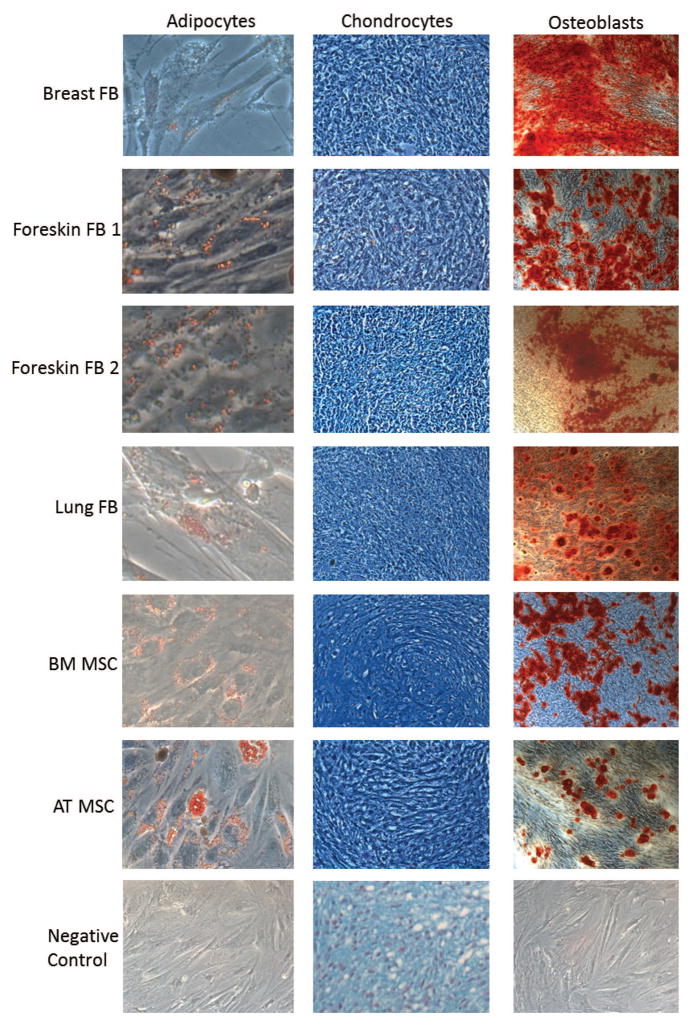
Background/aims: Human mesenchymal stromal/stem cells (MSCs), derived from many different tissues, are characterized by a fibroblast-like morphology, the expression of certain cell surface markers and their ability to differentiate into adipocytes, chondrocytes and osteoblasts. A number of studies have shown that MSCs share many characteristics with fibroblasts; however, there is no well-defined set of phenotypic characteristics that could distinguish between these 2 types of cells.
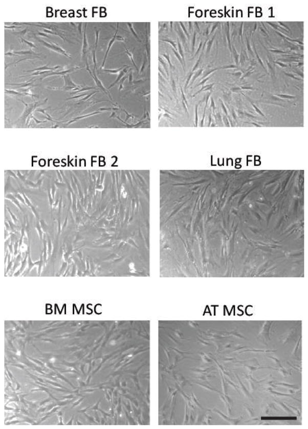
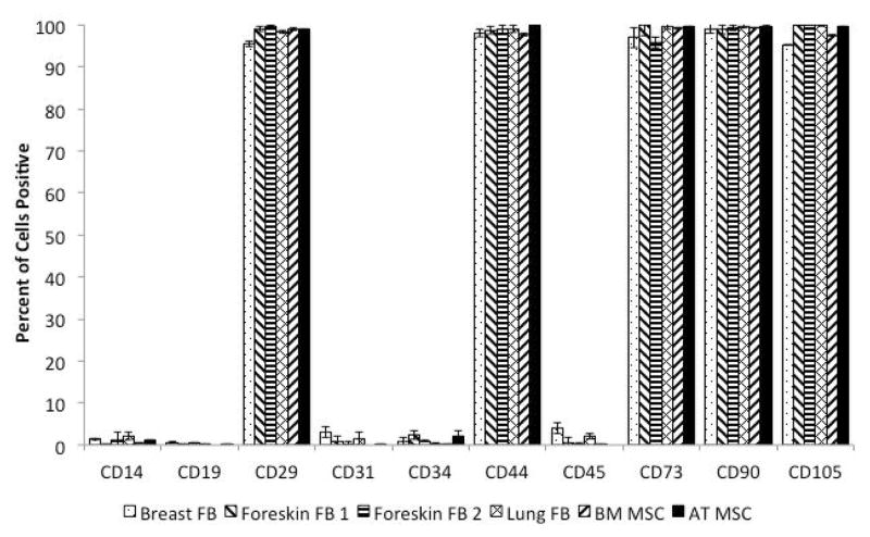
Methods: We used 4 well-established human fibroblast strains from 3 different tissue sources and several human MSC strains from 2 different tissue sources to compare the phenotypic and immunological characteristics of these cells.
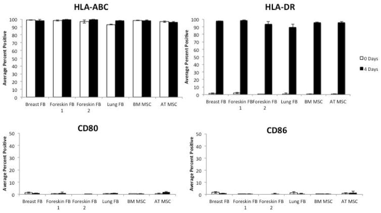
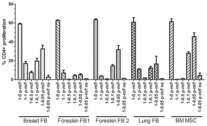
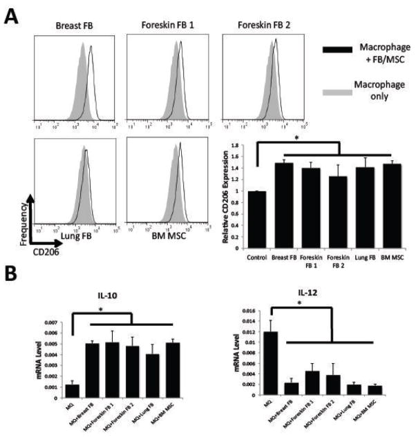
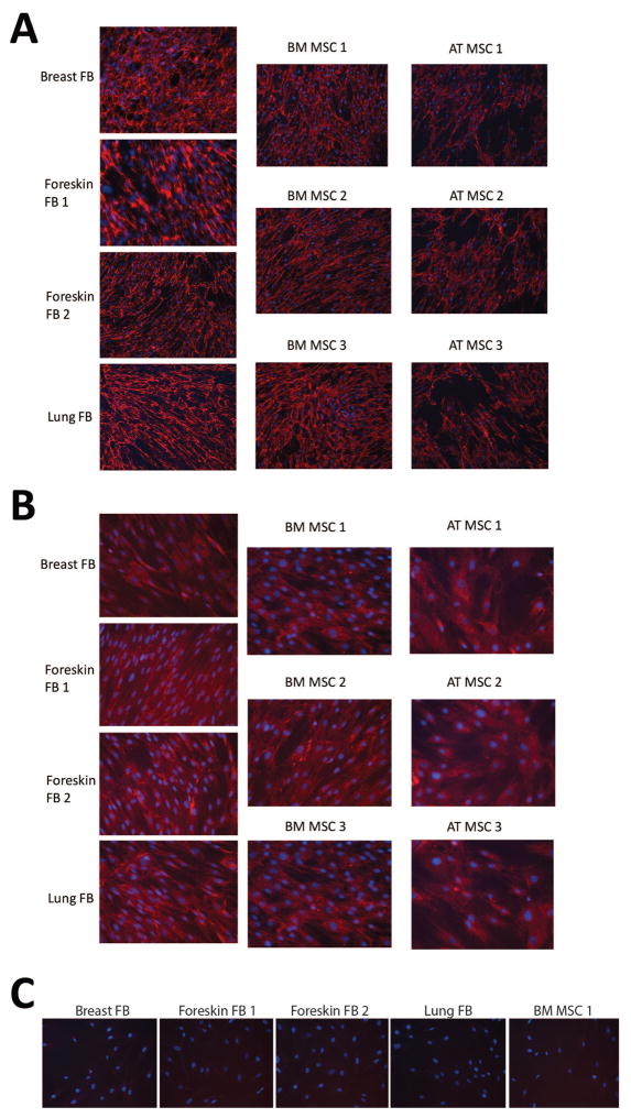
Results: Fibroblast strains had a similar morphology to MSCs, expressed the same cell surface markers as MSCs and could also differentiate into adipocytes, chondrocytes and osteoblasts. Also, similar to MSCs, these fibroblasts were capable of suppressing T cell proliferation and modulating the immunophenotype of macrophages. We also show that MSCs deposit extracellular matrices of collagen type I and fibronectin, and express FSP1 in patterns similar to fibroblasts.
Conclusions: Based on currently accepted definitions for cultured human MSCs and fibroblasts, we could not find any immunophenotypic property that could make a characteristic distinction between MSCs and fibroblasts.
© 2016 S. Karger AG, Basel.
Conflict of interest statement
The authors declare that there is no conflict of interests regarding the publication of this paper.
Figures
Comment on
-
Multipotent mesenchymal stromal cells obtained from diverse human tissues share functional properties and gene-expression profile with CD146+ perivascular cells and fibroblasts.Exp Hematol. 2008 May;36(5):642-54. doi: 10.1016/j.exphem.2007.12.015. Epub 2008 Mar 4. Exp Hematol. 2008. PMID: 18295964
-
Human fibroblasts share immunosuppressive properties with bone marrow mesenchymal stem cells.J Clin Immunol. 2010 Jul;30(4):607-19. doi: 10.1007/s10875-010-9415-4. Epub 2010 Apr 20. J Clin Immunol. 2010. PMID: 20405178
References
-
- Horwitz EM, Le Blanc K, Dominici M, Mueller I, Slaper-Cortenbach I, Marini FC, Deans RJ, Krause DS, Keating A. Clarification of the nomenclature for msc: The international society for cellular therapy position statement. Cytotherapy. 2005;7:393–395. - PubMed
-
- Friedenstein AJ, Chailakhjan RK, Lalykina KS. The development of fibroblast colonies in monolayer cultures of guinea-pig bone marrow and spleen cells. Cell and tissue kinetics. 1970;3:393–403. - PubMed
-
- Friedenstein AJ. Marrow stromal fibroblasts. Calcif Tissue Int. 1995;56(Suppl 1):S17.
-
- Friedenstein AJ, Chailakhyan RK, Latsinik NV, Panasyuk AF, Keiliss-Borok IV. Stromal cells responsible for transferring the microenvironment of the hemopoietic tissues. Cloning in vitro and retransplantation in vivo Transplantation. 1974;17:331–340. - PubMed
-
- Friedenstein AJ. Precursor cells of mechanocytes. International review of cytology. 1976;47:327–359. - PubMed
Publication types
MeSH terms
Grants and funding
LinkOut - more resources
Full Text Sources
Other Literature Sources
Research Materials
