Review
. 2016 Jun;9(6):e002558.
doi: 10.1161/CIRCHEARTFAILURE.115.002558.

Transthyretin Cardiac Amyloidosis in Black Americans

Affiliations
Review

Transthyretin Cardiac Amyloidosis in Black Americans

Keyur B Shah et al. Circ Heart Fail. 2016 Jun.

Abstract

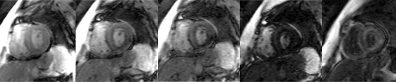
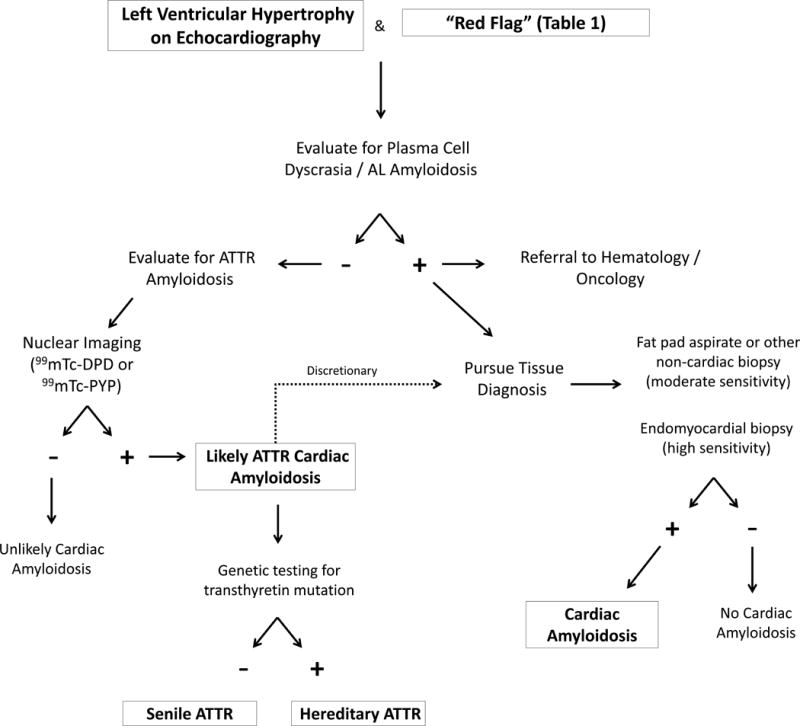
Transthyretin-related cardiac amyloidosis is a progressive infiltrative cardiomyopathy that mimics hypertensive and hypertrophic heart disease and often goes undiagnosed. In the United States, the hereditary form disproportionately afflicts black Americans, who when compared with whites with wild-type transthyretin amyloidosis, a phenotypically similar condition, present with more advanced disease despite having a noninvasive method for early identification (genetic testing). Although reasons for this are unclear, this begs to consider the inadequate access to care, societal factors, or a biological basis. In an effort to improve awareness and explore unique characteristics, we review the pathophysiology, epidemiology, and therapeutic strategies for transthyretin amyloidosis and highlight diagnostic pitfalls and clinical pearls for identifying patients with amyloid heart disease.

Keywords: African Continental Ancestry Group; amyloidosis; cardiomyopathy, restrictive; continental population groups; heart failure; prealbumin.


Figures

Figure 1
Figure 1
A surface twelve-lead electrocardiograms from a patient with ATTRmt (Val122Ile). The patient had severe left ventricular hypertrophy on two-dimensional echocardiography and no coronary artery disease on coronary angiography. The electrocardiogram displayed lower than expected QRS voltage and suggestion of an anterior wall myocardial infarction (poor R-wave progression).
Figure 2
Figure 2
Transthoracic echocardiography of a patient with ATTRmt (Val122Ile) cardiac amyloidosis with classic features of restrictive heart disease. (A) Two-dimensional imaging obtained in the apical four chamber view shows left and right ventricular wall hypertrophy, bi-atrial dilation and increased myocardial echogenicity.(B) Measurements of Doppler inflow velocities across the mitral valve show an increased E/A ratio and decreased deceleration time of early diastolic inflow. (C) Tissue Doppler imaging quantifying the mitral annular velocities are depressed.
Figure 2
Figure 2
Transthoracic echocardiography of a patient with ATTRmt (Val122Ile) cardiac amyloidosis with classic features of restrictive heart disease. (A) Two-dimensional imaging obtained in the apical four chamber view shows left and right ventricular wall hypertrophy, bi-atrial dilation and increased myocardial echogenicity.(B) Measurements of Doppler inflow velocities across the mitral valve show an increased E/A ratio and decreased deceleration time of early diastolic inflow. (C) Tissue Doppler imaging quantifying the mitral annular velocities are depressed.
Figure 3
Figure 3
Summary of the advanced imaging modalities for diagnosis and monitoring of cardiac amyloidosis.
Figure 4
Figure 4
Four chamber and mid-ventricular short axis views of patients with ATTRmt (Val122Ile) and (A,B) mild and (D,E) severe left ventricular hypertrophy. (C) The patient with mild ventricular hypertrophy exhibits only mild late gadolinium enhancement (LGE) in a subendocardial distribution. (F) The patient with severe hypertrophy has a transmural pattern of LGE. Not shown is an example of patchy LGE. (Courtesy of Dr. John D. Grizzard)
Figure 4
Figure 4
Four chamber and mid-ventricular short axis views of patients with ATTRmt (Val122Ile) and (A,B) mild and (D,E) severe left ventricular hypertrophy. (C) The patient with mild ventricular hypertrophy exhibits only mild late gadolinium enhancement (LGE) in a subendocardial distribution. (F) The patient with severe hypertrophy has a transmural pattern of LGE. Not shown is an example of patchy LGE. (Courtesy of Dr. John D. Grizzard)
Figure 5
Figure 5
T1 inversion scout sequences of (A) a normal heart and (B) a patient with cardiac amyloidosis. (A) In the normal heart, the blood pool “nulls” or blackens before the surrounding myocardium and nearby spleen. (B) In the patient with amyloid heart disease, the order of “nulling” between the myocardium and blood pool is inversed, where the blood pool “nulls” after the spleen after the myocardium. (Courtesy of Dr. John D. Grizzard)
Figure 5
Figure 5
T1 inversion scout sequences of (A) a normal heart and (B) a patient with cardiac amyloidosis. (A) In the normal heart, the blood pool “nulls” or blackens before the surrounding myocardium and nearby spleen. (B) In the patient with amyloid heart disease, the order of “nulling” between the myocardium and blood pool is inversed, where the blood pool “nulls” after the spleen after the myocardium. (Courtesy of Dr. John D. Grizzard)
Figure 6
Figure 6
An algorithm for evaluating patients for amyloid heart disease. The first step is to exclude any evidence of a plasma cell disorder.

References

    1. Ayanian JZ, Weissman JS, Chasan-Taber S, Epstein AM. Quality of care by race and gender for congestive heart failure and pneumonia. Med Care. 1999;37:1260–1269. - PubMed
    1. Dickson VV, McCarthy MM, Howe A, Schipper J, Katz SM. Sociocultural influences on heart failure self-care among an ethnic minority black population. J Cardiovasc Nurs. 2013;28:111–118. - PubMed
    1. Chaudhry SI, Herrin J, Phillips C, Butler J, Mukerjhee S, Murillo J, Onwuanyi A, Seto TB, Spertus J, Krumholz HM. Racial disparities in health literacy and access to care among patients with heart failure. J Card Fail. 2011;17:122–127. - PMC - PubMed
    1. Yancy CW, Abraham WT, Albert NM, Clare R, Stough WG, Gheorghiade M, Greenberg BH, O’Connor CM, She L, Sun JL, Young JB, Fonarow GC. Quality of care of and outcomes for African Americans hospitalized with heart failure: findings from the OPTIMIZE-HF (Organized Program to Initiate Lifesaving Treatment in Hospitalized Patients With Heart Failure) registry. J Am Coll Cardiol. 2008;51:1675–1684. - PubMed
    1. Kamath SA, Drazner MH, Wynne J, Fonarow GC, Yancy CW. Characteristics and outcomes in African American patients with decompensated heart failure. Arch Intern Med. 2008;168:1152–1158. - PubMed

Publication types

MeSH terms

Supplementary concepts