

doi: 10.3349/ymj.2016.57.4.1022.
Surgical Treatment for Falcotentorial Meningiomas
Affiliations
- PMID: 27189300
- PMCID: PMC4951445
- DOI: 10.3349/ymj.2016.57.4.1022
Item in Clipboard
Surgical Treatment for Falcotentorial Meningiomas
Yonsei Med J.
2016 Jul.
Abstract
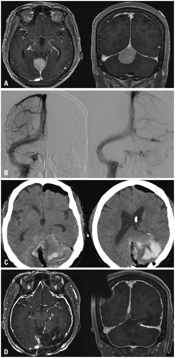
Among intracranial meningiomas, falcotentorial meningiomas, occurring at the junction of the falx cerebri and tentorial dural folds, are extremely rare. Because of their deep location, they are surrounded by critical structures, and have been regarded as one of the most challenging lesions for surgical treatment. In this study, we describe our surgical strategy for falcotentorial meningiomas and provide a review of our experience.
Keywords: Meningiomas; craniotomy; operations; venous infarction.
Conflict of interest statement
The authors have no financial conflicts of interest.
Figures


References
-
- Bassiouni H, Asgari S, König HJ, Stolke D. Meningiomas of the falcotentorial junction: selection of the surgical approach according to the tumor type. Surg Neurol. 2008;69:339–349. - PubMed
-
- Okami N, Kawamata T, Hori T, Takakura K. Surgical treatment of falcotentorial meningioma. J Clin Neurosci. 2001;8(Suppl 1):15–18. - PubMed
-
- Raco A, Agrillo A, Ruggeri A, Gagliardi FM, Cantore G. Surgical options in the management of falcotentorial meningiomas: report of 13 cases. Surg Neurol. 2004;61:157–164. - PubMed
-
- Obrador S, Soto M, Gutierrez-Diaz JA. Surgical management of tumours of the pineal region. Acta Neurochir (Wien) 1976;34:159–171. - PubMed
-
- Rozario R, Adelman L, Prager RJ, Stein BM. Meningiomas of the pineal region and third ventricle. Neurosurgery. 1979;5:489–485. - PubMed
MeSH terms
LinkOut - more resources
Full Text Sources
Other Literature Sources
