. 2016 Aug;33(8):1921-36.
doi: 10.1093/molbev/msw066. Epub 2016 Apr 4.

Searching the Evolutionary Origin of Epithelial Mucus Protein Components-Mucins and FCGBP

Affiliations

Searching the Evolutionary Origin of Epithelial Mucus Protein Components-Mucins and FCGBP

Tiange Lang et al. Mol Biol Evol. 2016 Aug.

Abstract

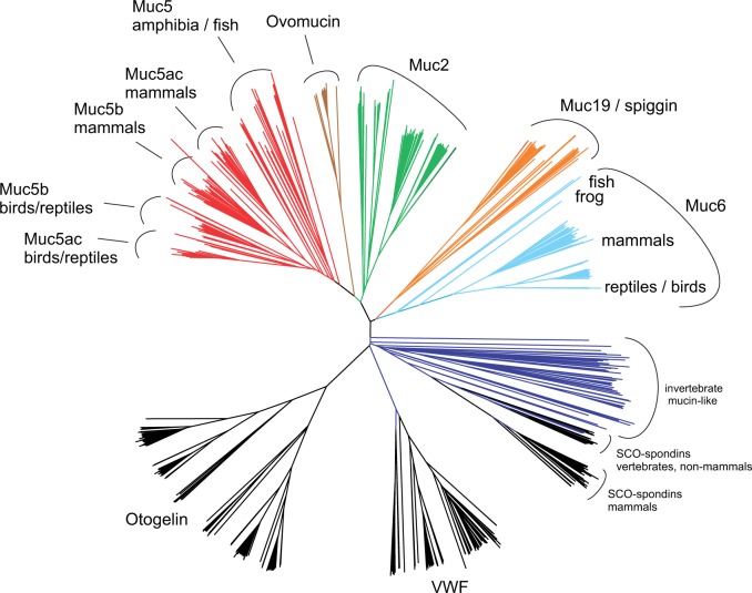
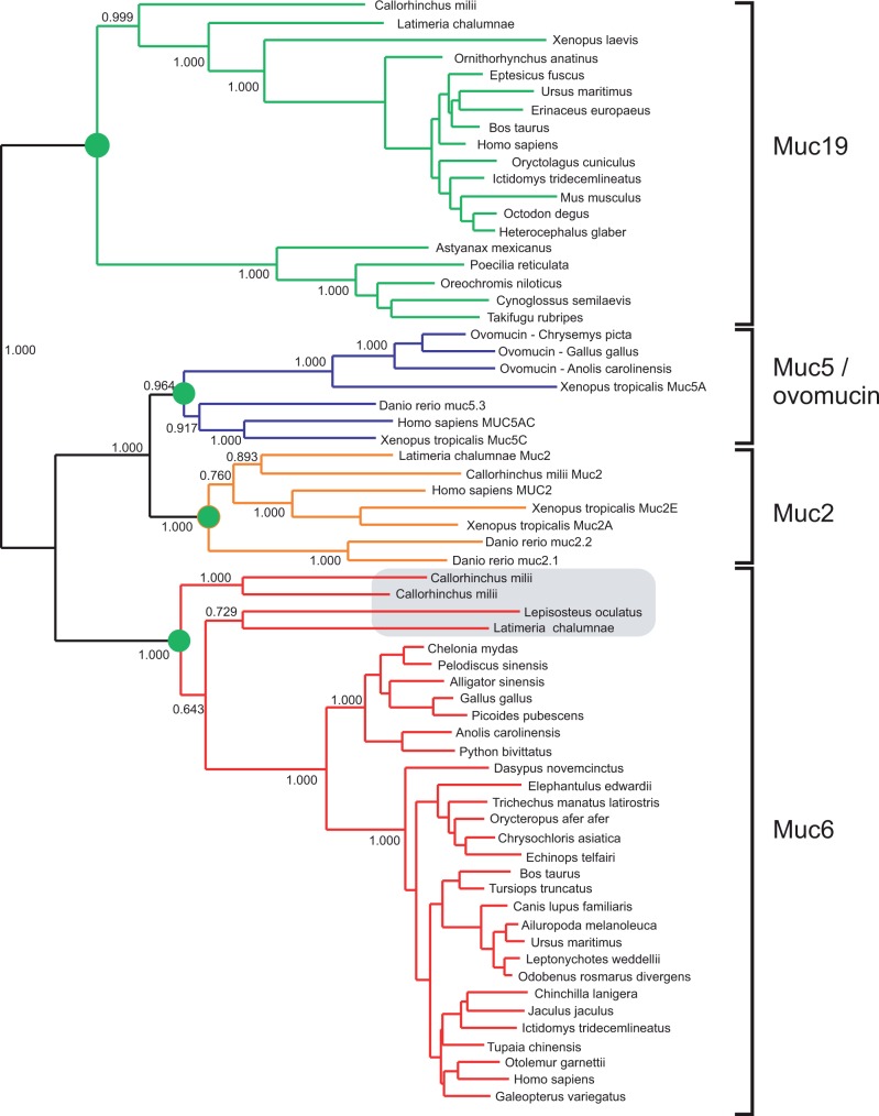
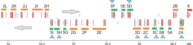
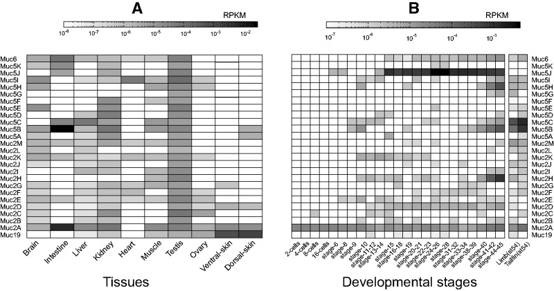
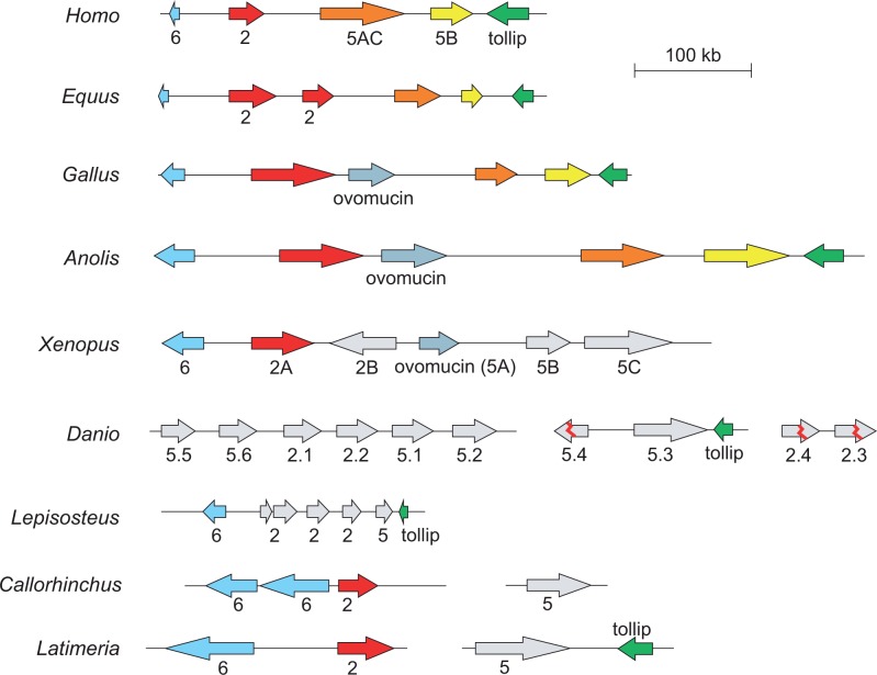
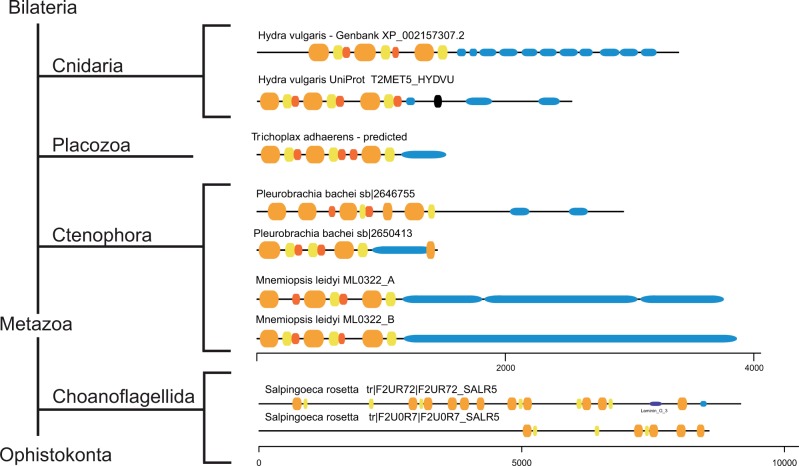
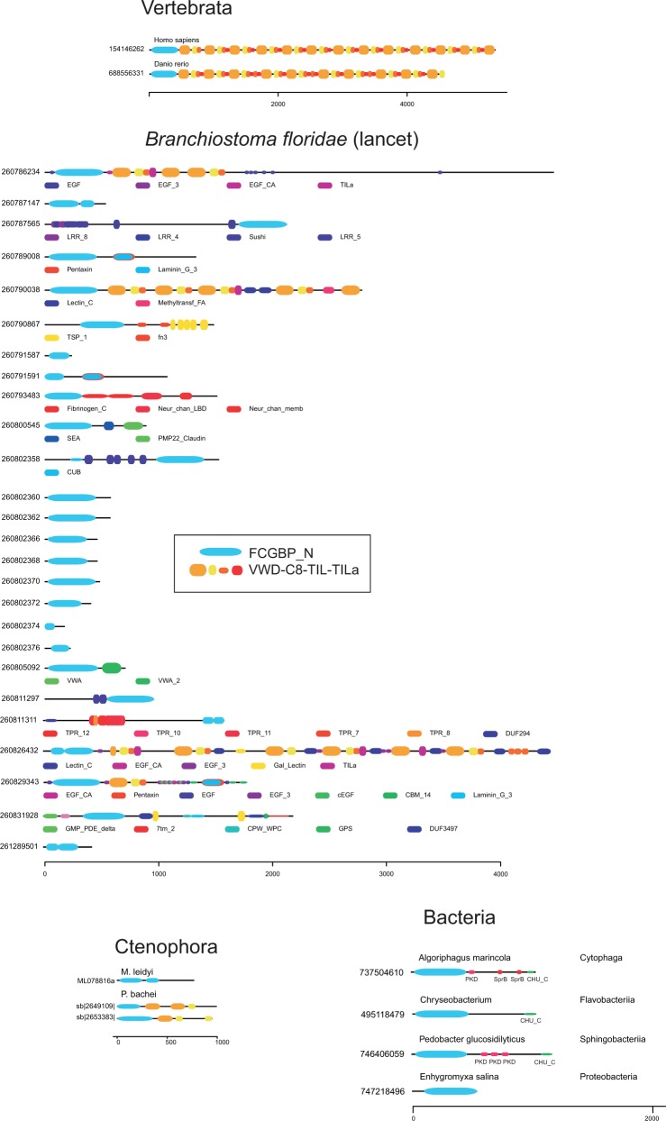
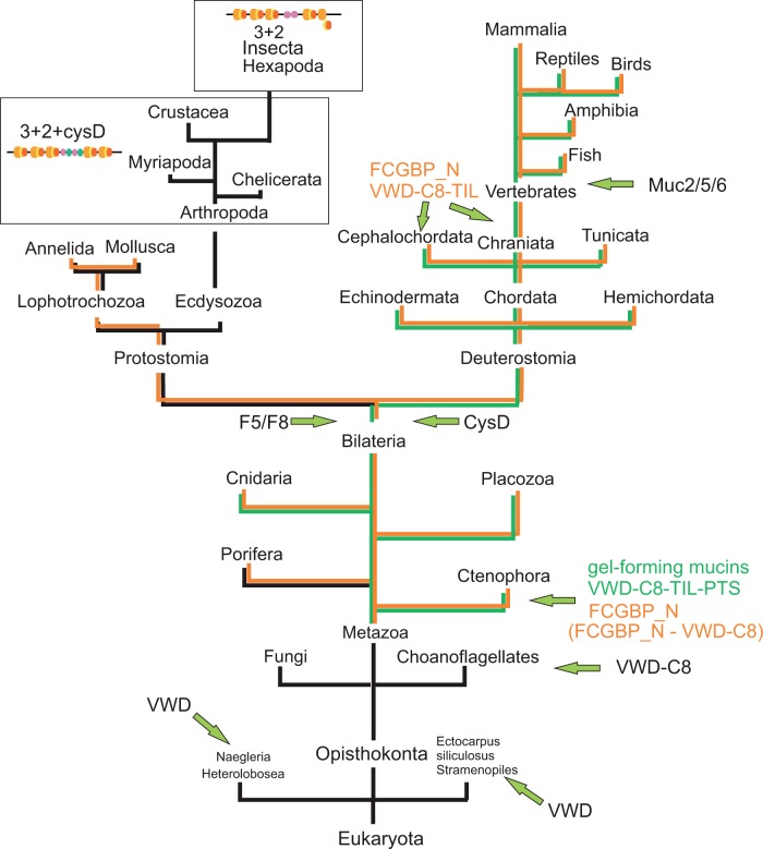
The gel-forming mucins are large glycosylated proteins that are essential components of the mucus layers covering epithelial cells. Using novel methods of identifying mucins based on profile hidden Markov models, we have found a large number of such proteins in Metazoa, aiding in their classification and allowing evolutionary studies. Most vertebrates have 5-6 gel-forming mucin genes and the genomic arrangement of these genes is well conserved throughout vertebrates. An exception is the frog Xenopus tropicalis with an expanded repertoire of at least 26 mucins of this type. Furthermore, we found that the ovomucin protein, originally identified in chicken, is characteristic of reptiles, birds, and amphibians. Muc6 is absent in teleost fish, but we now show that it is present in animals such as ghost sharks, demonstrating an early origin in vertebrate evolution. Public RNA-Seq data were analyzed with respect to mucins in zebrafish, frog, and chicken, thus allowing comparison in regard of tissue and developmental specificity. Analyses of invertebrate proteins reveal that gel-forming-mucin type of proteins is widely distributed also in this group. Their presence in Cnidaria, Porifera, and in Ctenophora (comb jellies) shows that these proteins were present early in metazoan evolution. Finally, we examined the evolution of the FCGBP protein, abundant in mucus and related to gel-forming mucins in terms of structure and localization. We demonstrate that FCGBP, ubiquitous in vertebrates, has a conserved N-terminal domain. Interestingly, this domain is also present as an N-terminal sequence in a number of bacterial proteins.

Keywords: bioinformatics; evolution; mucin; mucus; von Willebrand D domain..


Figures

F<sc>ig.</sc> 1
Fig. 1
Phylogenetic classification of mucins. Muc2, Muc5, Muc6, Muc19, ovomucin, otogelin, and VWF protein sequences were aligned with Clustal Omega (Sievers and Higgins 2014) and a neighbor-joining tree with 100 bootstrap replicates was obtained with ClustalW (Thompson et al. 2002). Highlighted in color are Muc5 (red), ovomucin (brown), Muc2 (green), Muc19/spiggin (orange), Muc6 (cyan), and invertebrate mucin-like proteins (blue). Classification of Muc5b and Muc5ac is also possible although all bird/reptile Muc5 are in one group and all mammalian Muc5 in another. Invertebrate mucin-like sequences seem to be most similar to the SCO-spondins, proteins previously known to be present in vertebrates only.
F<sc>ig.</sc> 2
Fig. 2
The mucin Muc6 was present early in vertebrate evolution. Tree was constructed with MrBayes using an alignment of the N-terminal parts of mucins that include the three VWD–C8–TIL units. Homologs of Muc19, Muc6, Muc5, ovomucin, and Muc2 are shown. Critical nodes are labeled with green circles. The four Muc6 homologs of C. milii, L. chalumnae, and Le. oculatus clearly belong to the group of Muc6.
F<sc>ig.</sc> 3
Fig. 3
Genomic organization of X. tropicalis mucins. Genomic region from assembly version 7, accession KB021653.1, region 53,600,000–57,000,000 is shown. Genes are shown for Muc5 paralogs (green) and Muc2 paralogs (orange). Naming of genes was based on their location from right to left. Location of VWD domains, PTS domains, and CysD domains (red, blue, and green vertical bars, respectively) are based on the analysis of the genomic sequence with genewise and an in-house Perl script. Genes where a CysD domain is predicted are indicated with cyan triangles.
F<sc>ig.</sc> 4
Fig. 4
X. tropicalis mucin gene transcription. A total of 26 different mucin genes were analyzed with respect to gene expression. Expression levels (RPKM) were estimated from public RNA-Seq data as listed in supplementary dataset S2, Supplementary Material online, and as described in more detail in Materials and Methods section. (A) Expression in different tissues. (B) Expression at different developmental stages.
F<sc>ig.</sc> 5
Fig. 5
A conserved structure of mucin genes in vertebrates. Arrows reflect the strand polarity. In the case of X. tropicalis only a part of a larger cluster of mucin genes is shown (see also fig. 3).
F<sc>ig.</sc> 6
Fig. 6
Gel-forming mucins evolved early during metazoan evolution. Phylogenetic distribution of proteins in selected basal Metazoa with a domain structure characteristic of gel-forming mucins. VWD, C8, TIL, and PTS domains are shown in orange, yellow, red, and blue, respectively.
F<sc>ig.</sc> 7
Fig. 7
Domain structures of selected proteins with a domain characteristic of FCGBP. In vertebrates, FCGBP proteins have a large number of VWD–C8–TIL units, in addition to the FCGBP_N domain. In the invertebrate B. floridae, there is a large number of proteins with the FCGBP_N domain. All domains that are not FCGBP_N, VWD, C8, TIL, TILa, or PTS domains are explained below each representation of protein domain architecture. For a complete collection of all proteins with the FCGBP domain, see supplementary figure S5, Supplementary Material online.
F<sc>ig.</sc> 8
Fig. 8
Summary of mucin and FCGBP protein evolution. The VWD domain seems to occur in a restricted number of protists, but not in combination with the C8 or TIL domains. The combination of VWD/C8 appears in choanoflagellates and the structure characteristic of gel-forming mucins as well as the FCGBP_N-containing proteins appear in Ctenophora. The F5_F8 and CysD domains occur in Bilateria. A protein with FCGBP_N and multiple VWD–C8–TIL domains appears later in evolution as it is observed only in cephalochordates and vertebrates.

Similar articles

Cited by

References

    1. Altschul SF, Gish W, Miller W, Myers EW, Lipman DJ. 1990. Basic local alignment search tool. J Mol Biol. 215:403–410. - PubMed
    1. Altschul SF, Madden TL, Schaffer AA, Zhang J, Zhang Z, Miller W, Lipman DJ. 1997. Gapped BLAST and PSI-BLAST: a new generation of protein database search programs. Nucleic Acids Res. 25:3389–3402. - PMC - PubMed
    1. Ambort D, Johansson ME, Gustafsson JK, Ermund A, Hansson GC. 2012. Perspectives on mucus properties and formation—lessons from the biochemical world. Cold Spring Harb Perspect Med. 2:a014159. - PMC - PubMed
    1. Anders S, Pyl PT, Huber W. 2015. HTSeq—a Python framework to work with high-throughput sequencing data. Bioinformatics 31:166–169. - PMC - PubMed
    1. Benson DA, Clark K, Karsch-Mizrachi I, Lipman DJ, Ostell J, Sayers EW. 2015. GenBank. Nucleic Acids Res. 43:D30–D35. - PMC - PubMed

Publication types

MeSH terms