Review
. 2016;22(26):3950-70.
doi: 10.2174/1381612822666160518141911.

AFM-Based Single Molecule Techniques: Unraveling the Amyloid Pathogenic Species

Affiliations
Review

AFM-Based Single Molecule Techniques: Unraveling the Amyloid Pathogenic Species

Francesco Simone Ruggeri et al. Curr Pharm Des. 2016.

Abstract

Background: A wide class of human diseases and neurodegenerative disorders, such as Alzheimer's disease, is due to the failure of a specific peptide or protein to keep its native functional conformational state and to undergo a conformational change into a misfolded state, triggering the formation of fibrillar cross-β sheet amyloid aggregates. During the fibrillization, several coexisting species are formed, giving rise to a highly heterogeneous mixture. Despite its fundamental role in biological function and malfunction, the mechanism of protein self-assembly and the fundamental origins of the connection between aggregation, cellular toxicity and the biochemistry of neurodegeneration remains challenging to elucidate in molecular detail. In particular, the nature of the specific state of proteins that is most prone to cause cytotoxicity is not established.

Methods: In the present review, we present the latest advances obtained by Atomic Force Microscopy (AFM) based techniques to unravel the biophysical properties of amyloid aggregates at the nanoscale. Unraveling amyloid single species biophysical properties still represents a formidable experimental challenge, mainly because of their nanoscale dimensions and heterogeneous nature. Bulk techniques, such as circular dichroism or infrared spectroscopy, are not able to characterize the heterogeneity and inner properties of amyloid aggregates at the single species level, preventing a profound investigation of the correlation between the biophysical properties and toxicity of the individual species.

Conclusion: The information delivered by AFM based techniques could be central to study the aggregation pathway of proteins and to design molecules that could interfere with amyloid aggregation delaying the onset of misfolding diseases.


Figures

Fig. (1)
Fig. (1)
Schematic nucleation dependent process of amyloid fibrils formation.
Fig. (2)
Fig. (2)
Possible processes of amyloid nucleation. A) Primary nucleation when monomers interact to form an oligomeric nucleus. B) Fragmentation whereby a nucleus breaks down into two nuclei. C) Surface-catalyzed secondary nucleation whereby fibrils surface catalyze the formation of more nuclei.
Fig. (3)
Fig. (3)
Schematic representation of distinct protein conformational states that can lead to the formation of amyloid structures. From [5].
Fig. (4)
Fig. (4)
Typical CD spectra of proteins. Signal indicating α-helical (red), random coil (green) and β-sheet (light blue) structures.
Fig. (5)
Fig. (5)
Schematic depiction of Scanning Probe Techniques. From Ref. [143].
Fig. (6)
Fig. (6)
AFM tip-sample potential energy representation and related AFM scanning modes.
Fig. (7)
Fig. (7)
AFM Schematic illustration of the convolution of the shape of the AFM tip with the shape of the feature or particle being scanned. (a) Blunt tip, (b) sharp tip.
Fig. (8)
Fig. (8)
Statistical characterization of α-synuclein amyloid aggregates cross-sectional dimensions. A) Monomeric and early oligomeric structures. B) Protofibrillar and fibrillar aggregates. Adapted from ref. [151].
Fig. (9)
Fig. (9)
Fibrils Hierarchical self-assembly and polymorphism. Hierarchical self-assembly of β-lactoglobulin. Polymorphism can be due to different number, arrangement and structure of protofilaments composing the fibril. From Ref. [150].
Fig. (10)
Fig. (10)
Persistence length of a flexible amyloid fibril. A) Schematic depiction of a semi-flexible polymer and the parameter used to calculate its persistence length. B) Example of flexible (top, α-lactalbumin) and rigid (bottom, TTR 105-115) amyloid fibrils. Adapted from ref [170].
Fig. (11)
Fig. (11)
The mechanistic pathway of Aβ42 aggregation. a-d) AFM images describing the aggregation process. The arrows indicate the secondary nucleation events. e) The proposed model of Aβ42 fibrillogenesis. Adapted from Ref. [54].
Fig. (12)
Fig. (12)
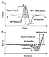
Ideal force-displacement curve depiction. Ideal (A) force-distance and (B) force-time curves representation. Adapted from Ref. [179].
Fig. (13)
Fig. (13)
AFM-based methods to investigate the nanomechanical properties of biomolecules and amyloid aggregates. From Ref. [168].
Fig. (14)
Fig. (14)
Nanoindentation measurements. (A) Schematic depiction of the indentation mechanism, (B) force-displacement curves on the sample and on an undeformable sample, where δ is the indentation depth. Adapted from Ref. [189].
Fig. (15)
Fig. (15)
Principle of operation of PF-QNM. (A) Principles of peak force tapping. (B) Force-distance curve and indicative representation of the calculated mechanical properties. From Ref. [191].
Fig. (16)
Fig. (16)
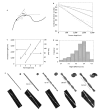
Young’s modulus evolution of Aβ42 species present during its fibrillization process. A-C) Morphology AFM maps. D-F) DMT Young’s modulus maps. G-I) Young’s modulus histograms. From Ref. [194].
Fig. (17)
Fig. (17)
Schematic depiction of the working principle of the nanoscale infrared spectroscopy [197].
Fig. (18)
Fig. (18)
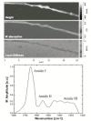
AFM-IR images and spectra of a bundle of collagen fibrils. In the figure are shown on the left the morphology, local stiffness and IR absorption maps and on the right an IR spectrum in the protein range (1800-1200 cm-1) is acquired at a specific location of the maps (indicated by a circle) [139].
Fig. (19)
Fig. (19)
AFM-IR can reconstruct the pathway of protein misfolding and aggregation. a) Young modulus of Josephin protein aggregates as a function of the maturation time. b) Spectra of native oligomers, misfolded oligomers and fibrils showing an increase of the β-sheet content in time. c) Sketch of the two proposed pathways of Josephin aggregation. From Ref. [140].

Similar articles

Cited by

References

    1. Hardy J., Selkoe D.J. The amyloid hypothesis of Alzheimer's disease: progress and problems on the road to therapeutics. Science. 2002;297(5580):353–356. - PubMed
    1. Goedert M. Familial Parkinson's disease. The awakening of alpha-synuclein. Nature. 1997;388(6639):232–233. - PubMed
    1. Zoghbi H.Y., Orr H.T. Glutamine repeats and neurodegeneration. Annu. Rev. Neurosci. 2000;23:217–247. - PubMed
    1. Chiti F., Dobson C.M. Protein misfolding, functional amyloid, and human disease. Annu. Rev. Biochem. 2006;75:333–366. - PubMed
    1. Knowles T.P., Vendruscolo M., Dobson C.M. The amyloid state and its association with protein misfolding diseases. Nat. Rev. Mol. Cell Biol. 2014;15(6):384–396. - PubMed

MeSH terms