Review
. 2015 Sep 10;1(1):15025.
doi: 10.1038/nrdp.2015.25.

Asthma

Affiliations
Review

Asthma

Stephen T Holgate et al. Nat Rev Dis Primers. .

Abstract

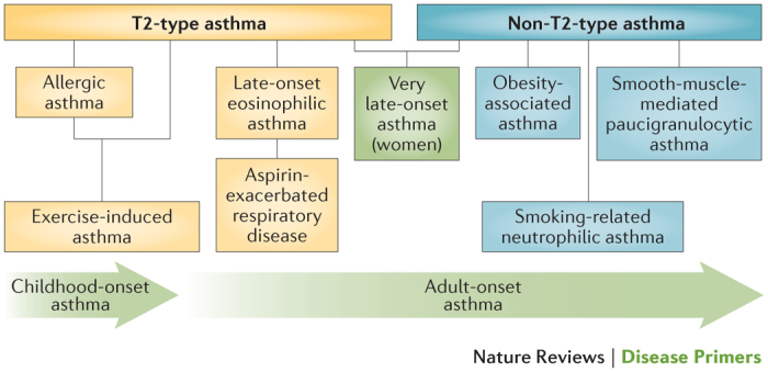
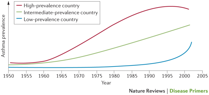
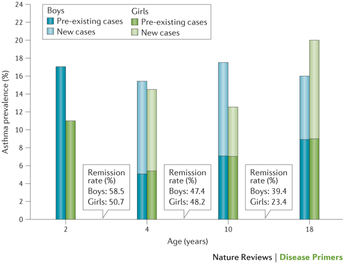
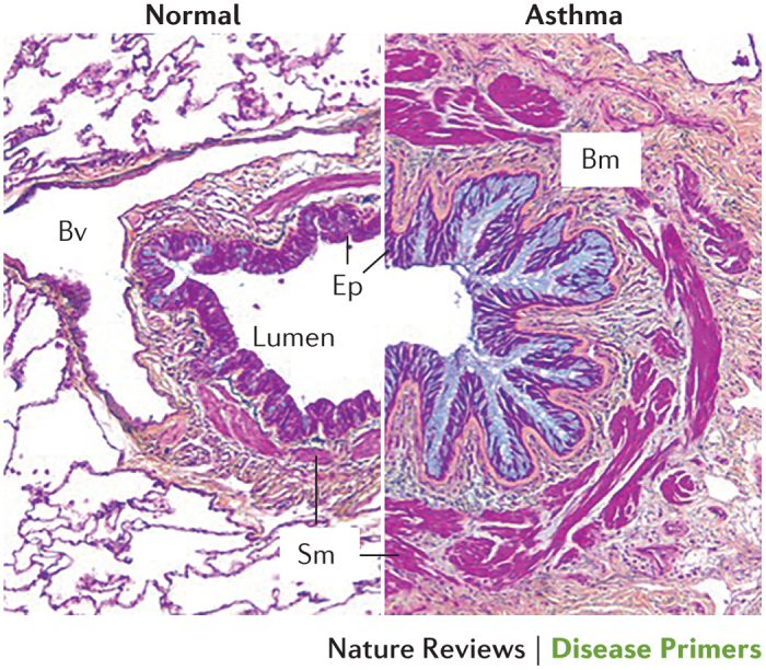
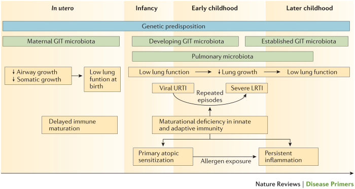
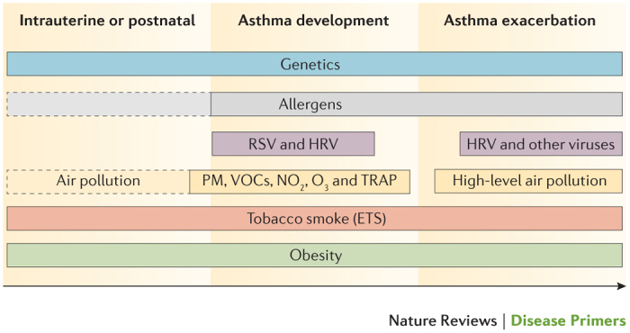
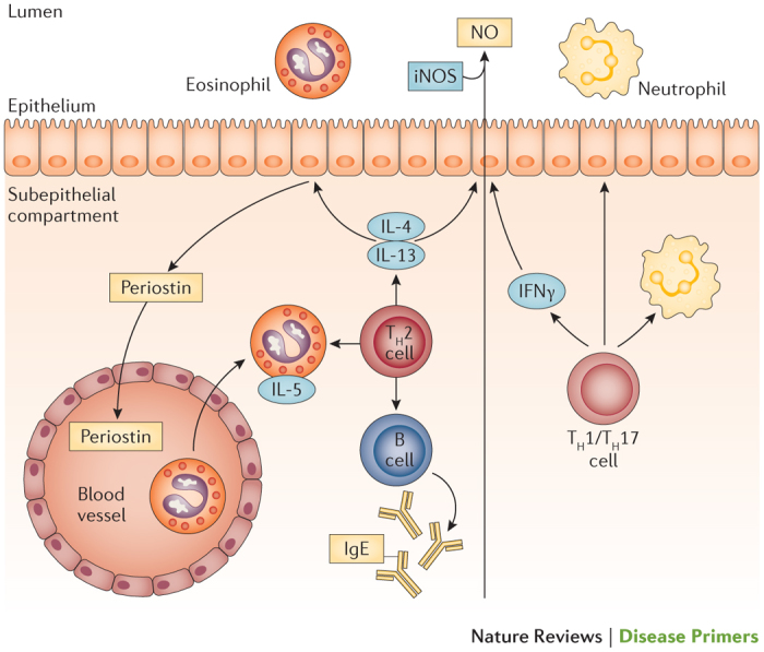
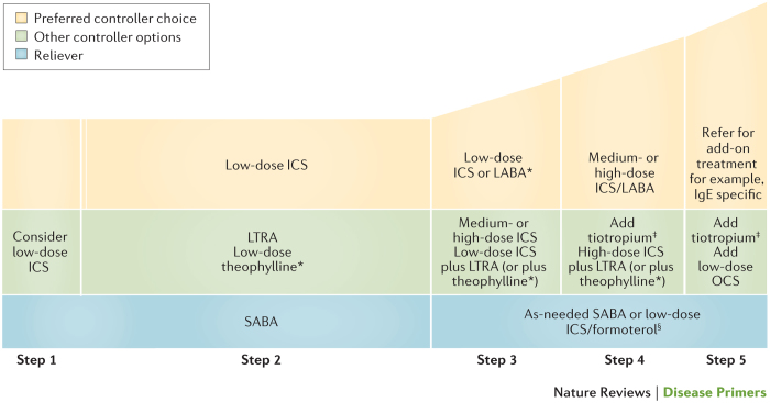
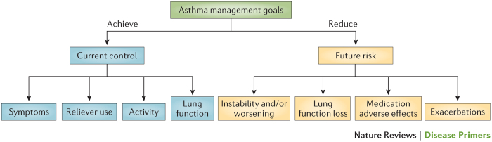
Asthma is the most common inflammatory disease of the lungs. The prevalence of asthma is increasing in many parts of the world that have adopted aspects of the Western lifestyle, and the disease poses a substantial global health and economic burden. Asthma involves both the large-conducting and the small-conducting airways, and is characterized by a combination of inflammation and structural remodelling that might begin in utero. Disease progression occurs in the context of a developmental background in which the postnatal acquisition of asthma is strongly linked with allergic sensitization. Most asthma cases follow a variable course, involving viral-induced wheezing and allergen sensitization, that is associated with various underlying mechanisms (or endotypes) that can differ between individuals. Each set of endotypes, in turn, produces specific asthma characteristics that evolve across the lifecourse of the patient. Strong genetic and environmental drivers of asthma interconnect through novel epigenetic mechanisms that operate prenatally and throughout childhood. Asthma can spontaneously remit or begin de novo in adulthood, and the factors that lead to the emergence and regression of asthma, irrespective of age, are poorly understood. Nonetheless, there is mounting evidence that supports a primary role for structural changes in the airways with asthma acquisition, on which altered innate immune mechanisms and microbiota interactions are superimposed. On the basis of the identification of new causative pathways, the subphenotyping of asthma across the lifecourse of patients is paving the way for more-personalized and precise pathway-specific approaches for the prevention and treatment of asthma, creating the real possibility of total prevention and cure for this chronic inflammatory disease.


Conflict of interest statement

S.T.H. is a non-executive director and a consultant for Synairgen, and a consultant for AstraZeneca and Novartis. He is in receipt of a UK Medical Research Council (MRC) programme grant and receives salary support as an MRC clinical professor. S.W. has served as a consultant for Aerocrine, GlaxoSmithKline and AstraZeneca. She has received research funding (paid to her institution) from GlaxoSmithKline, AstraZeneca, Genentech and Sanofi-Aventis. She has also received research support from the NIH National Heart, Lung and Blood Institute (NHLBI) and the National Institute of Allergy and Infectious Diseases (NIAID), which provides her salary support. D.S.P. has received an unrestricted educational grant for research (paid to the University of Groningen) from AstraZeneca. Her travel to the European Respiratory Society (ERS) and/or the American Thoracic Society (ATS) meetings has been partially funded by AstraZeneca, Chiesi, GlaxoSmithKline and Takeda. Fees for consultancies were given to the University of Groningen by AstraZeneca, Boehringer Ingelheim, Chiesi, GlaxoSmithKline, Takeda and Teva. Her travel and lectures in China have been funded by Chiesi. S.T.W. is funded by the NIH NHLBI and is a consultant for the TENOR study for Novartis. H.R. has received research support from the German Research Foundation (DFG), the Federal Ministry of Education and Research (BMBF), the European Union, Land Hessen, the German Academic Exchange Service (DAAD), ALK, Stiftung Pathobiochemie, Ernst-Wendt-Stiftung, Mead Johnson Nutritional and Beckman Coulter. He has also received speakers honoraria from Allergopharma, Novartis, Thermo Fisher Scientific, Danone, Mead Johnson Nutritional and Bencard Allergie. H.R. also serves as a consultant for Bencard Allergie and Sterna Biologicals (for which he is also a cofounder). P.D.S. declares no competing interests.

Figures

Figure 1
Figure 1. Selected asthma subphenotypes.
New subphenotypes and associated causal pathways, or endotypes, of asthma are being discovered through the application of non-hierarchical statistical analyses of clinical, physiological and laboratory characteristics. Figure from Ref. , Nature Publishing Group. PowerPoint slide
Figure 2
Figure 2. Changing trends in the prevalence of asthma according to gross domestic product.
The prevalence of asthma has plateaued in recent decades in high-prevalence countries, which have a high gross domestic product (GDP). Conversely, there has been a steep increase in the prevalence of asthma in low-prevalence and intermediate-prevalence countries, which have low-ranking and middle-ranking GDPs, respectively. These steep increases have occurred alongside acquisition aspects of the western lifestyle in these countries. Reprinted from Bulletin of World Health Organisation, 83, Bousquet, J., Bousquet, P. J., Godard, P. & Dyres, J.-P., The public health implications of asthma, 548–554, Copyright (2005). PowerPoint slide
Figure 3
Figure 3. Proportions of children and adolescents with asthma in the Isle of Wight birth cohort.
Bars represent the proportion of either boys or girls at particular ages who have asthma, with light-shaded sections representing the proportion of new cases that did not carry over from the previous age group (positive transition); the boxes between bars indicate the percentage of children who grew out of asthma in the intervening years (negative transition or remission). Data from Ref. . PowerPoint slide
Figure 4
Figure 4. Histopathology of the asthmatic airway.
Cross section of a severe asthmatic airway (right) compared with a normal airway (left). Asthma involves mucosal inflammation that most frequently consists of activated eosinophils, mast cells and T lymphocytes within the context of a remodelled airway with mucous metaplasia, an increase in smooth muscle (Sm), fibrosis and angiogenesis. Bm, basement membrane; Bv, blood vessel; Ep, epithelium. Republished with permission of Dove Medical Press, from Clinical update on the use of biomarkers of airway inflammation in the management of asthma. Wadsworth, S., Sin, D. & Dorscheid, D., 4, 2011; permission conveyed through Copyright Clearance Center, Inc. PowerPoint slide
Figure 5
Figure 5. Involvement of various T cell subtypes in asthma pathogenesis and asthma endotypes.
Dendritic cells (DCs) and the thymic epithelium together trigger the immune response that drives the development of asthma. In response to a combination of signals transmitted by cytokines and direct contact, the DCs and thymic epithelium promote the differentiation of an array of different leukocyte subsets (inner, yellow circle). Before differentiation, these subsets either augment or protect the airways from inflammatory responses linked to asthma, whereas following differentiation, they generate cytokines that promote the development of asthma. These cytokines influence various different cell types and the attendant inflammatory responses to drive allergic airway inflammation and airway hyper-responsiveness. GM-CSF, granulocyte–macrophage colony-stimulating factor; ILC2, group 2 innate lymphoid cell; iTReg, inhibitory regulatory T; NKT, natural killer T; TGFβ, transforming growth factor-β TH, T helper; TNF, tumour necrosis factor. Figure from Ref. , Nature Publishing Group. PowerPoint slide
Figure 6
Figure 6. Allergen sensitization of the airways during the induction of allergic-type asthma.
Infection (bacterial and viral) and pollutants perturb the airway epithelium, which leads to initial danger signalling and activation of innate signalling receptors. This signalling causes airway epithelial cells (ECs) to secrete chemokines and leads to trafficking of immature dendritic cells (DCs) to the mucosal epithelium. These DCs respond to danger signals through pattern recognition receptors (PRRs), which leads to their maturation into competent antigen-presenting myeloid-type DCs. Allergen detection and processing by these activated DCs is mediated by the extension of cellular processes into the airways or by the capture of allergens that have breached the epithelium. Allergen-loaded DCs then drive T cell differentiation by migrating to local lymph nodes where they interact with naive T cells (TN) via the T cell receptor (TCR), major histocompatibility complex (MHC) class II and co-stimulatory molecules. DC activation and T helper 2 (TH2) cell maturation and migration into the mucosa are influenced by additional epithelial-derived cytokines and chemokines, including IL-25, IL-33, CC-chemokine ligand 17 (CCL17) and CCL22. CCR, CC-chemokine receptor; GM-CSF, granulocyte–macrophage colony-stimulating factor; mDC, mucosal DC; TNF, tumour necrosis factor; TSLP, thymic stromal lymphopoietin. Figure from Ref. , Nature Publishing Group. PowerPoint slide
Figure 7
Figure 7. Epithelial–mesenchymal trophic unit in asthma.
In chronic moderate-to-severe asthma, the behaviour of the epithelium resembles that in chronic wound scenarios: it is more susceptible to environmental and viral injury than usual and exhibits impaired repair. In addition, the epithelium no longer undergoes healing by ‘primary intention’ but instead undergoes ‘secondary intention’ — a process that involves the production of growth factors that drive remodelling responses in the underlying airway wall. Similarly, the damaged and stimulated epithelium generates growth factors that contribute to goblet cell metaplasia. The augmented communication between the epithelium and the underlying mesenchyme resembles the activation of the epithelium–mesenchymal trophic unit that drives airway morphogenesis in the developing fetal lung. The resulting cytokine milieu also provides a favourable environment for sustaining chronic inflammation. This research was originally published in Clin. Sci. (Lond.). Holgate, S. T., Arshad, H. S., Roberts, G. C., Howarth, P. H., Thurner, P. & Davies, D. E., A new look at the pathogenesis of asthma. Clin. Sci. (Lond.). 2009; 118: 439–450 © Portland Press. PowerPoint slide
Figure 8
Figure 8. Asthma as a developmental disease.
Asthma is caused by failure of the respiratory and immune systems to develop normally. This schematic represents asthma risk factors that operate at different stages of life. Asthma risk at birth is influence by genetic predispositions, impaired lung function and delayed immune maturation. Postnatal risk factors that increase asthma risk include reduced lung growth resulting in low lung function, the timing of acquisition of specific components of the pulmonary microbiota, repeated episodes of viral upper respiratory tract infections (URTIs) that spread to the lower airway and result in severe lower respiratory tract infections (LRTIs), maturational deficiencies in the innate and adaptive immune systems that increase the risk of severe LRTIs and favour primary allergic sensitization and repeated allergen exposure, resulting in persistent airway inflammation. The maternal gastrointestinal tract (GIT) microbiota is thought to influence priming of the fetal immune system, and the postnatal development of the infant GIT microbiota is influenced by early-life exposures. Although each individual pathway increases the risk of asthma, the major risk is produced when a child progresses through multiple risk pathways simultaneously. PowerPoint slide
Figure 9
Figure 9. Contribution of risk factors to the development and/or exacerbation of asthma.
Solid outline indicates that the role of these risk factors has been firmly established. Dotted outline indicates that there are controversial and/or preliminary results for the contribution of these risk factors. ETS, environmental tobacco smoke; HRV, human rhinovirus; NO2, nitric dioxide; O3, ozone; PM, particulate matter; RSV, respiratory syncytial virus; TRAP, traffic-related air pollution; VOCs, volatile organic compounds. PowerPoint slide
Figure 10
Figure 10. Biomarkers for the assessment of T2-type asthma.
This molecular phenotype, or endotype, of asthma can be identified using biomarkers. These include already established biomarkers (blue boxes) and markers currently under clinical evaluation (yellow boxes). iNOS, inducible nitric oxide synthase; NO, nitric oxide; TH, T helper. PowerPoint slide
Figure 11
Figure 11. Global Initiative for Asthma-based steps for treatment of asthma at all stages of severity.
The Global Initiative for Asthma (GINA) recommends that asthma is classified and treated according to 5 steps that reflect symptom severity and patient characteristics. The relative widths of each step reflect the number of patients who receive each treatment. In particular, dose–response studies demonstrate that the majority of patients with asthma should be able to be treated with low-dose inhaled corticosteroids (ICSs; step 2). The relative heights of each step reflect disease severity, with treatment in step 5 reserved for those with the most severe disease. *For children 6–11 years of age, theophylline is not recommended and the preferred step 3 treatment with is medium-dose ICSs. Tiotropium by soft-mist inhaler is indicated as add-on treatment for patients with a history of exacerbations; it is not indicated in patients <18 years of age. §For patients prescribed inhaled beclomethasone dipropionate/formoterol or budesonid/formoterol maintenance and reliever therapy. LABA, long-acting β2-adrenergic receptor agonist; LTRA, leukotriene receptor antagonist; OCS, oral corticosteroid; SABA, short-acting β2-receptor agonist. Adapted from the Global Strategy for Asthma Management and Prevention 2015, © Global Initiative for Asthma (GINA) all rights reserved. Available from http://www.ginasthma.org. PowerPoint slide
Figure 12
Figure 12. Overall goals of asthma management.
Asthma management should aim to both control the current symptoms of asthma and reduce the risk of future adverse outcomes for patients. Adapted with permission from Ref. , Elsevier. PowerPoint slide

References

    1. Global Initiative for Asthma. Global strategy for asthma management and prevention GINA[online], (2015).
    1. Lange P, Parner J, Vestbo J, Schnohr P, Jensen G. A 15-year follow-up study of ventilatory function in adults with asthma. N. Engl. J. Med. 1998;339:1194–1200. doi: 10.1056/NEJM199810223391703. - DOI - PubMed
    1. Porsbjerg C, Lange P, Ulrik CS. Lung function impairment increases with age of diagnosis in adult onset asthma. Respir. Med. 2015;109:821–827. doi: 10.1016/j.rmed.2015.04.012. - DOI - PubMed
    1. Custovic A. To what extent is allergen exposure a risk factor for the development of allergic disease? Clin. Exp. Allergy. 2015;45:54–62. doi: 10.1111/cea.12450. - DOI - PubMed
    1. Amelink M. Severe adult-onset asthma: a distinct phenotype. J. Allergy Clin. Immunol. 2013;132:336–341. doi: 10.1016/j.jaci.2013.04.052. - DOI - PubMed