Review
. 2015 Nov 19:1:15071.
doi: 10.1038/nrdp.2015.71.

22q11.2 deletion syndrome

Affiliations
Review

22q11.2 deletion syndrome

Donna M McDonald-McGinn et al. Nat Rev Dis Primers. .

Abstract

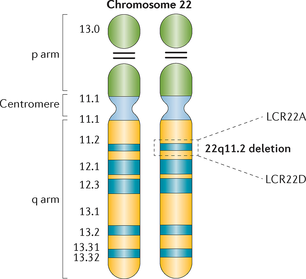
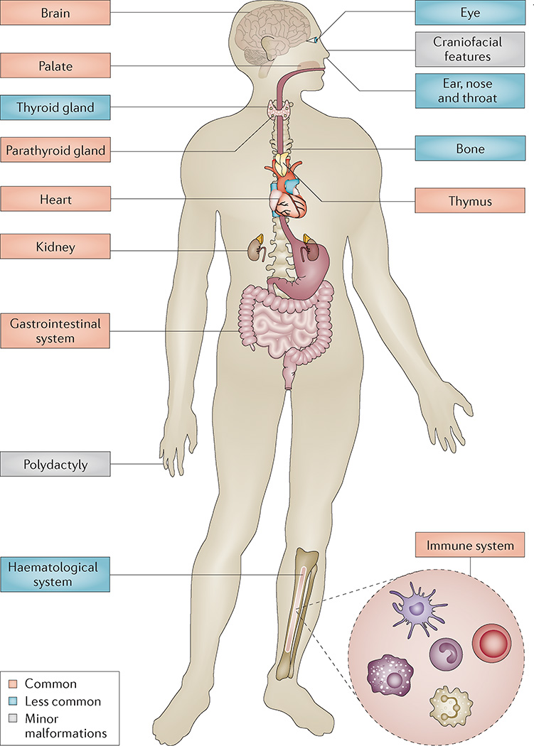
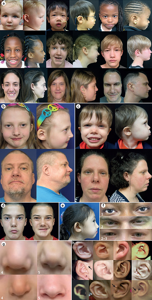
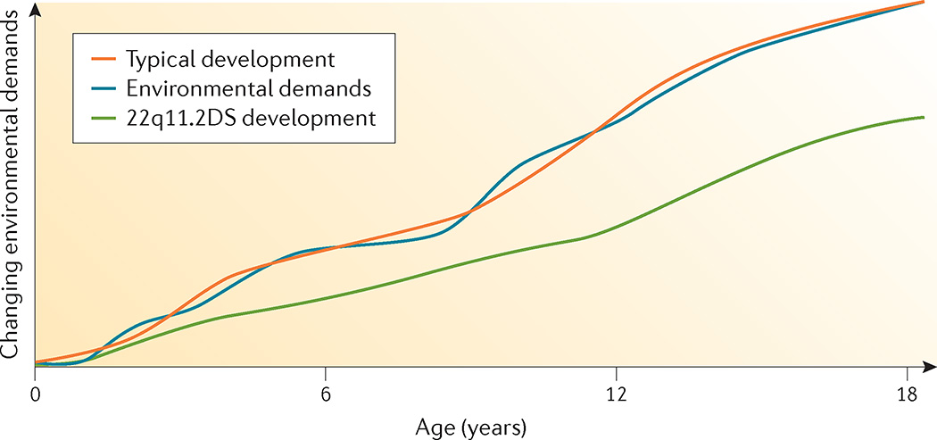
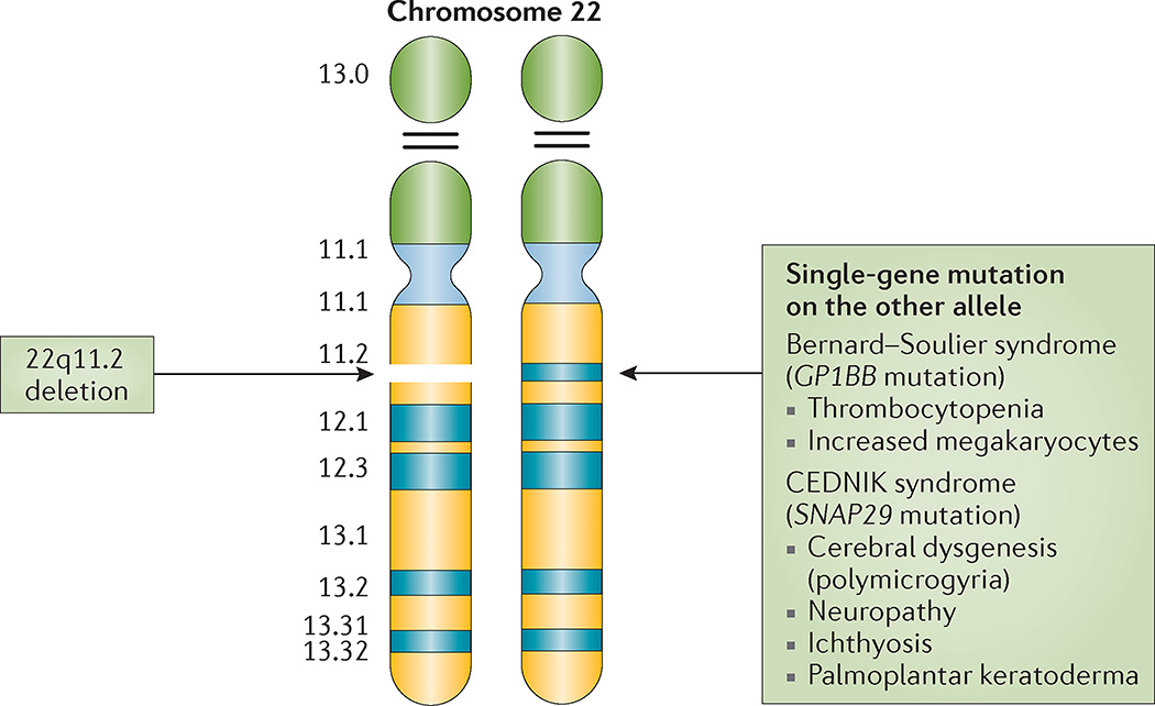
22q11.2 deletion syndrome (22q11.2DS) is the most common chromosomal microdeletion disorder, estimated to result mainly from de novo non-homologous meiotic recombination events occurring in approximately 1 in every 1,000 fetuses. The first description in the English language of the constellation of findings now known to be due to this chromosomal difference was made in the 1960s in children with DiGeorge syndrome, who presented with the clinical triad of immunodeficiency, hypoparathyroidism and congenital heart disease. The syndrome is now known to have a heterogeneous presentation that includes multiple additional congenital anomalies and later-onset conditions, such as palatal, gastrointestinal and renal abnormalities, autoimmune disease, variable cognitive delays, behavioural phenotypes and psychiatric illness - all far extending the original description of DiGeorge syndrome. Management requires a multidisciplinary approach involving paediatrics, general medicine, surgery, psychiatry, psychology, interventional therapies (physical, occupational, speech, language and behavioural) and genetic counselling. Although common, lack of recognition of the condition and/or lack of familiarity with genetic testing methods, together with the wide variability of clinical presentation, delays diagnosis. Early diagnosis, preferably prenatally or neonatally, could improve outcomes, thus stressing the importance of universal screening. Equally important, 22q11.2DS has become a model for understanding rare and frequent congenital anomalies, medical conditions, psychiatric and developmental disorders, and may provide a platform to better understand these disorders while affording opportunities for translational strategies across the lifespan for both patients with 22q11.2DS and those with these associated features in the general population.


Figures

Figure 1
Figure 1. Chromosome 22 idiogram
Cytogenetic representation of chromosome 22 showing the short (p) and long (q) arms along with the centromere, which functions to separate both arms. Chromosome 22 is an acrocentric chromosome, as indicated by the two horizontal lines in the p arm. The 22q11.2 deletion occurs on the long arm of one of the two chromosomes, depicted by dashed lines in the 22q11.2 band. The position of the two low copy repeats (LCRs) on 22q11.2 (LCR22A and LCR22D), which flank the typical 3-Mb deletion, are indicated.
Figure 2
Figure 2. Low copy repeats and genes within the 22q11.2 deletion
Schematic representation of the 3-Mb 22q11.2 region that is commonly deleted in 22q11.2 deletion syndrome, including the four low copy repeats (LCR22s) that span this region (LCR22A, LCR22B, LCR22C and LCR22D). Common commercial probes for fluorescence in situ hybridization (FISH) are indicated (N25 and TUPLE). The protein-coding and selected non-coding (*) genes are indicated with respect to their relative position along chromosome 22 (Chr22). T-box 1 (TBX1; green box) is highlighted as the most widely studied gene within the 22q11.2 region. Mutations in this gene have resulted in conotruncal cardiac anomalies in animal models and humans. Known human disease-causing genes that map to the region are indicated in grey boxes. These include proline dehydrogenase 1 (PRODH; associated with type I hyperprolinaemia), solute carrier family 25 member 1 (SLC25A1; encoding the tricarboxylate transport protein and is associated with combined D-2- and L-2-hydroxyglutaric aciduria), platelet glycoprotein Ib β-polypeptide (GP1BB; associated with Bernard–Soulier syndrome), scavenger receptor class F member 2 (SCARF2; associated with Van den Ende–Gupta syndrome), synaptosomal-associated protein 29 kDa (SNAP29; associated with cerebral dysgenesis, neuropathy, ichthyosis and palmoplantar keratoderma (CEDNIK) syndrome), and leucine-zipper-like transcription regulator 1 (LZTR1; associated with schwannomatosis 2). Further details on the location of non-coding RNAs and pseudogenes in the 22q11.2 region may be found in Guna et al.. Common 22q11.2 deletions are shown, with the typical 3-Mb deletion flanked by LCR22A and LCR22D (LCR22A– LCR22D) on top and the nested deletions, with their respective deletion sizes indicated below. Each of the deletions portrayed is flanked by a particular LCR22. Those rare deletions not mediated by LCRs are not shown. AIF3M, apoptosis-inducing factor mitochondrion-associated 3; ARVCF, armadillo repeat gene deleted in velocardiofacial syndrome; CDC45, cell division cycle 45; Cen, centromere; CLDN5, claudin 5; CLTCL1, clathrin heavy chain-like 1; COMT, catechol-O-methyltransferase; CRKL, v-crk avian sarcoma virus CT10 oncogene homologue-like; DGCR, DiGeorge syndrome critical region; GNB1L, guanine nucleotide-binding protein (G protein), β-polypeptide 1-like; GSC2, goosecoid homeobox 2; HIC2, hypermethylated in cancer 2; HIRA, histone cell cycle regulator; KLHL22, kelch-like family member 22; LINC00896, long intergenic non-protein-coding RNA 896; LOC101927859, serine/arginine repetitive matrix protein 2-like; CCDC188, coiled-coil domain-containing 188; LRRC74B, leucine-rich repeat-containing 74B; MED15, mediator complex subunit 15; mir, microRNA; MRPL40, mitochondrial ribosomal protein L40; P2RX6, purinergic receptor P2X ligand-gated ion channel 6; PI4KA, phosphatidylinositol 4-kinase catalytic-α; RANBP1, Ran-binding protein 1; RTN4R, reticulon 4 receptor; SEPT7, septin 7; SERPIND1, serpin peptidase inhibitor clade D (heparin co-factor) member 1; TANGO2, transport and golgi organization 2 homologue; THAP7, THAP domain-containing 7; TRMT2A, tRNA methyltransferase 2 homologue A; TSSK2, testis-specific serine kinase 2; TXNRD2, thioredoxin reductase 2; UFD1L, ubiquitin fusion degradation 1-like; USP41, ubiquitin-specific peptidase 41; ZDHHC8, zinc-finger DHHC-type-containing 8; ZNF74, zinc-finger protein 74.
Figure 3
Figure 3. 22q11.2 non-allelic homologous recombination
Diagram of two different types of meiotic non-allelic homologous recombination events that can occur between low copy repeats on chromosome 22 (LCR22s). Rearrangements between LCR22A and LCR22D are indicated (A and D) on each allele (blue versus yellow). Interchromosomal events (left) occur between paralogous LCR22s (A and D) in two different alleles owing to >99% sequence identity of direct repeats (‘X’ shows the crossover of the two chromosomes). The hybrid LCR22 is shown as half yellow and half blue. This process results in a duplication or deletion of intervening genes in resulting gametes. Intrachromosomal recombination events (right) result from crossing over (indicated by ‘X’) within one allele, resulting in a deletion (left) or a ring chromosome (right); the ring chromosome is not viable.
Figure 4
Figure 4. Development of the cardiovascular and pharyngeal structures affected in 22q11.2 deletion syndrome
a | The schematic ventral view of an embryonic day 7.5 (E7.5) mouse embryo shows the relationship of the cardiac crescent to the head folds and also depicts the distinct cellular fields termed the first heart field and the second heart field (SHF). Within the second heart field, the anterior segment (aSHF) contributes to the outflow tract (OFT) and the right ventricle (RV) of the heart, whereas the posterior segment (pSHF) contributes to the inflow of the heart. b | Cardiac neural crest cells (NCCs) delaminate from the hindbrain and migrate ventrolaterally to populate the pharyngeal arches. The T-box transcription factor TBX1 is required within the pharyngeal surface ectoderm to regulate as yet unknown signalling pathways, which pattern the cardiac NCCs (dashed arrow). The pharyngeal endoderm and cardiac NCCs interact in the formation of the thymus and parathyroid glands. c | Lateral view of an E10.5 mouse embryo. The cardiac neural crest arises from the neural tube at the level between the otic placode and somite three, and migrates ventrolaterally to populate the pharyngeal arches, interacting with the core mesoderm and ultimately contributing the smooth muscle cells to the remodelling arch arteries. The caudal stream enters the OFT. d | Schematic presentation of the cell lineage that contributes to the OFT of the heart at approximately E10.5. (1) Cells derived from the aSHF enter the OFT where they contribute to the myocardium and endocardium. (2) These cells interact with cardiac NCCs that migrate in from the pharyngeal arch region. Signals to the cardiac neural crest are also received from the pharyngeal epithelium. Disruption to these cellular contributions or interactions can result in a common arterial trunk, alignment defects or ventricular septation defects.
Figure 5
Figure 5. Organ and system involvement in 22q11.2 deletion syndrome
22q11.2 deletion syndrome leads to significant morbidity (and some premature mortality), with frequent multi-organ system involvement, such as cardiac and palatal abnormalities, immune differences, endocrine and gastrointestinal problems, and later-onset conditions across the lifespan including variable cognitive deficits and psychiatric illness that is attributable to functional brain changes. Less-frequent manifestations, when present, contribute to substantial morbidity (examples include: idiopathic seizures; polymicrogyria; sclerocornea; coloboma; deafness; choanal atresia; laryngeal cleft or web; tracheo-oesophageal fistula; hypothyroidism or hyperthyroidism; juvenile rheumatoid arthritis; idiopathic thrombocytopenia; autoimmune haemolytic anaemia; craniosynostosis; scoliosis; intestinal malrotation; Hirschsprung disease; and imperforate anus). Minor malformations generally confer little indisposition but may enhance ascertainment. These generally include: mild dysmorphic craniofacial features, such as hooded eyelids, auricular anomalies, nasal differences including a dimple or crease, and asymmetric crying facies; and, cervical and thoracic vertebral anomalies or butterfly vertebrae, arachnodactyly, camptodactyly, 2–3 toe syndactyly and polydactyly (preaxial and postaxial of the hands and postaxial of the feet).
Figure 6
Figure 6. Craniofacial features associated with 22q11.2 deletion syndrome
Patients with 22q11.2 deletion syndrome (22q11.2DS), shown here from infancy through to adulthood, demonstrate variability of associated craniofacial features — most with few recognizable dysmorphia (part a). A person with 22q11.2DS has a 50% recurrence risk with each pregnancy for this microdeletion syndrome, but some adults only come to attention following the diagnosis in a child with associated features, as in these unrelated nuclear families (daughter and father (part b) and son and mother (part c)). When viewed individually, some craniofacial features provide important clues to the diagnosis, for example, microstomia and asymmetric crying facies (part d), and malar flatness and micrognathia (part e). External eye findings (part f) may include upslanting palpebral fissures and hypertelorism (1), hooded eyelids and/or ptosis (2) and mild epicanthal folds (3). Nasal features (part g) may include a bulbous nasal tip with hypoplastic alae nasi (4) often with a nasal dimple or crease with or without a faint haemangioma (5). Auricular differences (part h) frequently include thick overfolded, squared-off and crumpled helices, microtic, cupped or posteriorly rotated ears, attached lobes and preauricular pits or tags (arrows).
Figure 7
Figure 7. Developmental trajectory
As the child with 22q11.2 deletion syndrome (22q11.2DS) ages, the discrepancy between developmental level (based on chronological age) and environmental demands widens owing to associated neurocognitive and behavioural developmental deficits. Note that IQ decline observed in 22q11.2DS may not only be due to a relative but also to an absolute decline in cognitive abilities.
Figure 8
Figure 8. Associated autosomal recessive conditions on 22q11.2
A deletion on 22q11.2 in combination with a mutation in a single gene on the other allele can unmask an autosomal recessive condition, for example, Bernard–Souilier syndrome (platelet glycoprotein Ib β-polypeptide (GP1BB)) and cerebral dysgenesis, neuropathy, ichthyosis and palmoplantar keratoderma (CEDNIK) syndrome (synaptosomal-associated protein 29 kDa (SNAP29)).

References

    1. DiGeorge A. Discussion on a new concept of the cellular immunology. J. Pediatr. 1965;67:907–908.
    1. Takao A, Ando M, Cho K, Kinouchi A, Murakami Y. In: Etiology and Morphogenesis of Congenital Heart Disease. Van Praagh R, Takao A, editors. Futura Pub. Co.; 1980. pp. 253–269.
    1. Digilio MC, Marino B, Formigari R, Giannotti A. Maternal diabetes causing DiGeorge anomaly and renal agenesis. Am. J. Med. Genet. 1995;55:513–514. - PubMed
    1. Sulik KK, Johnston MC, Daft PA, Russell WE, Dehart DB. Fetal alcohol syndrome and DiGeorge anomaly: critical ethanol exposure periods for craniofacial malformations as illustrated in an animal model. Am. J. Med. Genet. Suppl. 1986;2:97–112. - PubMed
    1. Coberly S, Lammer E, Alashari M. Retinoic acid embryopathy: case report and review of literature. Pediatr. Pathol. Lab. Med. 1996;16:823–836. - PubMed

Publication types