Review
. 2017 Jan;65(1):5-18.
doi: 10.1002/glia.23006. Epub 2016 May 18.

Microglia-Neuron Communication in Epilepsy

Affiliations
Review

Microglia-Neuron Communication in Epilepsy

Ukpong B Eyo et al. Glia. 2017 Jan.

Abstract

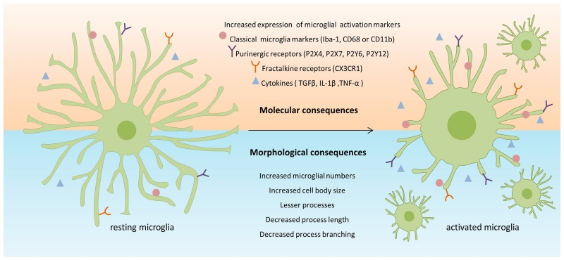
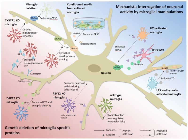
Epilepsy has remained a significant social concern and financial burden globally. Current therapeutic strategies are based primarily on neurocentric mechanisms that have not proven successful in at least a third of patients, raising the need for novel alternative and complementary approaches. Recent evidence implicates glial cells and neuroinflammation in the pathogenesis of epilepsy with the promise of targeting these cells to complement existing strategies. Specifically, microglial involvement, as a major inflammatory cell in the epileptic brain, has been poorly studied. In this review, we highlight microglial reaction to experimental seizures, discuss microglial control of neuronal activities, and propose the functions of microglia during acute epileptic phenotypes, delayed neurodegeneration, and aberrant neurogenesis. Future research that would help fill in the current gaps in our knowledge includes epilepsy-induced alterations in basic microglial functions, neuro-microglial interactions during chronic epilepsy, and microglial contribution to developmental seizures. Studying the role of microglia in epilepsy could inform therapies to better alleviate the disease. GLIA 2016;65:5-18.

Keywords: epilepsy; kainic acid; microglia; pilocarpine; seizures.


Figures

Figure 1
Figure 1. Schematic diagram depicting molecular and morphological changes of microglial activation following seizures
The molecular consequences of seizures on microglial activation (above) include changes in the expression pattern of an array of microglial molecules such as classical microglial markers, purinergic receptors, fractalkine receptor and cytokines. The morphological consequences of seizures on microglial activation (below) include changes in microglial cell body size, process length, process numbers and complexity of branching. Please refer to “[2] Microglial Morphological and Molecular Activation in Response to Seizures” for references.
Figure 2
Figure 2. Microglial influence on neuronal activity
This schematic summarizes the current literature on the effects of microglia on neuronal activity. The studies were broadly classified into two categories based on the experimental approach: (1) genetic deletion of microglial-specific proteins (lower left, pink) and (2) mechanistic interrogation of neuronal activity by microglial manipulations (upper right, blue). Arrowheads indicate an enhancing effect, while rounded head indicate an inhibitory effect. Proven pathways and proposed pathways are represented by solid and dashed lines, respectively. Please refer to “[3] Microglial Regulation of Neuronal Activities in Epilepsy” for references.
Figure 3
Figure 3. Microglia at different stages after seizures
This figure highlights three keynote studies that investigated microglial activation at different time points following seizures. In the acute phase (1-3 hours), microglial P2Y12 receptor-mediated process extension attenuated seizure outcome, playing a neuroprotective role. In the sub-acute phase (48-72 hours), fractalkine signaling is one signaling axis that has been identified that mediates microglial activation resulting in neuronal degeneration. Finally, in the chronic phase (several weeks), microglia was shown to be capable of recognizing DNA from degenerating neurons via TLR9 and TLR9 signaling prevented aberrant neurogenesis following seizures. Please refer to “[4] Microglial Function in Acute Seizures, Neurodegeneration, and Neurogenesis in Epilepsy” for references.

References

    1. Abraham J, Fox PD, Condello C, Bartolini A, Koh S. Minocycline attenuates microglia activation and blocks the long-term epileptogenic effects of early-life seizures. Neurobiol Dis. 2012;46:425–30. - PMC - PubMed
    1. Ahlers KE, Karacay B, Fuller L, Bonthius DJ, Dailey ME. Transient activation of microglia following acute alcohol exposure in developing mouse neocortex is primarily driven by BAX-dependent neurodegeneration. Glia. 2015;63:1694–713. - PMC - PubMed
    1. Ajami B, Bennett JL, Krieger C, McNagny KM, Rossi FM. Infiltrating monocytes trigger EAE progression, but do not contribute to the resident microglia pool. Nat Neurosci. 2011;14:1142–9. - PubMed
    1. Ajami B, Bennett JL, Krieger C, Tetzlaff W, Rossi FM. Local self-renewal can sustain CNS microglia maintenance and function throughout adult life. Nat Neurosci. 2007;10:1538–43. - PubMed
    1. Akahoshi N, Murashima YL, Himi T, Ishizaki Y, Ishii I. Increased expression of the lysosomal protease cathepsin S in hippocampal microglia following kainate-induced seizures. Neurosci Lett. 2007;429:136–41. - PubMed

Publication types