. 2016 Jun 24;291(26):13689-98.
doi: 10.1074/jbc.M116.726034. Epub 2016 May 4.

Type II Turn of Receptor-bound Salmon Calcitonin Revealed by X-ray Crystallography

Affiliations

Type II Turn of Receptor-bound Salmon Calcitonin Revealed by X-ray Crystallography

Eva Johansson et al. J Biol Chem. .

Abstract

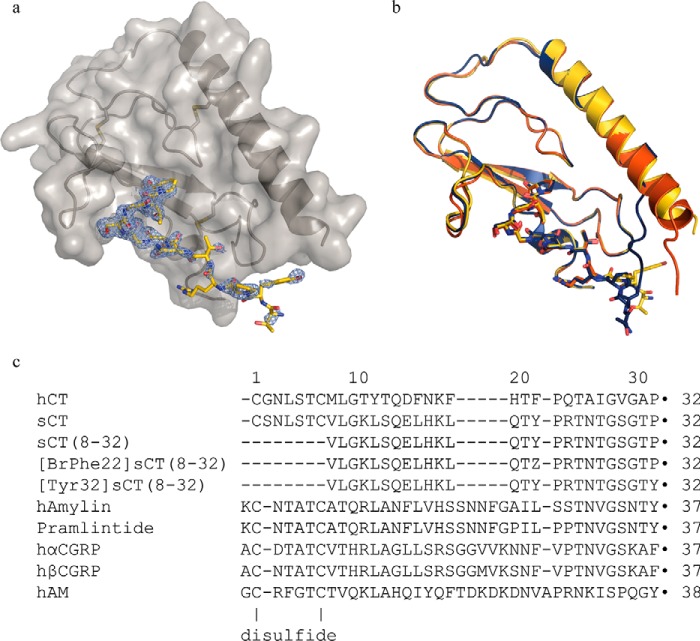
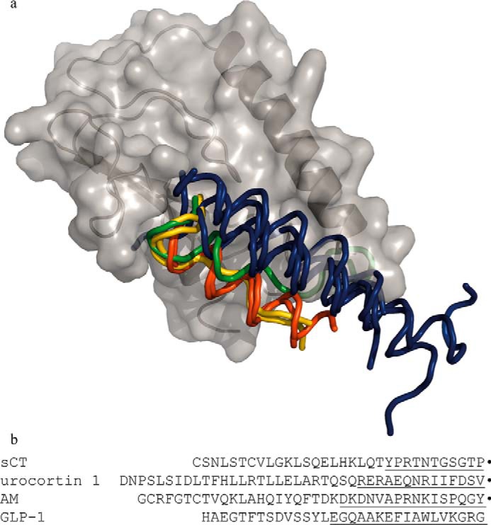
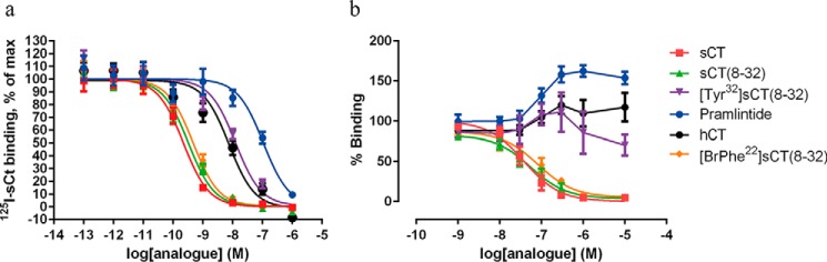
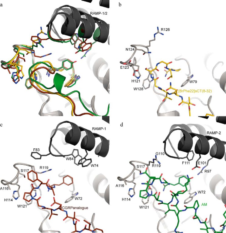
Calcitonin is a peptide hormone consisting of 32 amino acid residues and the calcitonin receptor is a Class B G protein-coupled receptor (GPCR). The crystal structure of the human calcitonin receptor ectodomain (CTR ECD) in complex with a truncated analogue of salmon calcitonin ([BrPhe(22)]sCT(8-32)) has been determined to 2.1-Å resolution. Parallel analysis of a series of peptide ligands showed that the rank order of binding of the CTR ECD is identical to the rank order of binding of the full-length CTR, confirming the structural integrity and relevance of the isolated CTR ECD. The structure of the CTR ECD is similar to other Class B GPCRs and the ligand binding site is similar to the binding site of the homologous receptors for the calcitonin gene-related peptide (CGRP) and adrenomedulin (AM) recently published (Booe, J. M., Walker, C. S., Barwell, J., Kuteyi, G., Simms, J., Jamaluddin, M. A., Warner, M. L., Bill, R. M., Harris, P. W., Brimble, M. A., Poyner, D. R., Hay, D. L., and Pioszak, A. A. (2015) Mol. Cell 58, 1040-1052). Interestingly the receptor-bound structure of the ligand [BrPhe(22)]sCT(8-32) differs from the receptor-bound structure of the homologous ligands CGRP and AM. They all adopt an extended conformation followed by a C-terminal β turn, however, [BrPhe(22)]sCT(8-32) adopts a type II turn (Gly(28)-Thr(31)), whereas CGRP and AM adopt type I turns. Our results suggest that a type II turn is the preferred conformation of calcitonin, whereas a type I turn is the preferred conformation of peptides that require RAMPs; CGRP, AM, and amylin. In addition the structure provides a detailed molecular explanation and hypothesis regarding ligand binding properties of CTR and the amylin receptors.

Keywords: G protein-coupled receptor (GPCR); amylin; calcitonin; crystal structure; peptide conformation; peptide hormone; peptide interaction.


Figures

FIGURE 1.
FIGURE 1.
Schematic of CTR ECD in complex with [BrPhe22]sCT(8–32) and peptide ligand sequences. a, CTR ECD in complex with [BrPhe22]sCT(8–32), chains A (gray schematic and surface representation) and D (yellow stick representation) displayed with PHENIX 2FoFc composite omit map (light blue) at σ = 1 level for chain D. b, the three complexes in the asymmetric unit superimposed (chains A and D in yellow, chains B and E in red, and chains C and F in dark blue) illustrating the overall similarity and the amino-terminal differences. c, sequence alignment using the MUSCLE algoritm of the CT peptides used in this work and other members of the calcitonin family of peptides. All peptides contain a carboxyl-terminal amide (marked by ●) and [BrPhe22]sCT(8–32) furthermore, contains a 4-bromo-l-phenylalanine residue, which is marked as Z. In full-length calcitonin the first and the 7th residues are forming a disulfide and in the amylins, CGRPs, and AM, the 2nd and 7th residues are forming a disulfide.
FIGURE 2.
FIGURE 2.
Structural alignment of GPCR Class B ECDs illustrating the variety of peptide ligand fold and binding. a, schematic displaying CTR ECD (gray) and a ribbon representation of the [BrPhe22]sCT(8–32) ligand (yellow). The ECDs of the following structures have been superposed on the CTR ECD and the peptide ligands are displayed as ribbons and peptides binding in a similar mode are colored in the same color. The other Class B GPCR ECDs used for structural alignment are: CLR in complex with AM (green) (PDB 4RWF) and a variant of CGRP (yellow) (PDB 4RWG, RAMP1 and -2 have been left out) (16), glucose-dependent insulinotropic polypeptide receptor in complex with glucose-dependent insulinotropic polypeptide (dark blue) (PDB 2QKH) (7), parathyroid hormone receptor in complex with parathyroid hormone-related protein (dark blue) (PDB 3C4M) (8), parathyroid hormone receptor in complex with parathyroid hormone (dark blue) (PDB 3H3G) (35), glucagon-like peptide-1 receptor in complex with glucagon-like peptide-1 (dark blue) (PDB 3IOL) (36), corticotropin-releasing factor receptor 2α in complex with urocortin 1 (red) (PDB 3N96) (32), and corticotropin releasing factor receptor 1 in complex with corticotropin-releasing factor (red) (PDB 3EHU) (5). The codes within parentheses are the Protein Data Bank accession numbers of the displayed coordinates. b, full-length sequences of the peptide ligands of one representative from each binding mode where the part within 4 Å from their respective ECD are underlined. Carboxyl-terminal amide residues are marked by ●.
FIGURE 3.
FIGURE 3.
Interactions between CTR ECD and [BrPhe22]sCT(8–32). a, schematic of CTR ECD (gray) with the side chains of amino acid residues within 4 Å from [BrPhe22]sCT(8–32) (yellow) in stick representation. Hydrogen bonds are marked with dotted lines. b, closeup on the β turn and the carboxyl-terminal prolineamide of [BrPhe22]sCT(8–32). The hydrogen bonds between the carboxyl-terminal amide and peptide backbone of residue Ser129 from CTR ECD are marked with black dotted lines, the hydrogen bond between the Thr31 hydroxyl group and the carboxyl-terminal amide is marked with a red dotted line and the internal β turn hydrogen bond is marked with a cyan dotted line. Residues are labeled with their respective residue number, the ones belonging to [BrPhe22]sCT(8–32) are in italics.
FIGURE 4.
FIGURE 4.
125I-sCT whole cell competition binding and CTR ECD binding. a, normalized competition binding curves from all three experiments using 25 pm 125I-sCT as the tracer ligand and increasing amounts of unlabeled CT analogue. Experiments were conducted on intact BHK cells stably transfected with the human CTR. For each experiment each analogue curve was normalized in GraphPad Prism by defining the fitted bottom value as 0% and top value as 100%. b, binding to the CTR ECD was assessed in a bead-based proximity assay (AlphaScreen®) with His6-CTR ECD binding to nickel chelate acceptor beads and biotinylated sCT binding to the streptavidin donor beads. The binding of compounds to CTR ECD is assessed indirectly by the ability to displace the binding between CTR ECD and biotinylated sCT. The experiments were repeated 3 times and the experimental details can be found under ”Experimental Procedures.“ Data are depicted as mean ± S.E. The summarized data of all experiments are reported in Table 2.
FIGURE 5.
FIGURE 5.
Hypothetical models of CTR-bound carboxyl-terminal part of hCT and CTR ECD-bound antagonistic peptide sCT(8–32). a, the sCT residues Arg24, Asn26, Thr27, Ser29, and Thr31 of the CTR ECD bound [BrPhe22]sCT(8–32) (chain D in this work) have been replaced by the corresponding hCT residues (Gln24, Ala26, Ile27, Val29, and Ala31) and these side chains are shown in green stick representation together with the CTR ECD·[BrPhe22]sCT(8–32) structure in gray schematic and surface for the ECD and yellow for the stick representation of the peptide. Hydrogen bonds likely to be conserved compared with Fig. 3b are shown as dotted lines. b, hybrid model of CTR ECD-bound antagonistic peptide sCT(8–32) (yellow schematic) based on residues 8–23 from a sCT NMR structure (PDB entry 2GLH) and residues 24–32 from [BrPhe22]sCT(8–32) (chain D in this work). Residues implied in receptor interaction by cross-linking studies (Val8, Leu16, Leu19, and Asn26) are shown with side chains represented in red sticks. CTR ECD (Chain A) is shown as a gray schematic with a transparent surface and residues Met49 and Thr138 are shown as blue sticks marking sites of interactions on the CTR as determined by cross-linking studies.
FIGURE 6.
FIGURE 6.
Comparison of the carboxyl-terminal parts of [BrPhe22]sCT(8–32) (yellow), the CGRP analogue (brown; PDB entry 4RWF), and AM (green; PDB entry 4RWG) binding to their respective receptor ECDs. a, peptide ligands are displayed as colored schematics with the carboxyl-terminal amide residues in stick representation. Selected side chains from the receptor ECD are displayed in stick representation in the color belonging to the respective peptide and the ECD schematics are displayed in gray with darker gray for the RAMP-1 and RAMP-2 ECDs in PDB 4RWF and 4RWG complexes, respectively. In b–d one complex at the time is shown and here the colors of the peptide sticks follow their respective receptor ECD.

References

    1. Copp D. H., Cameron E. C., Cheney B. A., Davidson A. G. F., and Henze K. G. (1962) Evidence for calcitonin: a new hormone from the parathyroid that lowers blood calcium. Endocrinology 70, 638–649 - PubMed
    1. Poyner D. R., Sexton P. M., Marshall I., Smith D. M., Quirion R., Born W., Muff R., Fischer J. A., and Foord S. M. (2002) International Union of Pharmacology: XXXII, the mammalian calcitonin gene-related peptides, adrenomedullin, amylin, and calcitonin receptors. Pharmacol. Rev. 54, 233–246 - PubMed
    1. Findlay D. M., and Sexton P. M. (2004) Calcitonin. Growth Factors 22, 217–224 - PubMed
    1. Hoare S. R. (2005) Mechanisms of peptide and nonpeptide ligand binding to Class B G-protein-coupled receptors. Drug Discov. Today 10, 417–427 - PubMed
    1. Pioszak A. A., Parker N. R., Suino-Powell K., and Xu H. E. (2008) Molecular recognition of corticotropin-releasing factor by its G-protein-coupled receptor CRFR1. J. Biol. Chem. 283, 32900–32912 - PMC - PubMed

LinkOut - more resources