. 2016 May 22;8(5):1459-66.
doi: 10.1093/gbe/evw093.

C-Terminal Region of Sulfite Reductase Is Important to Localize to Chloroplast Nucleoids in Land Plants

Affiliations

C-Terminal Region of Sulfite Reductase Is Important to Localize to Chloroplast Nucleoids in Land Plants

Yusuke Kobayashi et al. Genome Biol Evol. .

Abstract

Chloroplast (cp) DNA is compacted into cpDNA-protein complexes, called cp nucleoids. An abundant and extensively studied component of cp nucleoids is the bifunctional protein sulfite reductase (SiR). The preconceived role of SiR as the core cp nucleoid protein, however, is becoming less likely because of the recent findings that SiRs do not associate with cp nucleoids in some plant species, such as Zea mays and Arabidopsis thaliana To address this discrepancy, we have performed a detailed phylogenetic analysis of SiRs, which shows that cp nucleoid-type SiRs share conserved C-terminally encoded peptides (CEPs). The CEPs are likely to form a bacterial ribbon-helix-helix DNA-binding motif, implying a potential role in attaching SiRs onto cp nucleoids. A proof-of-concept experiment was conducted by fusing the nonnucleoid-type SiR from A. thaliana (AtSiR) with the CEP from the cp nucleoid-type SiR of Phaseolus vulgaris The addition of the CEP drastically altered the intra-cp localization of AtSiR to cp nucleoids. Our analysis supports the possible functions of CEPs in determining the localization of SiRs to cp nucleoids and illuminates a possible evolutionary scenario for SiR as a cp nucleoid protein.

Keywords: evolution; nucleoid; sulfite reductase.


Figures

F<sc>ig</sc>. 1.—
Fig. 1.—
Multiple sequence alignment of SiRs in land plants. GmSiR, Glycine max SiR; PsSiR, Pisum sativum SiR; PvSiR, Phaseolus vulgaris SiR; NtSiR, Nicotiana tabacum SiR; AtSiR, Arabidopsis thaliana SiR; ZmSiR, Zea mays SiR.
F<sc>ig</sc>. 2.—
Fig. 2.—
Homology model of the CEP in PsSiR. (A) C-terminal region of PsSiR. The blue arrow and green bar indicate the regions predicted to form Ribbon and Helix structures, respectively. (B) The homology model of the CEP in PsSiR was constructed using Swiss Model with an E. coli Transcriptional Repressor COPG homolog (Protein Data Bank: 1B01) as the template. CEP was predicted to form a bacterial Ribbon–Helix–Helix DNA-binding motif. The arrow indicates the N-terminus of the CEP. (C) Homology model of the CEP in PsSiR. The template was the E. coli Transcriptional Repressor COPG-homodimer/DNA complex (PDB: 1B01). Green indicates the PsCEP. The arrow indicates the N-terminus of the CEP. Red indicates basic amino acids.
F<sc>ig</sc>. 3.—
Fig. 3.—
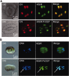
Localization of AtSiR and AtSiR-CEP. (A) Confocal microscopy of guard cells of A. thaliana expressing AtSiR-YFP or AtSiR-PvCEP-YFP under the control of the 35S promoter. Differential interference contrast microscopy shows the guard cells. Dot-lines trace the outline of the guard cells. Chl indicates the autofluorescence emitted by the chlorophyll. (B) Epifluorescence microscopy of cps isolated from A. thaliana expressing AtSiR-YFP or AtSiR-PvCEP-YFP under the control of the 35S promoter. Cps were stained with the DNA-specific fluorochrome DAPI. Arrows indicate a cp nucleoid.
F<sc>ig</sc>. 4.—
Fig. 4.—
Phylogenetic points at which CEP was lost. A phylogenetic tree of SiRs based on Bayesian inference, maximum likelihood and maximum parsimony methods. Posterior probabilities for Bayesian inference (≥0.90) and Bootstrap values (≥50%) for the maximum likelihood and maximum parsimony, respectively, are indicated at the appropriate nodes. Blue indicates SiRs containing CEPs. Green indicates SiRs not containing CEPs.
F<sc>ig</sc>. 5.—
Fig. 5.—
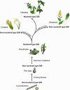
Schematic model representing the evolutionary history of the nucleoid-type SiR. Branch lengths do not represent phylogenetic distances.

References

    1. Allen JF. 2003. The function of genomes in bioenergetic organelles. Philos Trans R Soc Lond B Biol Sci. 358:19–37. - PMC - PubMed
    1. Biasini M, et al. 2014. SWISS-MODEL: modelling protein tertiary and quaternary structure using evolutionary information. Nucleic Acids Res. 42:W252–W258. - PMC - PubMed
    1. Bogorad L. 2008. Evolution of early eukaryotic cells: genomes, proteomes, and compartments. Photosynth Res. 95:11–21. - PubMed
    1. Bohrer A-S, et al. 2015. Alternative translational initiation of ATP sulfurylase underlying dual localization of sulfate assimilation pathways in plastids and cytosol in Arabidopsis thaliana. Front Plant Sci. 5:750. - PMC - PubMed
    1. Bowman JL, Floyd SK, Sakakibara K. 2007. Green genes-comparative genomics of the green branch of life. Cell 129:229–234. - PubMed

MeSH terms

Associated data