. 2016 May 15;143(10):1663-73.
doi: 10.1242/dev.136358.

The transcriptional repressor Blimp1 is expressed in rare luminal progenitors and is essential for mammary gland development

Affiliations

The transcriptional repressor Blimp1 is expressed in rare luminal progenitors and is essential for mammary gland development

Mohammed I Ahmed et al. Development. .

Abstract

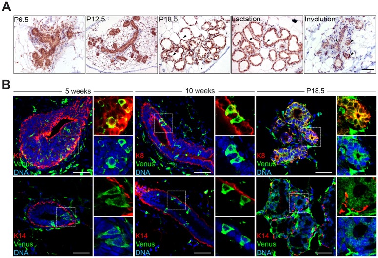
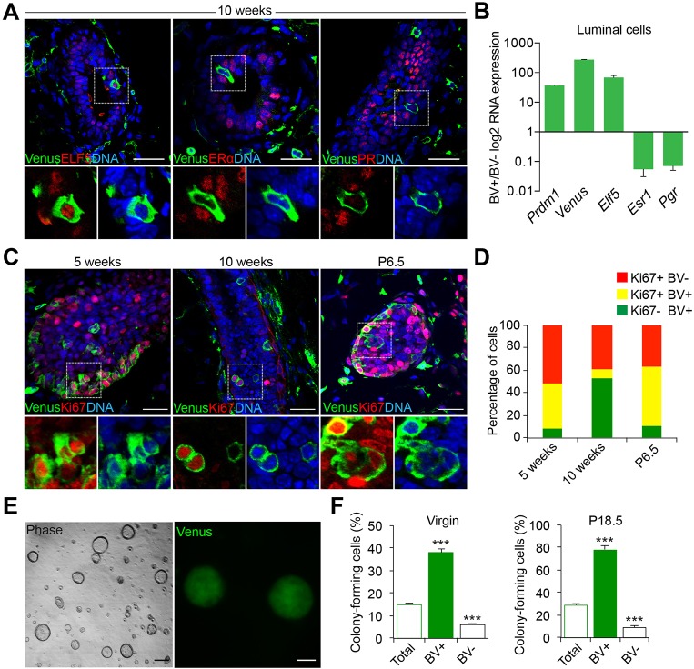
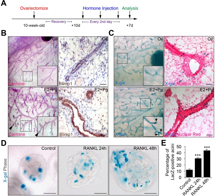
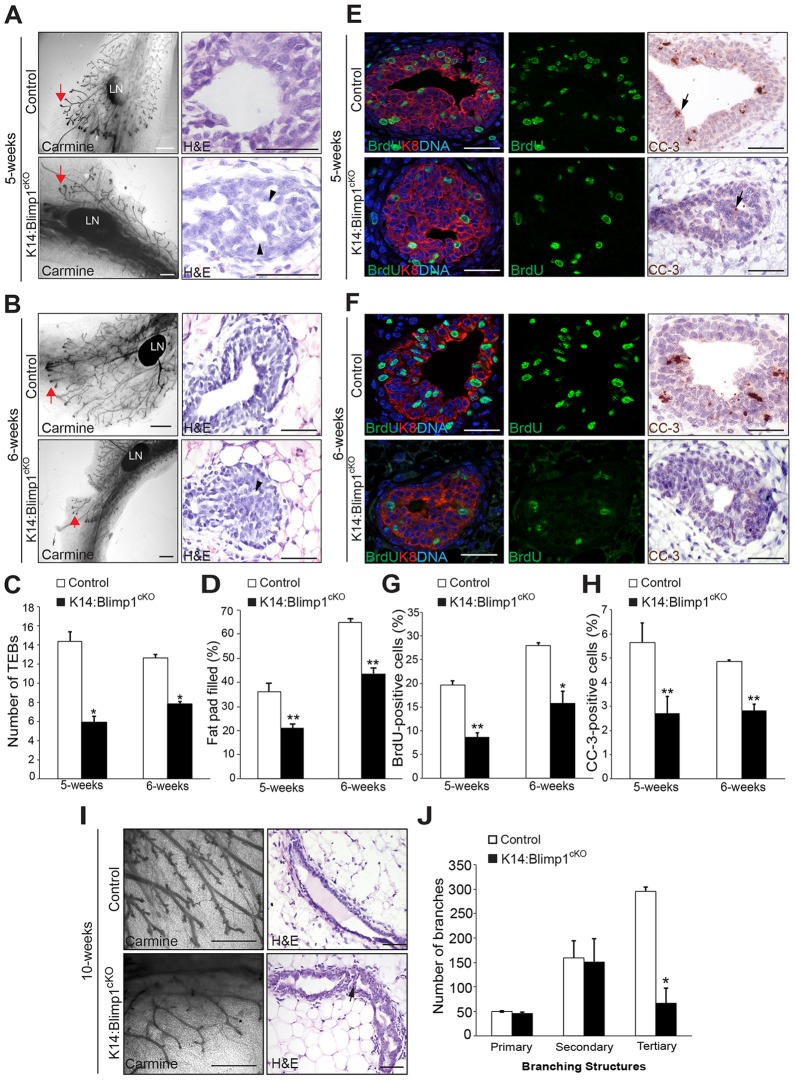
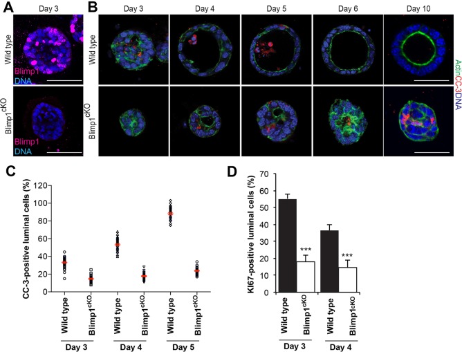
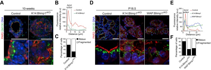
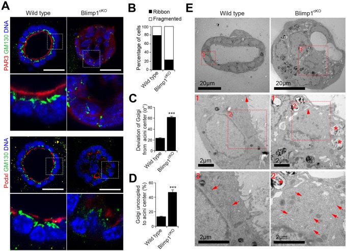
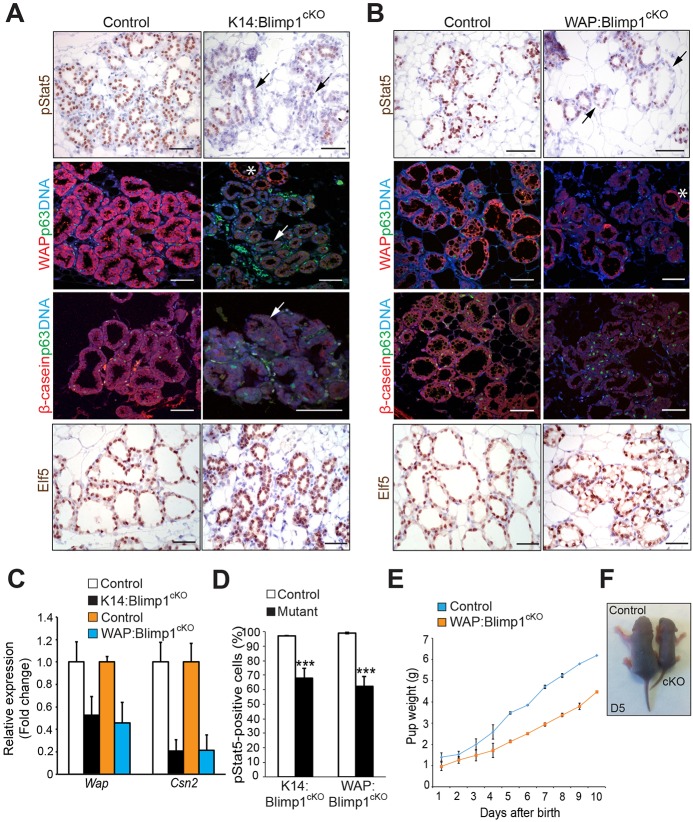
Mammary gland morphogenesis depends on a tight balance between cell proliferation, differentiation and apoptosis, to create a defined functional hierarchy within the epithelia. The limited availability of stem cell/progenitor markers has made it challenging to decipher lineage relationships. Here, we identify a rare subset of luminal progenitors that express the zinc finger transcriptional repressor Blimp1, and demonstrate that this subset of highly clonogenic luminal progenitors is required for mammary gland development. Conditional inactivation experiments using K14-Cre and WAPi-Cre deleter strains revealed essential functions at multiple developmental stages. Thus, Blimp1 regulates proliferation, apoptosis and alveolar cell maturation during puberty and pregnancy. Loss of Blimp1 disrupts epithelial architecture and lumen formation both in vivo and in three-dimensional (3D) primary cell cultures. Collectively, these results demonstrate that Blimp1 is required to maintain a highly proliferative luminal subset necessary for mammary gland development and homeostasis.

Keywords: 3D culture; Blimp1; Lumen formation; Luminal progenitors; Mammary gland morphogenesis; Mouse; Polarity; Prdm1; Transcriptional repressor.


Conflict of interest statement

Competing interests

The authors declare no competing or financial interests.

Figures

Fig. 1.
Fig. 1.
Blimp1 expression is restricted to the luminal cell compartment of the developing mammary gland. (A) Blimp1 immunohistochemical staining of mammary gland tissue during pregnancy (P6.5, P12.5 and P18.5) and at day 1 of lactation and day 5 of involution. During pregnancy, lactation and involution, Blimp1+ cells are confined to the luminal cell compartment of alveolar structures (arrowheads). (B) Cryosections from 5- and 10-week-old virgins and P18.5 Blimp1-Venus (BV) mammary glands stained for GFP (green) and Krt8 (K8; red) or Krt14 (K14; red). Blimp1-expressing cells (green) are Krt8+ and Krt14. High-magnification images of the boxed areas are shown to the right of each image. Scale bars: 50 μm.
Fig. 2.
Fig. 2.
Blimp1 expression marks highly clonogenic luminal progenitors. (A) Cryosections from 10-week-old virgin BV mammary glands stained for GFP and Elf5, ERα or PR. Blimp1-expressing cells (green) are Elf5+ and ERα/PR. (B) FACS-sorted BV+ luminal cells isolated from 12-week-old virgin BV mice were analysed by qRT-PCR for Elf5, Esr1 and Pgr expression. The values were normalized to Hprt expression. Data are presented as mean±s.e.m. from n=3 independent experiments. (C) Cryosections from 5- and 10-week-old virgin (TEBs and ductal structures) and P6.5 (alveoli) BV mice stained for GFP (green) and Ki67 (red). High-magnification images of the boxed areas in A,C are shown below each image. (D) Histogram showing the percentages of Ki67+BV (red), Ki67+BV+ (yellow) and Ki67BV+ (green) at 5 and 10 weeks of age and at P6.5. BV+ cells are highly proliferative in 5-week TEBs and P6.5 alveoli, but display reduced proliferation in 10-week ducts. Data are presented as mean±s.e.m. from n=3 independent experiments. (E) Representative images of colonies formed by BV+ luminal cells isolated from 12-week-old virgin mice (n=3 independent experiments). (F) Colony-forming efficiency of BV+, BV and unsorted luminal cells (total) from 12-week-old virgin and P18.5 mice. BV+ luminal cells form significantly higher numbers of colonies compared with BV and unsorted luminal cells. Data are presented as mean±s.e.m. from n=3 independent experiments. ***P<0.001. Scale bars: 50 μm.
Fig. 3.
Fig. 3.
Expansion of Blimp1+ luminal cells in response to hormone treatment. (A) Wild-type and Prdm1Cre.IRES.nLacZ 10-week-old virgin mice were ovariectomized, allowed to recover for 10 days (d) and injected over 6 days with a cocktail of 17β-oestradiol and progesterone (E2+Pg) or mineral oil as a control. Mammary glands were recovered for analysis 7 days later. (B) Whole-mount Carmine staining and Blimp1 immunostaining reveal mammary gland expansion and enhanced Blimp1 expression in response to E2+Pg treatment. Arrowheads indicate the formation of alveolar buds (images representative from two independent experiments; n=5 mice per treatment). (C) X-gal-stained whole-mount and tissue sections of control (oil) or E2+Pg-treated Prdm1Cre.IRES.nLacZ mammary glands. Sections were counterstained with Nuclear Fast Red. Arrows indicate increased numbers of Blimp1-lacZ+ cells in the alveolar buds (images representative from two independent experiments; n=5 mice per treatment). Insets in B,C show high-magnification images of the boxed areas. (D) RANKL treatment results in significantly increased numbers of Blimp1-lacZ+ cells. Day 6 3D MECs from Prdm1Cre.IRES.nLacZ P15.5-P16.5 females were treated with RANKL (24 h or 48 h) prior to staining. (E) Percentage of Blimp1-lacZ+ acini in control versus RANKL-treated cultures (control: n=30 acini; RANKL 24 h: n=30 acini; RANKL 48 h: n=30). Error bars represent s.e.m. ***P<0.001. Scale bars: 50 μm.
Fig. 4.
Fig. 4.
Blimp1 conditional inactivation results in defective mammary gland morphogenesis. (A,B) Whole-mount Carmine-stained and histological sections of mammary glands from 5- and 6-week-old virgin K14:Blimp1cKO or littermate controls (representative images from n=3 mice of each genotype). Red arrows indicate TEBs. Black arrowheads indicate defective lumen formation within the TEBs. (C,D) Histograms showing the number of TEBs (C), and the percentage of the fat pad filled by the invading mammary epithelial front (D) past the mid-point of the lymph node, assessed by whole-mount staining. Histograms are presented as mean±s.e.m. from n=3 mice of each genotype. *P<0.05; **P<0.01. (E,F) Decreased proliferation and apoptosis. TEBs from 5- and 6-week-old Blimp1-deficient virgins stained for BrdU (green) and Krt8 (K8; red), or cleaved caspase-3 (CC-3) show markedly reduced numbers of BrdU+ and CC-3+ cells (arrows) (representative images from n=3 mice of each genotype). (G,H) Significantly reduced percentages of BrdU+ (G) and CC-3+ (H) cells within TEBs from 5- and 6-week-old virgin Blimp1 mutants. Histograms are presented as mean±s.e.m. from n=3 mice of each genotype. (I) Whole-mount Carmine-stained and histological sections of mammary glands from 10-week-old virgin K14:Blimp1cKO or littermate controls (representative images from n=3 mice of each genotype). Arrow indicates defective lumen formation within ducts. (J) Histograms showing the number of primary, secondary and tertiary branches within 10-week-old mammary glands from K14:Blimp1cKO and control littermates. *P<0.05; **P<0.01. LN, lymph node. Scale bars: 1 mm (whole mount); 50 μm (sections).
Fig. 5.
Fig. 5.
Loss of Blimp1 delays lumen formation in MEC 3D cultures. (A) Immunostaining of day 3 wild-type and Prdm1BEH/CA;ROSA26:CreERT2 (Blimp1cKO) MEC 3D acini for Blimp1. Treatment with 100 nM 4OHT (to induce Cre) results in elimination of Blimp1 in Blimp1cKO acini (representative images from n=3 independent experiments). (B) Immunostaining at day 3, 4, 5, 6 and 10 of wild-type and Blimp1cKO MEC 3D acini for CC-3+ (red) and actin (green) (representative images from three independent experiments). Loss of Blimp1 results in altered apoptosis-mediated clearing of intraluminal cells. (C) Percentage of CC-3+ luminal cells. Data are presented as mean±s.e.m. values from day 3 (wild type: n=30 acini; Blimp1cKO: n=31 acini); day 4 (wild type: n=33 acini; Blimp1cKO: n=30 acini); and day 5 (wild type: n=32 acini; Blimp1cKO: n=32 acini) 3D cultures. (D) Percentage of Ki67+ luminal cells was significantly reduced in Blimp1cKO MECs at day 3 and 4 compared with wild type. Data are presented as mean±s.e.m. values from day 3 (wild type: n=30 acini; Blimp1cKO: n=30 acini) and day 4 (wild type: n=30 acini; Blimp1cKO: n=30 acini) 3D culture. Error bars represent s.e.m. ***P<0.001. Scale bars: 50 μm.
Fig. 6.
Fig. 6.
Blimp1 inactivation causes epithelial cell polarity defects during puberty, pregnancy and lactation. (A) Representative images of 10-week-old mammary glands from K14:Blimp1cKO and control littermates stained for GM130 (green) and Par3 (red) (n=3 mice of each genotype). (B) Representative line-scan analysis (relative fluorescence intensity; minimum of 20 cells were analysed per genotype), reveals a failure of Par3 apical accumulation in Blimp1-deficient ductal cells. (C) Percentages of ductal cells showing ribbon-like and fragmented localization of GM130 Golgi in control and Blimp1-mutant mammary glands. (D) Representative images of mammary glands (P18.5) from K14:Blimp1cKO, WAP:Blimp1cKO and control littermates stained for GM130 (green) and Par3 (red) (n=3 mice of each genotype). High-magnification images of the boxed areas in A,D are shown below. (E) Representative line-scan analysis (relative fluorescence intensity; minimum of 20 cells were analysed per genotype), reveals lack of Par3 apical accumulation in Blimp1-deficient alveolar cells. (F) Percentages of alveolar cells showing ribbon-like and fragmented localization of GM130 Golgi in control and Blimp1-mutant mammary glands. Scale bars: 50 μm.
Fig. 7.
Fig. 7.
Loss of Blimp1 alters lumen formation and maturation in MEC 3D cultures. (A) Wild-type and Prdm1CA/CA:ROSA26:CreERT2 (Blimp1cKO) day 6 4OHT-treated MEC 3D acini stained for GM130 (green) and Par3 (red; top panel) or podocalyxin (Podxl, red; bottom panel). MECs were treated with 4OHT for 24 h following plating. Scale bars: 50 μm. (B) Percentage of acini with ribbon-like and fragmented localization of GM130 Golgi in wild-type and Blimp1cKO day 6 4OHT-treated MEC 3D acini (wild type: n=30 acini; Blimp1cKO: n=30 acini). (C) Deviation of the Golgi from acini centre (α°) (wild type: n=30 acini; Blimp1cKO: n=30 acini). (D) Percentage of acini with Golgi uncoupled to centre (wild type: n=30 acini; Blimp1cKO: n=30 acini). Data are presented as mean±s.e.m. ***P<0.001. (E) Representative electron micrographs of wild-type and Blimp1cKO day 6 4OHT-treated MEC 3D acini revealing alterations in the establishment of an apical-basal polarity in mutant acini compared with wild type, accompanied by dismantled tight junctions (1′ vs 1; arrowheads), fragmentation of the Golgi apparatus (2′ vs 2; arrows) and loss of apical villi.
Fig. 8.
Fig. 8.
Blimp1 function is essential for alveolar maturation during pregnancy and lactation. (A,B) Representative images of mammary glands (P18.5) stained for pStat5, p63 (green), Wap (red) or β-casein (red), and Elf5 (representative images from n=3 mice of each genotype); black arrows indicate a dramatic reduction of pStat5+ cells and white arrows indicate markedly reduced expression of milk proteins Wap and β-casein in mutant alveoli. Asterisks indicate areas showing normal expression levels of Wap. The numbers of p63+ basal cells are similar in control and mutant tissues. (C) qRT-PCR analysis demonstrates markedly reduced expression of Csn2 (β-casein) and Wap transcripts in total mammary gland mRNA samples from Blimp1 mutants. The values were normalized to Krt8 expression. Data are presented as mean values from n=2 mice of each genotype. (D) Quantification of pStat5+ cells from Blimp1 mutants and littermate controls. Data are presented as mean±s.e.m. values from n=3 mice of each genotype. ***P<0.001. (E) Pups nursed by lactating WAP:Blimp1cKO mutant females display growth retardation. Pups were weighed daily for 10 days. Body weights of pups are shown as mean±s.e.m. per litter per day, n=2 lactating females nursing identical litter sizes (P=0.0002). A total of 15 pups for each genotype were analysed. (F) Representative photographs of day 5 pups nursed by control and WAP:Blimp1cKO females. Scale bars: 50 μm.

Similar articles

Cited by

References

    1. Akhtar N. and Streuli C. H. (2013). An integrin-ILK-microtubule network orients cell polarity and lumen formation in glandular epithelium. Nat. Cell Biol. 15, 17-27. 10.1038/ncb2646 - DOI - PMC - PubMed
    1. Arulanandam R., Batenchuk C., Angarita F. A., Ottolino-Perry K., Cousineau S., Mottashed A., Burgess E., Falls T. J., De Silva N., Tsang J. et al. (2015). VEGF-mediated induction of PRD1-BF1/Blimp1 expression sensitizes tumor vasculature to oncolytic virus infection. Cancer Cell 28, 210-224. 10.1016/j.ccell.2015.06.009 - DOI - PubMed
    1. Asselin-Labat M.-L., Vaillant F., Sheridan J. M., Pal B., Wu D., Simpson E. R., Yasuda H., Smyth G. K., Martin T. J., Lindeman G. J. et al. (2010). Control of mammary stem cell function by steroid hormone signalling. Nature 465, 798-802. 10.1038/nature09027 - DOI - PubMed
    1. Bernardo G. M., Lozada K. L., Miedler J. D., Harburg G., Hewitt S. C., Mosley J. D., Godwin A. K., Korach K. S., Visvader J. E., Kaestner K. H. et al. (2010). FOXA1 is an essential determinant of ERalpha expression and mammary ductal morphogenesis. Development 137, 2045-2054. 10.1242/dev.043299 - DOI - PMC - PubMed
    1. Bikoff E. K., Morgan M. A. and Robertson E. J. (2009). An expanding job description for Blimp-1/PRDM1. Curr. Opin. Genet. Dev. 19, 379-385. 10.1016/j.gde.2009.05.005 - DOI - PubMed

Publication types

MeSH terms