

Editorial
doi: 10.1093/cvr/cvw098.
Epub 2016 May 17.
Myosin light chain phosphorylation, novel targets to repair a broken heart?
Affiliations
- PMID: 27190055
- PMCID: PMC4909165
- DOI: 10.1093/cvr/cvw098
Item in Clipboard
Editorial
Myosin light chain phosphorylation, novel targets to repair a broken heart?
Cardiovasc Res.
.
No abstract available
Figures
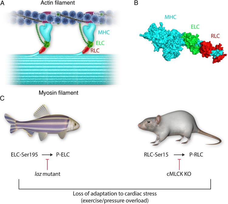
Cardiac myosin light chains in health and disease. The cardiac sarcomere contains myosin located in the thick filament and actin located in the thin filament (A). Cardiac muscle contraction is controlled by access of the myosin heads to actin binding sites on the thin filament. This process starts with the binding of calcium to troponin (positioned on the thin filament), which causes a structural repositioning of the coiled-coil tropomyosin molecule within the grooves of the double-stranded actin filament, thereby exposing actin-binding sites on the thin filament for the myosin heads to bind and develop contractile force. Moreover, it has been suggested that the N-terminus of the ELC may interact with thin-filament actin and functionally modulate thin-filament regulation and muscle contraction (A). Each myosin head is composed of the myosin heavy chain (MHC; cyan), one regulatory light chain (RLC; red), and one essential light chain (ELC; green) (B). Both myosin light chains can be phosphorylated at ELC-Ser195 and RLC-Ser-15 (C), and this process has been postulated to play a role in the normal function of the heart. Diminished phosphorylation of either light chain may negatively impact cardiac function, particularly under conditions of haemodynamic stress (C). Modulation of the light chain phosphorylation (RLC and/or ELC) may provide a pathway for the development of novel treatment strategies to combat heart failure, an entity of ever increasing clinical significance.
Comment on
-
Essential light chain S195 phosphorylation is required for cardiac adaptation under physical stress.Cardiovasc Res. 2016 Jul 1;111(1):44-55. doi: 10.1093/cvr/cvw066. Epub 2016 Mar 24. Cardiovasc Res. 2016. PMID: 27013636
-
Acute heart failure with cardiomyocyte atrophy induced in adult mice by ablation of cardiac myosin light chain kinase.Cardiovasc Res. 2016 Jul 1;111(1):34-43. doi: 10.1093/cvr/cvw069. Epub 2016 Mar 29. Cardiovasc Res. 2016. PMID: 27025239 Free PMC article.
References
-
- Geeves MA, Holmes KC. The molecular mechanism of muscle contraction. Adv Protein Chem 2005;71:161–193. - PubMed
-
- Rayment I, Rypniewski WR, Schmidt-Base K, Smith R, Tomchick DR, Benning MM et al. . Three-dimensional structure of myosin subfragment-1: a molecular motor. Science 1993;261:50–58. - PubMed
-
- Arrell DK, Neverova I, Fraser H, Marban E, Van Eyk JE. Proteomic analysis of pharmacologically preconditioned cardiomyocytes reveals novel phosphorylation of myosin light chain 1. Circ Res 2001;89:480–487. - PubMed
-
- Scheid LM, Mosqueira M, Hein S, Kossack M, Juergensen L, Mueller M et al. . Essential light chain S195 phosphorylation is required for cardiac adaptation under physical stress. Cardiovasc Res 2016;111:44–55. - PubMed
Publication types
MeSH terms
Substances
Grants and funding
LinkOut - more resources
Full Text Sources