. 2016 Jul 1;311(1):L74-86.
doi: 10.1152/ajplung.00429.2015. Epub 2016 May 17.

Early coagulation events induce acute lung injury in a rat model of blunt traumatic brain injury

Affiliations

Early coagulation events induce acute lung injury in a rat model of blunt traumatic brain injury

Hideki Yasui et al. Am J Physiol Lung Cell Mol Physiol. .

Abstract

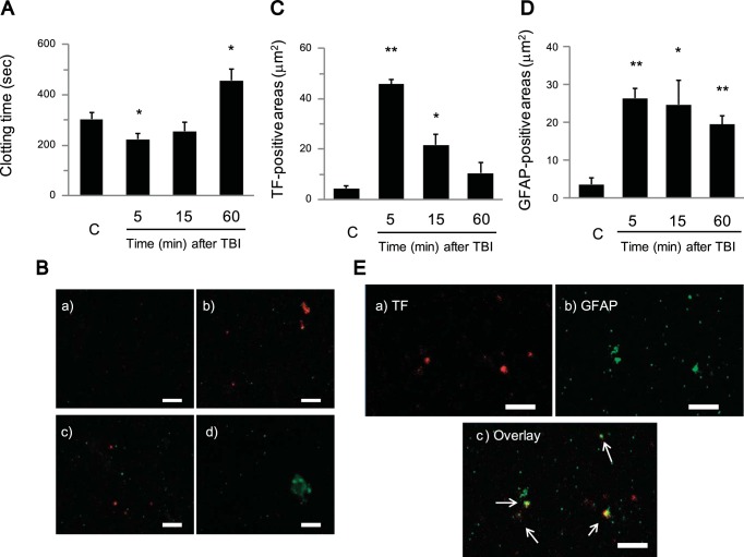
Acute lung injury (ALI) and systemic coagulopathy are serious complications of traumatic brain injury (TBI) that frequently lead to poor clinical outcomes. Although the release of tissue factor (TF), a potent initiator of the extrinsic pathway of coagulation, from the injured brain is thought to play a key role in coagulopathy after TBI, its function in ALI following TBI remains unclear. In this study, we investigated whether the systemic appearance of TF correlated with the ensuing coagulopathy that follows TBI in ALI using an anesthetized rat blunt trauma TBI model. Blood and lung samples were obtained after TBI. Compared with controls, pulmonary edema and increased pulmonary permeability were observed as early as 5 min after TBI without evidence of norepinephrine involvement. Systemic TF increased at 5 min and then diminished 60 min after TBI. Lung injury and alveolar hemorrhaging were also observed as early as 5 min after TBI. A biphasic elevation of TF was observed in the lungs after TBI, and TF-positive microparticles (MPs) were detected in the alveolar spaces. Fibrin(ogen) deposition was also observed in the lungs within 60 min after TBI. Additionally, preadministration of a direct thrombin inhibitor, Refludan, attenuated lung injuries, thus implicating thrombin as a direct participant in ALI after TBI. The results from this study demonstrated that enhanced systemic TF may be an initiator of coagulation activation that contributes to ALI after TBI.

Keywords: acute lung injury; coagulation pathway; coagulopathy; tissue factor; traumatic brain injury.


Figures

Fig. 1.
Fig. 1.
Analysis of lung wet/dry weight ratios, pulmonary capillary permeability, and plasma norepinephrine levels after traumatic brain injury (TBI). A: lung wet/dry weight ratio of control (C) and 5 and 60 min after TBI. B: capillary permeability in lungs using the Evans blue assay of C and 5 and 60 min after TBI. C: concentration of plasma norepinephrine of the C and 5, 15, and 60 min after TBI groups. Data are represented as the mean ± SE (n = 4 to 6 per group). *P < 0.05, **P < 0.01, compared with the control.
Fig. 2.
Fig. 2.
Analysis of plasma hemostasis parameters, platelet counts, and platelet mapping after TBI. A, B, and C: hemostasis parameters using a Diagnostica Stago STArt4 Hemostasis Analyzer of control (C) and 5, 15, and 60 min after TBI [A: prothrombin time (PT); B: activated partial thromboplastin time (aPTT); C: fibrinogen]. D: platelet counts of C and 5, 15, and 60 min after TBI groups. E and F: TEG platelet mapping kit was used to assess platelet function. The response of platelets to agonists for C and 5, 15, and 60 min after TBI [E: arachidonic acid (AA); F: adenosine diphosphate (ADP)]. Data are represented as the mean ± SE. *P < 0.05, **P < 0.01, compared with the control.
Fig. 3.
Fig. 3.
Analysis of systemic Tissue Factor (TF) as measured by clotting time and confocal microscopy of plasma after TBI. A: clotting time using plasma samples of control (C) and 5, 15, and 60 min after TBI. Clotting time was determined after recalcification of platelet-poor plasma (PPP). B: representative confocal microscopic images of plasma smears from C and 5, 15, and 60 min after TBI and stained with Alexa Fluor 647 (red) conjugated to anti-human TF and Alexa Fluor 488 (green) conjugated to anti-human glial fibrillary acidic protein (GFAP) (a: C; b: 5 min after TBI; c: 15 min after TBI; d: 60 min after TBI; scale bars = 5 μm). On the plasma smears, TF (red) was observed, especially 5 min after TBI, and GFAP (green) was detected after TBI. C and D: quantitative immunofluorescent analysis of TF (red) (C) and GFAP (green) (D)-positive areas after TBI on plasma smears using the captured confocal microscopic images and analyzed by Imaris software. E: confocal microscopic image of plasma smears 5 min after TBI [a: TF (red); b: GFAP (green); c: overlay; scale bars = 5 μm]. Colocalization of TF with GFAP was observed on some particles (arrows). Data represented as the mean ± SE (n = 5 to 6 per group). *P < 0.05, **P < 0.01, compared with the control.
Fig. 4.
Fig. 4.
Histopathological analysis of the lungs after TBI. A: gross appearance after TBI showing some bleeding on the lung surface (arrows). B: representative photomicrographs of lung tissue stained with hematoxylin and eosin (H&E) (a and b: control; c and d: 15 min after TBI; original magnification a and c: ×100; b and d: ×400). H&E staining of control lung tissue showing normal lung architecture. H&E staining of lung tissue after TBI showing alveolar wall thickening, alveolar hemorrhage, and exudates in the alveolar spaces. C: quantitative analysis of acute lung injury (ALI) using the lung injury score for control (C) and 5, 15, and 60 min after TBI. ALI was scored according to a standardized protocol. Data are represented as the mean ± SE (n = 5 to 6 per group). **P < 0.01, compared with the control.
Fig. 5.
Fig. 5.
Analysis of alveolar hemorrhage and inflammatory cells in bronchoalveolar fluid (BALF) after TBI. Leukocytes and red blood cells (RBCs) in BALF were determined using a hemocytometer under a light microscope, and BALF was centrifuged onto microscope slides using a cytospin. A: Quantitative analysis of alveolar hemorrhage in BALF using the RBC score [1) <10 × 104/ml; 2) 10–35 × 104/ml; 3) 35–60 × 104/ml; 4) 60–85 × 104/ml; and 5) >85 × 104/ml]. B: representative photomicrographs of the BALF stained with Wright-Giemsa (a: control; b: 5 min; c: 60 min after TBI; original magnification ac: ×200) showing a large number of RBC after TBI. C: Total leukocyte (Total), macrophage (Mac), lymphocyte (Lym), and neutrophil (Neu) counts in BALF. Data are represented as the mean ± SE (n = 5 to 6 per group). *P < 0.05, **P < 0.01, compared with control.
Fig. 6.
Fig. 6.
Analysis of TNF-α levels and activated macrophages in BALF after TBI. A: concentration of TNF-α in BALF of the control (C) and 5, 15, and 60 min after TBI groups. B: photomicrograph of the BALF stained with Wright-Giemsa showing activated macrophages (arrows) 60 min after TBI (original magnification: ×400). C: quantitation of activated macrophages of C and 5, 15, and 60 min after TBI. Activated macrophages were identified by size and granular appearance. Data are represented as the mean ± SE (n = 4 to 6 per group). *P < 0.05, **P < 0.01, compared with control.
Fig. 7.
Fig. 7.
Immunohistochemical analysis of TF in the lung after TBI. Representative photomicrographs of lung tissue immunostained for TF after TBI (A: control; B: 60 min after TBI; original magnification: ×100, brown = positive staining). A: in control lung tissue, TF was detected in the bronchial epithelial region; B: while it was detected entirely in the alveolar region 60 min after TBI. Inset: some macrophages stained positive for TF (arrowheads) (original magnification: ×400). C: TF-positive granules (arrows) were observed at 5 min after TBI (original magnification: ×400). D: quantitative immunohistochemical analysis of TF in the lung of control (C) and 5, 15, and 60 min after TBI. Data are represented as the mean ± SE (n = 5 to 6 per group). *P < 0.05, **P < 0.01, compared with the control.
Fig. 8.
Fig. 8.
Immunohistochemical analysis of GFAP in the lung after TBI. A: photomicrograph of lung tissue immunostained for GFAP 5 min after TBI (original magnification: ×400, brown = positive staining). GFAP stain of the lungs shows some GFAP-positive particles in lung tissues 5 min after TBI (arrows). B: representative photomicrographs of lung tissue immunostained for GFAP after TBI (a: control; b: 5 min; c: 15 min; d: 60 min after TBI; original magnification: ×400). GFAP-positive particles were diminished at later time points (15 and 60 min after TBI).
Fig. 9.
Fig. 9.
Immunofluorescent analysis of GFAP and TF in lung tissue after TBI. Representative confocal microscopic images of lung tissues from C and 5, 15, and 60 min after TBI and stained with Alexa Fluor 647 (red) conjugated to anti-human TF and Alexa Fluor 488 (green) conjugated to anti-human GFAP (A: C; B: 5 min after TBI; C: 15 min after TBI; D: 60 min after TBI; scale bars = 50 μm). On the lung tissues, TF (red) was observed on the alveolar walls after TBI, and TF-positive particles were detected 5 min after TBI (arrows). GFAP (green)-positive particles were also observed especially 5 min after TBI. Some particles appear to present with both TF and GFAP (arrowheads).
Fig. 10.
Fig. 10.
Immunohistochemical analysis of fibrino(gen) in the lung after TBI. Representative photomicrographs of lung tissue immunostained for fibrin(ogen) after TBI (A: control; B: 60 min after TBI; original magnification: ×200, brown = positive staining). A: in the control lung sample, fibrin(ogen) was not observed in the alveolar region; B: while fibrin(ogen) deposition was detected in the alveolar wall 60 min after TBI. C: faint fibrin(ogen) staining was identified in the alveolar region 5 min after TBI (original magnification: ×400). D: quantitative immunohistochemical analysis of fibrin(ogen) in the lung of control (C) and 5, 15, and 60 min after TBI. Data are represented as the mean ± SE (n = 5 to 6 per group). **P < 0.01, compared with the control.
Fig. 11.
Fig. 11.
Effects of preadministration of Refludan on ALI after TBI. A: gross appearance of lungs after TBI pretreated with Refludan showing a small blood spot on the lung surface (arrow). B: representative photomicrographs of the lung tissues stained with H&E after TBI with or without pretreatment with Refludan [control (a), control pretreated with Refludan (b), 5 min after TBI (c), 5 min after TBI pretreated with Refludan (d), 60 min after TBI (e), 60 min after TBI pretreated with Refludan (f); original magnification: ×200]. a And b: control lungs from rats with or without pretreatment with Refludan showing no major histological abnormalities. c And e: lung tissue from rats 5 and 60 min after TBI showing alveolar hemorrhage and alveolar wall thickening. d And f: lung tissue from rats 5 and 60 min after TBI pretreated with Refludan showing less hemorrhage and alveolar congestion. C: quantitative analysis of ALI after TBI with or without Refludan pretreament. Data are represented as the mean ± SE (n = 5 to 6 per group). **P < 0.01, compared with injured rats pretreated with Refludan.

Similar articles

Cited by

References

    1. Carlson RW, Schaeffer RC Jr, Michaels SG, Weil MH. Pulmonary edema following intracranial hemorrhage. Chest 75: 731–734, 1979. - PubMed
    1. Castellino FJ, Chapman MP, Donahue DL, Thomas S, Moore EE, Wohlauer MV, Fritz B, Yount R, Ploplis V, Davis P, Evans E, Walsh M. Traumatic brain injury causes platelet adenosine diphosphate and arachidonic acid receptor inhibition independent of hemorrhagic shock in humans and rats. J Trauma Acute Care Surg 76: 1169–1176, 2014. - PMC - PubMed
    1. Chapman HA Jr, Allen CL, Stone OL, Fair DS. Human alveolar macrophages synthesize factor VII in vitro. Possible role in interstitial lung disease. J Clin Invest 75: 2030–2037, 1985. - PMC - PubMed
    1. Chen HI, Sun SC, Chai CY. Pulmonary edema and hemorrhage resulting from cerebral compression. Am J Physiol 224: 223–229, 1973. - PubMed
    1. Cohen MJ, Brohi K, Ganter MT, Manley GT, Mackersie RC, Pittet JF. Early coagulopathy after traumatic brain injury: the role of hypoperfusion and the protein C pathway. J Trauma 63: 1254–1261; discussion 1261–1252, 2007. - PubMed

MeSH terms