Comparative Study
. 2016 May 31;113(22):6271-6.
doi: 10.1073/pnas.1606113113. Epub 2016 May 17.

Role of Plasmodium vivax Duffy-binding protein 1 in invasion of Duffy-null Africans

Affiliations
Comparative Study

Role of Plasmodium vivax Duffy-binding protein 1 in invasion of Duffy-null Africans

Karthigayan Gunalan et al. Proc Natl Acad Sci U S A. .

Abstract

The ability of the malaria parasite Plasmodium vivax to invade erythrocytes is dependent on the expression of the Duffy blood group antigen on erythrocytes. Consequently, Africans who are null for the Duffy antigen are not susceptible to P. vivax infections. Recently, P. vivax infections in Duffy-null Africans have been documented, raising the possibility that P. vivax, a virulent pathogen in other parts of the world, may expand malarial disease in Africa. P. vivax binds the Duffy blood group antigen through its Duffy-binding protein 1 (DBP1). To determine if mutations in DBP1 resulted in the ability of P. vivax to bind Duffy-null erythrocytes, we analyzed P. vivax parasites obtained from two Duffy-null individuals living in Ethiopia where Duffy-null and -positive Africans live side-by-side. We determined that, although the DBP1s from these parasites contained unique sequences, they failed to bind Duffy-null erythrocytes, indicating that mutations in DBP1 did not account for the ability of P. vivax to infect Duffy-null Africans. However, an unusual DNA expansion of DBP1 (three and eight copies) in the two Duffy-null P. vivax infections suggests that an expansion of DBP1 may have been selected to allow low-affinity binding to another receptor on Duffy-null erythrocytes. Indeed, we show that Salvador (Sal) I P. vivax infects Squirrel monkeys independently of DBP1 binding to Squirrel monkey erythrocytes. We conclude that P. vivax Sal I and perhaps P. vivax in Duffy-null patients may have adapted to use new ligand-receptor pairs for invasion.

Keywords: DNA expansion; Duffy blood group antigen; Duffy-binding protein; Plasmodium vivax.


Conflict of interest statement

The authors declare no conflict of interest.

Figures
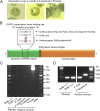
Fig. 1.
Fig. 1.
P. vivax infection in Duffy-null Ethiopians. (A) The blood film contains P. vivax ring-stage parasites in a Duffy-null Ethiopian. (Right) The third panel shows the irregular amoeboid shape of P. vivax in the erythrocyte. (B) GATA1 transcription factor binds to a specific sequence (CTTATCTT) in the upstream promoter region of the Duffy blood group antigen. Binding of GATA1 transcription factor is required for the expression of Duffy antigen Fya or Fyb on human erythrocytes. A similar GATA1-binding site is observed in both Squirrel and Aotus monkeys’ Duffy blood group antigen. The point mutation at the GATA1-binding site from T to C at position −33 leads to loss of GATA1 binding and results in a homozygous Duffy-null blood type. The heterozygous Duffy blood type individual will have this point mutation (T-33C) in one of the alleles whereas the other allele is wild type (T −33) and will have less Duffy blood group expression on the erythrocyte surface. (C) Nested PCR amplification using P. ovale-specific 18S rRNA gene primers. The gel image is representative of two independent experiments. The sample with P. ovale (control MR4-180) shows a band size of ∼700 bp. In the two Duffy-null samples as well as the remaining control samples, no band was observed for P. ovale. (D) P. vivax- and P. falciparum-specific primers were used in combination in PCR. P. vivax and P. falciparum infection corresponds to a band size of 100 and 200 bp, respectively. Both the Duffy-null Ethiopian patients were specifically infected by P. vivax and not by P. falciparum.
Fig. S1.
Fig. S1.
Alignment of Duffy blood group antigen. Multiple sequence alignment of Duffy blood group antigen of Squirrel (NCBI: XM_003937900.2), Aotus (NCBI: XM_012449733.1) monkeys, and humans (NCBI: NP_002027.2) shows that they are the same length with minor differences in sequence. The red block shows the epitope region detected by anti-Fy6. The first tyrosine (Y at the 30th position) is important for chemokine binding, and the second tyrosine at the 41st position is sulfated for DBP1 binding. The amino acid at position 42 determines Fya or Fyb blood group, G at the 42nd position determines human Fya blood group antigen, and D at the 42nd position determines Fyb. Duffy null in Africa has a D in position 42 (Fyb), but is not expressed because of a mutation in the GATA1 transcription factor-binding site (Fig. 1B). Squirrel and Aotus monkeys are the Fyb type. Four unique mutations (UM) are observed in the N terminus of the Squirrel monkey sequence. The blood group has seven transmembrane (TM) domains (underlined).
Fig. 2.
Fig. 2.
Mutations observed in region II of DBP1 from different isolates do not bind Duffy-null erythrocytes. (A) The observed mutations in DBP1 region II in Madagascar (22) and Ethiopia field isolates and in India VII (PVIIG_04680.1) and Brazil I (PVBG_05060.1) strains that were grown in Squirrel monkeys are compared with Sal I (PVX110810). Importantly, Ethiopia Duffy (+) is the P. vivax Duffy-positive sample, and Ethiopia Duffy (−) 1 and 2 are the P. vivax samples from two Duffy-null/negative (−) individuals. The amino acid number for each mutation site is given based on the Salvador I sequence from PlasmoDB. Between position 429 and 430 is an Indel for leucine (L) in the Indian VII and Brazil I sequence. (B) Mutated DBP1 sequences from these isolates were cloned and expressed in COS-7 cells using the pRE4 vector. The binding assay was performed using both Duffy-positive and Duffy-null erythrocytes. The binding assays show that, regardless of mutations in DBP1, the mutants still bind only Duffy-positive and not Duffy-null erythrocytes. EBP/DBP2 (GenBank: KC987954) binds both Duffy-positive and -null erythrocytes at low frequency. The PfRH5 (PlasmoDB ID: PF3D7_0424100) full-length extracellular domain (64–1578 bp), which is cloned in pRE4 vector, does not bind to either Duffy-positive or -null erythrocytes. In addition, no plasmid-binding control is also plotted. The data are plotted as a log scale on the y axis. At least three independent experiments with replicates were performed. The error bars indicate the SD. (C) Microscopy images show the erythrocyte rosette formation in COS-7 expressing DBP1 from Salvador 1, Ethiopia, Madagascar, India VII, Brazil I, and EBP/DBP2 from Cambodia. Note that the rosettes are smaller in EBP/DBP2. Blue (Hoechst) stain the COS-7 cell nuclei. (Scale bar, 10 µM.)
Fig. 3.
Fig. 3.
Copy number expansion in Duffy nulls from Ethiopia. Real-time quantitative PCR plot of relative fluorescent units against the number of threshold cycles (Ct) of P. vivax DBP1 (red) in relation to the single-copy P. vivax aldolase (blue). The green line represents the cutoff where the optimal Ct value of each gene for each sample is determined. The difference in the optimal Ct values (∆Ct; *) between the targeted and reference genes reflects variation in gene copy number among the samples. The tabular material shows the qPCR data for DBP1 gene copy number for all of the isolates from two independent runs (four replicates each). The double asterisk (**) indicates the DBP1 copy number from the whole-genome sequence (WGS) of the two Cambodian samples (24) and the Sal I parasites (25).
Fig. 4.
Fig. 4.
Salvador I P. vivax infection in the Squirrel monkey is independent of DBP1. (A) Salvador I DBP1 expressed in COS-7 cells does not bind Squirrel monkey erythrocytes but binds Aotus monkey erythrocytes. However, DBP1 mutations observed in India VII and Brazil I strains are capable of binding equally to both Squirrel and Aotus monkey erythrocytes. These data indicate that Salvador I infection in the Squirrel monkey is independent of DBP1 and can be a model system to study Duffy-null P. vivax infection that occurs in Africans. The data are plotted as a log scale on the y axis. At least three independent experiments with replicates were performed. The error bars indicate the SD. (B) Fluorescent microscopy images show the erythrocyte rosette formation on COS-7 expressing DBP1 from Salvador I, India VII, and Brazil I using Squirrel and Aotus monkey erythrocytes. Blue (Hoechst) stains the COS-7 cell nuclei. (Scale bar, 10 µM.)

References

    1. Miller LH, Mason SJ, Dvorak JA, McGinniss MH, Rothman IK. Erythrocyte receptors for (Plasmodium knowlesi) malaria: Duffy blood group determinants. Science. 1975;189(4202):561–563. - PubMed
    1. Miller LH, Mason SJ, Clyde DF, McGinniss MH. The resistance factor to Plasmodium vivax in blacks. The Duffy-blood-group genotype, FyFy. N Engl J Med. 1976;295(6):302–304. - PubMed
    1. Miller LH, McGinniss MH, Holland PV, Sigmon P. The Duffy blood group phenotype in American blacks infected with Plasmodium vivax in Vietnam. Am J Trop Med Hyg. 1978;27(6):1069–1072. - PubMed
    1. Spencer HC, et al. The Duffy blood group and resistance to Plasmodium vivax in Honduras. Am J Trop Med Hyg. 1978;27(4):664–670. - PubMed
    1. Tournamille C, Colin Y, Cartron JP, Le Van Kim C. Disruption of a GATA motif in the Duffy gene promoter abolishes erythroid gene expression in Duffy-negative individuals. Nat Genet. 1995;10(2):224–228. - PubMed

Publication types

MeSH terms

Substances